RANKL

Last updated
TNFSF11
Protein TNFSF11 PDB 1s55.png
Available structures
PDB Ortholog search: PDBe RCSB
Identifiers
Aliases TNFSF11 , CD254, ODF, OPGL, OPTB2, RANKL, TRANCE, hRANKL2, sOdf, TNLG6B, tumor necrosis factor superfamily member 11, TNF superfamily member 11
External IDs OMIM: 602642 MGI: 1100089 HomoloGene: 2744 GeneCards: TNFSF11
Orthologs
SpeciesHumanMouse
Entrez
Ensembl
UniProt
RefSeq (mRNA)

NM_003701
NM_033012

NM_011613

RefSeq (protein)

NP_003692
NP_143026

NP_035743

Location (UCSC) Chr 13: 42.56 – 42.61 Mb Chr 14: 78.51 – 78.55 Mb
PubMed search [3] [4]
Wikidata
View/Edit Human View/Edit Mouse

Receptor activator of nuclear factor kappa-Β ligand (RANKL), also known as tumor necrosis factor ligand superfamily member 11 (TNFSF11), TNF-related activation-induced cytokine (TRANCE), osteoprotegerin ligand (OPGL), and osteoclast differentiation factor (ODF), is a protein that in humans is encoded by the TNFSF11 gene. [5] [6]

Contents

RANKL is known as a type II membrane protein and is a member of the tumor necrosis factor (TNF) superfamily. [7] RANKL has been identified to affect the immune system and control bone regeneration and remodeling. RANKL is an apoptosis regulator gene, a binding partner of osteoprotegerin (OPG), a ligand for the receptor RANK and controls cell proliferation by modifying protein levels of Id4, Id2 and cyclin D1. [8] [9] RANKL is expressed in several tissues and organs including: skeletal muscle, thymus, liver, colon, small intestine, adrenal gland, osteoblast, mammary gland epithelial cells, prostate and pancreas. [9] Variation in concentration levels of RANKL throughout several organs reconfirms the importance of RANKL in tissue growth (particularly bone growth) and immune functions within the body.

Tissue expression

The level of RANKL expression does not linearly correlate to the effect of this ligand. High protein expression of RANKL is commonly detected in the lungs, thymus and lymph nodes. Low protein expression is found in bone marrow, the stomach, peripheral blood, the spleen, the placenta, leukocytes, the heart, the thyroid, and skeletal muscle. [9] While bone marrow expresses low levels of RANKL, RANKL plays a critical role for adequate bone metabolism. This surface-bound molecule (also known as CD254), found on osteoblasts, serves to activate osteoclasts, which are critically involved in bone resorption. Osteoclastic activity is triggered via the osteoblasts' surface-bound RANKL activating the osteoclasts' surface-bound receptor activator of nuclear factor kappa-B (RANK). Recent studies suggest that in postnatal bones, the osteocyte is the major source of RANKL regulating bone remodeling. [10] [11] [12] [13] RANKL derived from other cell types contributes to bone loss in conditions involving inflammation such as rheumatoid arthritis, and in lytic lesions caused by cancer, such as in multiple myeloma.

Gene and expression

RANKL can be expressed in three different molecular forms consisting of either a: (1) trimeric transmembrane protein, (2) primary secreted form, and (3) truncated ectodomain. [14] RANKL is identified as a part of the TNF family; RANKL is specifically categorized under the TNFSF11, the TNF ligand superfamily member. RANKL is composed of 314 amino acids and was originally described to have a gene sequence containing 5 exons. [15] [16] Among the exons, Exon 1 encoded the intracellular and transmembrane protein domains and Exon 2-5 encoded the extracellular domains. [15] RANKL’s extracellular domains are similar to other TNF family members in regards to the structural homology and are able to cleave from the cell surface. [15] While the function and significance of A kinase anchor protein 11(AKAP11) is presently unknown, AKAP11 is immediately upstream from RANKL for all species that has a RANKL gene. [16] The upstream of AKAP11 may suggest there is a complex regulator process that regulates the level of RANKL expression.

Function

RANKL is a member of the tumor necrosis factor (TNF) cytokine family. It binds to RANK on cells of the myeloid lineage and functions as a key factor for osteoclast differentiation and activation. RANKL may also bind to osteoprotegerin, a protein secreted mainly by cells of the osteoblast lineage which is a potent inhibitor of osteoclast formation by preventing binding of RANKL to RANK. RANKL also has a function in the immune system, where it is expressed by T helper cells and is thought to be involved in dendritic cell maturation. It is a dendritic cell survival factor and helps regulate T cell-dependent immune responses. T cell activation induces RANKL expression and can lead to an increase of osteoclastogenesis and bone loss. RANKL can also activate the antiapoptotic kinase AKT/PKB through a signaling complex involving SRC kinase and tumor necrosis factor receptor-associated factor 6 (TRAF6), indicating that RANKL may have a role in the regulation of apoptosis. [17] A further role for RANKL in immunity was found in sinusoidal macrophages in lymph nodes that alert the immune system to lymph-borne antigens. [18] In addition to directly signaling through RANK for macrophage differentiation, RANKL activates the adjacent lymphatic endothelial cells to create a niche environment for these specialized immune cells. [18]

Animal models

Targeted disruption of the related gene in mice led to severe osteopetrosis and a lack of osteoclasts. Deficient mice, with an inactivation of RANKL or its receptor RANK, exhibited defects in early differentiation of T and B lymphocytes, and failed to form lobulo-alveolar mammary structures during pregnancy. [9] [17] It was observed that during pregnancy, RANK-RANKL signaling played a critical role in regulating skeletal calcium release; in which contributed to the hormone response that stimulated proliferation in the mammary cells. [9] Ultimately, impaired lobuloalveolar mammary structures resulted in death of the fetus. [9] Those who suffer from osteoporosis often have a cardiovascular defect, such as heart failure. Some studies suggest, since RANK-RANKL pathway regulates calcium release and homeostasis, RANK-RANKL signal could invertedly affect the cardiovascular system; thus, an explanation for the positive correlation between osteoporosis and cardiovascular deficiencies. [9]

Role in cancer

Primary tumors will commonly metastasize into the bone. Breast and prostate cancers typically have a greater chance of inducing secondary cancers within bone. [19] Stephen Paget's seed and soil theory suggests, the microenvironment in bone creates a sufficient ‘soil’ for secondary tumors to grow in. Some studies suggest the expression of RANKL allows sufficient micro environmental conditions to influence cancer cell migration (i.e. chronic lymphocytic leukemia (CLL) and multiple myeloma). [20] Among patients with multiple myeloma, RANKL activity was greatly increased. In fact RANKL surface expression and secreted RANKL expression was reported to be increased, 80% and 50% respectively. [20] Therefore, RANKL is considered to be a key signal regulator for cancer-induced bone loss.

According to the vicious cycle hypothesis, after secondary tumors cells have migrated to bone, the tumor cell will secrete cytokines and growth factors that can act on osteoblast lineage cells. Since osteoblasts control the regulation of RANKL, the stimulation via cytokines and growth factors will then stimulate osteoblasts to increase the expression of RANKL, often while simultaneously reducing bone formation. The additional RANKL-mediated osteoclast frequency and activity will in turn increase secretion of growth factors, or matrix derived factors, which can ultimately increase tumor growth and bone destruction activity.

Clinical significance

RANKL, through its ability to stimulate osteoclast formation and activity, is a critical mediator of bone resorption and overall bone density. Overproduction of RANKL is implicated in a variety of degenerative bone diseases, such as rheumatoid arthritis and psoriatic arthritis. In addition to degenerative bone diseases, bone metastases can also induce pain and other abnormal health complexities that can significantly reduce a cancer patient’s quality of life. Some examples of these complications that are a consequence of bone metastasis are: hypercalcemia, pathological fractures and spinal cord compression. [21] Some findings also suggest that some cancer cells, particularly prostate cancer cells, can activate an increase in bone remodeling and ultimately increase overall bone production. [21] This increase in bone remodeling and bone production increases the overall growth of bone metastasizes. The overall control of bone remodeling is regulated by the binding of RANKL with its receptor or its decoy receptor, respectively, RANK and OPG. [21]

Denosumab

Denosumab is an FDA-approved fully human monoclonal antibody to RANKL and during pre-clinical trials was first used to treat postmenopausal patients suffering with osteoporosis (PMO). [21] [22] In denosumab's third stage of the FDA's clinical trial, it was shown to: (1) decrease bone turnover, (2) reduce fractures in the PMO population, and (3) increase bone mineral density. [21] The anti-RANKL antibody, denosumab, is also approved for use in cancer settings, and in those indications, it is branded as Xgeva. In both prostate and breast cancer, denosumab has been shown to reduce cancer treatment–induced bone loss. [21]

Prostate cancer

The HALT-prostate cancer trial (also known as NCT00089674) included 1468 non-metastatic prostate cancer patients who were currently receiving androgen deprivation therapy. [23] Randomly selected patients were given either 60 mg of denosumab or calcium and vitamin D supplements. This was done to measure the effectiveness of preventing treatment-induced bone loss. [21] The patients who received 60 mg of denosumab showed a +5.6% increased in bone mineral density and a 1.5% decrease in bone fracture rates. [21]

Another clinical trial (NCT00321620) was established to determine the safety and effectiveness of using denosumab compared to zoledronic acid. [24] In this trial, they used 1901 bone metastatic prostate patients whom were also suffering with other complication of bone diseases. Again, patients were randomized and some were given either 120 mg of denosumab or 4 mg of zoledronic acid. [21] Patients who were given 120 mg of denosumab (in comparison to those who were given 4 mg of zoledronic acid) showed a greater increase in hypocalcemia, a greater resistance to bone turnover markers uNTx, a delay response in both pathological fractures and spinal cord compression. [21] However, survival rates for both clinical groups were comparable. [21]

Breast cancer

Hormone receptor positive breast cancer patients have a significant increased risk of complications such as osteopenia and osteoporosis. About two out of every three breast cancer patients are hormone receptor positive. [25] In the past several years, denosumab has been used in clinical trials, primarily because a large population is affected by bone complication among those who have breast cancer.

252 patients enlisted in the HALT-BC clinical trial (also known as NCT00089661). In addition to receiving vitamin D and calcium supplements, half of the patients were randomly given 60 mg of denosumab while the other half were given a placebo. [21] [26] Patients given denosumab had an increase in lumbar spine bone mineral density, a decrease in bone turnover markers, with no significant change in survival rates. [21]

NCT00321464 was another phase III RCT. [27] Similar to NCT00321620 (prostate), this trial measured the safety and efficacy of denosumab versus zoledronic acid. Both groups showed similar survival rates and adverse event frequency. [21]

Multiple myeloma

Patients whom are diagnosed with multiple myeloma have approximately 80-100% chance of developing bone complications due to an increase in activity and/or formation of osteoclasts and a decrease activity of osteoblasts. [20] [21] In a stage II clinical trial, denosumab decreased bone turnover markers by blocking the RANKL/RANK pathway. [21] Once this trial was completed, 1176 patients with either multiple myeloma or progressed cancers were entered into the stage III clinical trial (known as NCT00330759). [28] The main objective of the NCT00330759 trial was to compare effects of patients who were given 120 mg of denosumab relative to patients give 4 mg of zoledronic acid. As a result of this trial, during a month period, patients who received denosumab had a decrease in pathological fractures and spinal cord compression; however, as time progressed it appear that denosumab had significantly delayed bone complications. [21] In both breast and prostate cancers, patients in either denosumab or zoledronic acid groups both appeared to have comparable adverse events and survival rates. [21]

Medroxyprogesterone acetate

Women with menopause have often been given various types of postmenopausal hormone therapies to prevent osteoporosis and reduce menopausal symptoms. [29] Medroxyprogesterone acetate (MPA) is a synthetic progestin and was commonly used as a contraceptive or used as a hormone therapy for endometriosis or osteoporosis. Recent studies suggest, using MPA increases patient risks of developing breast cancer due to an increase expression of RANKL. [29] MPA causes a substantial induction of RANKL in mammary-gland epithelial cells while deletion of RANKL decreases the incidence MPA-induced breast cancer. Hence inhibition of RANKL has potential for the prevention and treatment of breast cancer. [30] [31]

See also

Related Research Articles

Hypercalcemia, also spelled hypercalcaemia, is a high calcium (Ca2+) level in the blood serum. The normal range is 2.1–2.6 mmol/L (8.8–10.7 mg/dL, 4.3–5.2 mEq/L), with levels greater than 2.6 mmol/L defined as hypercalcemia. Those with a mild increase that has developed slowly typically have no symptoms. In those with greater levels or rapid onset, symptoms may include abdominal pain, bone pain, confusion, depression, weakness, kidney stones or an abnormal heart rhythm including cardiac arrest.

Autocrine signaling is a form of cell signaling in which a cell secretes a hormone or chemical messenger that binds to autocrine receptors on that same cell, leading to changes in the cell. This can be contrasted with paracrine signaling, intracrine signaling, or classical endocrine signaling.

<span class="mw-page-title-main">Osteoclast</span> Cell that breaks down bone tissue

An osteoclast is a type of bone cell that breaks down bone tissue. This function is critical in the maintenance, repair, and remodeling of bones of the vertebral skeleton. The osteoclast disassembles and digests the composite of hydrated protein and mineral at a molecular level by secreting acid and a collagenase, a process known as bone resorption. This process also helps regulate the level of blood calcium.

<span class="mw-page-title-main">Osteoprotegerin</span> Mammalian protein found in Homo sapiens

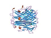
Osteoprotegerin (OPG), also known as osteoclastogenesis inhibitory factor (OCIF) or tumour necrosis factor receptor superfamily member 11B (TNFRSF11B), is a cytokine receptor of the tumour necrosis factor (TNF) receptor superfamily encoded by the TNFRSF11B gene.

<span class="mw-page-title-main">Amphiregulin</span> Protein-coding gene in the species Homo sapiens

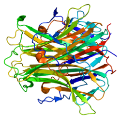
Amphiregulin, also known as AREG, is a protein synthesized as a transmembrane glycoprotein with 252 aminoacids and it is encoded by the AREG gene. in humans.

<span class="mw-page-title-main">LNCaP</span>

LNCaP cells are a cell line of human cells commonly used in the field of oncology. LNCaP cells are androgen-sensitive human prostate adenocarcinoma cells derived from the left supraclavicular lymph node metastasis from a 50-year-old caucasian male in 1977. They are adherent epithelial cells growing in aggregates and as single cells.

<span class="mw-page-title-main">Giant-cell tumor of bone</span> Medical condition

Giant-cell tumor of the bone (GCTOB), is a relatively uncommon tumor of the bone. It is characterized by the presence of multinucleated giant cells. Malignancy in giant-cell tumor is uncommon and occurs in about 2% of all cases. However, if malignant degeneration does occur, it is likely to metastasize to the lungs. Giant-cell tumors are normally benign, with unpredictable behavior. It is a heterogeneous tumor composed of three different cell populations. The giant-cell tumour stromal cells (GCTSC) constitute the neoplastic cells, which are from an osteoblastic origin and are classified based on expression of osteoblast cell markers such as alkaline phosphatase and osteocalcin. In contrast, the mononuclear histiocytic cells (MNHC) and multinucleated giant cell (MNGC) fractions are secondarily recruited and comprise the non-neoplastic cell population. They are derived from an osteoclast-monocyte lineage determined primarily by expression of CD68, a marker for monocytic precursor cells. In most patients, the tumors are slow to develop, but may recur locally in as many as 50% of cases.

<span class="mw-page-title-main">RANK</span> Mammalian protein found in Homo sapiens

Receptor activator of nuclear factor κ B (RANK), also known as TRANCE receptor or TNFRSF11A, is a member of the tumor necrosis factor receptor (TNFR) molecular sub-family. RANK is the receptor for RANK-Ligand (RANKL) and part of the RANK/RANKL/OPG signaling pathway that regulates osteoclast differentiation and activation. It is associated with bone remodeling and repair, immune cell function, lymph node development, thermal regulation, and mammary gland development. Osteoprotegerin (OPG) is a decoy receptor for RANKL, and regulates the stimulation of the RANK signaling pathway by competing for RANKL. The cytoplasmic domain of RANK binds TRAFs 1, 2, 3, 5, and 6 which transmit signals to downstream targets such as NF-κB and JNK.

<span class="mw-page-title-main">E-selectin</span> Mammalian protein found in Homo sapiens

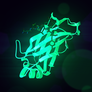
E-selectin, also known as CD62 antigen-like family member E (CD62E), endothelial-leukocyte adhesion molecule 1 (ELAM-1), or leukocyte-endothelial cell adhesion molecule 2 (LECAM2), is a selectin cell adhesion molecule expressed only on endothelial cells activated by cytokines. Like other selectins, it plays an important part in inflammation. In humans, E-selectin is encoded by the SELE gene.

<span class="mw-page-title-main">Macrophage colony-stimulating factor</span> Mammalian protein found in humans

The colony stimulating factor 1 (CSF1), also known as macrophage colony-stimulating factor (M-CSF), is a secreted cytokine which causes hematopoietic stem cells to differentiate into macrophages or other related cell types. Eukaryotic cells also produce M-CSF in order to combat intercellular viral infection. It is one of the three experimentally described colony-stimulating factors. M-CSF binds to the colony stimulating factor 1 receptor. It may also be involved in development of the placenta.

<span class="mw-page-title-main">Denosumab</span> Human monoclonal antibody

Denosumab, sold under the brand names Prolia and Xgeva among others, is a human monoclonal antibody used for the treatment of osteoporosis, treatment-induced bone loss, metastases to bone, and giant cell tumor of bone.

<span class="mw-page-title-main">Oncostatin M receptor</span> Protein-coding gene in the species Homo sapiens

Oncostatin-M specific receptor subunit beta also known as the Oncostatin M receptor (OSMR), is one of the receptor proteins for oncostatin M, that in humans is encoded by the OSMR gene.

Osteoimmunology is a field that emerged about 40 years ago that studies the interface between the skeletal system and the immune system, comprising the "osteo-immune system". Osteoimmunology also studies the shared components and mechanisms between the two systems in vertebrates, including ligands, receptors, signaling molecules and transcription factors. Over the past decade, osteoimmunology has been investigated clinically for the treatment of bone metastases, rheumatoid arthritis (RA), osteoporosis, osteopetrosis, and periodontitis. Studies in osteoimmunology reveal relationships between molecular communication among blood cells and structural pathologies in the body.

<span class="mw-page-title-main">STAT5A</span> Protein-coding gene in the species Homo sapiens

Signal transducer and activator of transcription 5A is a protein that in humans is encoded by the STAT5A gene. STAT5A orthologs have been identified in several placentals for which complete genome data are available.

<span class="mw-page-title-main">Colony stimulating factor 1 receptor</span> Protein found in humans

Colony stimulating factor 1 receptor (CSF1R), also known as macrophage colony-stimulating factor receptor (M-CSFR), and CD115, is a cell-surface protein encoded by the human CSF1R gene. CSF1R is a receptor that can be activated by two ligands: colony stimulating factor 1 (CSF-1) and interleukin-34 (IL-34). CSF1R is highly expressed in myeloid cells, and CSF1R signaling is necessary for the survival, proliferation, and differentiation of many myeloid cell types in vivo and in vitro. CSF1R signaling is involved in many diseases and is targeted in therapies for cancer, neurodegeneration, and inflammatory bone diseases.

Cixutumumab (IMC-A12) is a human monoclonal antibody for the treatment of solid tumors.

<span class="mw-page-title-main">Tooth resorption</span> Medical condition

Resorption of the root of the tooth, or root resorption, is the progressive loss of dentin and cementum by the action of odontoclasts. Root resorption is a normal physiological process that occurs in the exfoliation of the primary dentition. However, pathological root resorption occurs in the permanent or secondary dentition and sometimes in the primary dentition.

<span class="mw-page-title-main">Bone metastasis</span> Medical condition

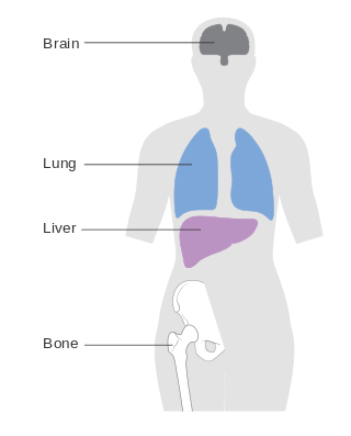
Bone metastasis, or osseous metastatic disease, is a category of cancer metastases that result from primary tumor invasions into bones. Bone-originating primary tumors such as osteosarcoma, chondrosarcoma, and Ewing sarcoma are rare; the most common bone tumor is a metastasis. Bone metastases can be classified as osteolytic, osteoblastic, or both. Unlike hematologic malignancies which originate in the blood and form non-solid tumors, bone metastases generally arise from epithelial tumors and form a solid mass inside the bone. Bone metastases, especially in a state of advanced disease, can cause severe pain, characterized by a dull, constant ache with periodic spikes of incident pain.

<span class="mw-page-title-main">Metastatic breast cancer</span> Type of cancer

Metastatic breast cancer, also referred to as metastases, advanced breast cancer, secondary tumors, secondaries or stage IV breast cancer, is a stage of breast cancer where the breast cancer cells have spread to distant sites beyond the axillary lymph nodes. There is no cure for metastatic breast cancer; there is no stage after IV.

<span class="mw-page-title-main">Osteolytic lesion</span>

An osteolytic lesion is a softened section of a patient's bone formed as a symptom of specific diseases, including breast cancer and multiple myeloma. This softened area appears as a hole on X-ray scans due to decreased bone density, although many other diseases are associated with this symptom. Osteolytic lesions can cause pain, increased risk of bone fracture, and spinal cord compression. These lesions can be treated using biophosphonates or radiation, though new solutions are being tested in clinical trials.

References

  1. 1 2 3 GRCh38: Ensembl release 89: ENSG00000120659 - Ensembl, May 2017
  2. 1 2 3 GRCm38: Ensembl release 89: ENSMUSG00000022015 - Ensembl, May 2017
  3. "Human PubMed Reference:". National Center for Biotechnology Information, U.S. National Library of Medicine.
  4. "Mouse PubMed Reference:". National Center for Biotechnology Information, U.S. National Library of Medicine.
  5. Wong BR, Rho J, Arron J, Robinson E, Orlinick J, Chao M, Kalachikov S, Cayani E, Bartlett FS, Frankel WN, Lee SY, Choi Y (October 1997). "TRANCE is a novel ligand of the tumor necrosis factor receptor family that activates c-Jun N-terminal kinase in T cells". J. Biol. Chem. 272 (40): 25190–4. doi: 10.1074/jbc.272.40.25190 . PMID   9312132.
  6. Anderson DM, Maraskovsky E, Billingsley WL, Dougall WC, Tometsko ME, Roux ER, Teepe MC, DuBose RF, Cosman D, Galibert L (November 1997). "A homologue of the TNF receptor and its ligand enhance T-cell growth and dendritic-cell function". Nature. 390 (6656): 175–9. Bibcode:1997Natur.390..175A. doi:10.1038/36593. PMID   9367155. S2CID   4373990.
  7. Hanada R, Hanada T, Sigl V, Schramek D, Penninger JM (2011). "RANKL/RANK-beyond bones". J. Mol. Med. 89 (7): 647–56. doi:10.1007/s00109-011-0749-z. PMID   21445556. S2CID   25285776.
  8. Mueller CG, Hess E (2012). "Emerging Functions of RANKL in Lymphoid Tissues". Front Immunol. 3: 261. doi: 10.3389/fimmu.2012.00261 . PMC   3432452 . PMID   22969763.
  9. 1 2 3 4 5 6 7 Wada T, Nakashima T, Hiroshi N, Penninger JM (2006). "RANKL-RANK signaling in osteoclastogenesis and bone disease". Trends Mol Med. 12 (1): 17–25. doi:10.1016/j.molmed.2005.11.007. PMID   16356770.
  10. Robling AG, Bonewald LF (10 February 2020). "The Osteocyte: New Insights". Annual Review of Physiology. 82 (1): 485–506. doi:10.1146/annurev-physiol-021119-034332. hdl:1805/30982. ISSN   0066-4278. PMC   8274561 . PMID   32040934 . Retrieved 8 March 2022.
  11. Xiong J, Onal M, Jilka RL, Weinstein RS, Manolagas SC, O'Brien CA (11 September 2011). "Matrix-embedded cells control osteoclast formation". Nature Medicine. 17 (10): 1235–1241. doi:10.1038/nm.2448. ISSN   1546-170X. PMC   3192296 . PMID   21909103.
  12. Xiong J, Piemontese M, Onal M, Campbell J, Goellner JJ, Dusevich V, Bonewald L, Manolagas SC, O'Brien CA (1 January 2015). "Osteocytes, not Osteoblasts or Lining Cells, are the Main Source of the RANKL Required for Osteoclast Formation in Remodeling Bone". PLOS ONE. 10 (9): e0138189. Bibcode:2015PLoSO..1038189X. doi: 10.1371/journal.pone.0138189 . ISSN   1932-6203. PMC   4578942 . PMID   26393791.
  13. Nakashima T, Hayashi M, Fukunaga T, Kurata K, Oh-Hora M, Feng JQ, Bonewald LF, Kodama T, Wutz A (11 September 2011). "Evidence for osteocyte regulation of bone homeostasis through RANKL expression". Nature Medicine. 17 (10): 1231–1234. doi:10.1038/nm.2452. ISSN   1546-170X. PMID   21909105. S2CID   21188945.
  14. Findlay DM, Atkins GJ (2011). "Relationship between serum RANKL and RANKL in bone". Osteoporos Int. 22 (10): 2597–602. doi:10.1007/s00198-011-1740-9. PMID   21850548. S2CID   908214.
  15. 1 2 3 Walsh NC, Alexander KA, Manning CA, Karmakar S, Karmakar SK, Wang JF, Weyand CM, Pettit AR, Gravallese EM (2013). "Activated human T cells express alternative mRNA transcripts encoding a secreted form of RANKL". Genes Immun. 14 (5): 336–45. doi:10.1038/gene.2013.29. PMC   3740552 . PMID   23698708.
  16. 1 2 O'Brien CA (2010). "Control of RANKL gene expression". Bone. 46 (4): 911–9. doi:10.1016/j.bone.2009.08.050. PMC   2842447 . PMID   19716455.
  17. 1 2 "Entrez Gene: TNFSF11 tumor necrosis factor (ligand) superfamily, member 11".
  18. 1 2 Camara A, Cordeiro OG, Alloush F, Sponsel J, Chypre M, Onder L, Asano K, Tanaka M, Yagita H, Ludewig B, Flacher V (18 June 2019). "Lymph Node Mesenchymal and Endothelial Stromal Cells Cooperate via the RANK-RANKL Cytokine Axis to Shape the Sinusoidal Macrophage Niche". Immunity. 50 (6): 1467–1481.e6. doi: 10.1016/j.immuni.2019.05.008 . ISSN   1074-7613. PMID   31201093.
  19. Coleman RE (2001). "Metastatic bone disease: clinical features, pathophysiology and treatment strategies". Cancer Treat. Rev. 27 (3): 165–76. doi:10.1053/ctrv.2000.0210. PMID   11417967.
  20. 1 2 3 Schmiedel BJ, Scheible CA, Nuebling T, Kopp HG, Wirths S, Azuma M, Schneider P, Jung G, Grosse-Hovest L, Salih HR (2013). "RANKL expression, function, and therapeutic targeting in multiple myeloma and chronic lymphocytic leukemia". Cancer Res. 73 (2): 683–94. doi: 10.1158/0008-5472.CAN-12-2280 . PMID   23139212.
  21. 1 2 3 4 5 6 7 8 9 10 11 12 13 14 15 16 17 18 Castellano D, Sepulveda JM, García-Escobar I, Rodriguez-Antolín A, Sundlöv A, Cortes-Funes H (2011). "The role of RANK-ligand inhibition in cancer: the story of denosumab". Oncologist. 16 (2): 136–45. doi:10.1634/theoncologist.2010-0154. PMC   3228090 . PMID   21285392.
  22. Miller PD (2009). "Denosumab: anti-RANKL antibody". Curr Osteoporos Rep. 7 (1): 18–22. doi:10.1007/s11914-009-0004-5. PMID   19239825. S2CID   10465712.
  23. "AMG 162 in the Treatment of Bone Loss in Subjects Undergoing Androgen-Deprivation Therapy for Non-metastatic Prostate Cancer". Nct00089674. ClinicalTrials.gov. 20 September 2018.
  24. "Study of Denosumab vs. Zoledronic Acid to Treat Bone Metastases in Men With Hormone-refractory Prostate Cancer". Nct00321620. ClinicalTrials.gov. August 2018.
  25. "Hormone therapy for breast cancer". Cancer.org. Archived from the original on 2 December 2016. Retrieved 31 January 2014.
  26. "AMG 162 in the Treatment of Bone Loss in Subjects Undergoing Aromatase Inhibitor Therapy for Non-metastatic Breast Cancer". Nct00089661. ClinicalTrials.gov. 20 September 2018.
  27. "A Study Comparing Denosumab vs. Zoledronic Acid for the Treatment of Bone Metastases in Breast Cancer Subjects". Nct00321464. ClinicalTrials.gov. 20 September 2018.
  28. "Study of Denosumab vs. Zoledronic Acid to Treat Bone Metastases in Subjects With Advanced Cancer or Multiple Myeloma". Nct00330759. ClinicalTrials.gov. August 2018.
  29. 1 2 Wood CE, Branstetter D, Jacob AP, Cline JM, Register TC, Rohrbach K, Huang LY, Borgerink H, Dougall WC (2013). "Progestin effects on cell proliferation pathways in the postmenopausal mammary gland". Breast Cancer Res. 15 (4): R62. doi: 10.1186/bcr3456 . PMC   3978455 . PMID   23938070.
  30. Schramek D, Leibbrandt A, Sigl V, Kenner L, Pospisilik JA, Lee HJ, Hanada R, Joshi PA, Aliprantis A, Glimcher L, Pasparakis M, Khokha R, Ormandy CJ, Widschwendter M, Schett G, Penninger JM (November 2010). "Osteoclast differentiation factor RANKL controls development of progestin-driven mammary cancer". Nature. 468 (7320): 98–102. Bibcode:2010Natur.468...98S. doi:10.1038/nature09387. PMC   3084017 . PMID   20881962.
  31. Gonzalez-Suarez E, Jacob AP, Jones J, Miller R, Roudier-Meyer MP, Erwert R, Pinkas J, Branstetter D, Dougall WC (November 2010). "RANK ligand mediates progestin-induced mammary epithelial proliferation and carcinogenesis". Nature. 468 (7320): 103–7. Bibcode:2010Natur.468..103G. doi:10.1038/nature09495. PMID   20881963. S2CID   4322105.

Further reading

This article incorporates text from the United States National Library of Medicine, which is in the public domain.