Bacterial adhesin

Last updated

Adhesins are cell-surface components or appendages of bacteria that facilitate adhesion or adherence to other cells or to surfaces, usually in the host they are infecting or living in. Adhesins are a type of virulence factor.

Contents

Adherence is an essential step in bacterial pathogenesis or infection, required for colonizing a new host. [1] Adhesion and bacterial adhesins are also a potential target either for prophylaxis or for the treatment of bacterial infections. [2]

Background

Bacteria are typically found attached to and living in close association with surfaces. During the bacterial lifespan, a bacterium is subjected to frequent shear-forces. In the crudest sense, bacterial adhesins serve as anchors allowing bacteria to overcome these environmental shear forces, thus remaining in their desired environment. However, bacterial adhesins do not serve as a sort of universal bacterial Velcro. Rather, they act as specific surface recognition molecules, allowing the targeting of a particular bacterium to a particular surface such as root tissue in plants, lacrimal duct tissues in mammals, or even tooth enamel. [3]

FimH is a bacterial adhesin that helps bacteria such as Escherichia coli to bind to host cells and their receptors (here: the human proteins CD48 and TLR4, or mannose residues). Bacterial Adhesin FimH - host interaction.png
FimH is a bacterial adhesin that helps bacteria such as Escherichia coli to bind to host cells and their receptors (here: the human proteins CD48 and TLR4, or mannose residues).

Most fimbria of gram-negative bacteria function as adhesins, but in many cases it is a minor subunit protein at the tip of the fimbriae that is the actual adhesin. In gram-positive bacteria, a protein or polysaccharide surface layer serves as the specific adhesin. To effectively achieve adherence to host surfaces, many bacteria produce multiple adherence factors called adhesins.

Bacterial adhesins provide species and tissue tropism. Adhesins are expressed by both pathogenic bacteria and saprophytic bacteria. This prevalence marks them as key microbial virulence factors in addition to a bacterium's ability to produce toxins and resist the immune defenses of the host.

Structures

Through the mechanisms of evolution, different species of bacteria have developed different solutions to the problem of attaching receptor specific proteins to the bacteria surface. Today many different types and subclasses of bacterial adhesins may be observed in the literature.

The typical structure of a bacterial adhesion is that of a fimbria or pilus. [3] The bacterial adhesion consists primarily of an intramembranous structural protein which provides a scaffold upon which several extracellular adhesins may be attached. [3] However, as in the case of the CFA1 fimbriae, the structural protein itself can sometimes act as an adhesion if a portion of the protein extends into the ECM.

FimH adhesin—structure

The best characterized bacterial adhesin is the type 1 fimbrial FimH adhesin. This adhesin is responsible for D-mannose sensitive adhesion. [3] The bacterium synthesizes a precursor protein consisting of 300 amino acids then processes the protein by removing several signal peptides ultimately leaving a 279 amino acid protein. [3] Mature FimH is displayed on the bacterial surface as a component of the type 1 fimbrial organelle. [3]

In 1999, the structure of FimH was resolved via x-ray crystallography. FimH is folded into two domains. The N terminal adhesive domain plays the main role in surface recognition while the C-terminal domain is responsible for organelle integration. [5] A tetra-peptide loop links the two domains. Additionally, a carbohydrate-binding pocket has been identified at the tip of the N-terminal adhesive domain. [5] This basic structure is conserved across type 1 fimbrial adhesins though recent studies have shown that in vitro induced mutations can lead to the addition of C-terminal domain specificity resulting in a bacterial adhesion with dual bending sites and related binding phenotypes. [6]

As virulence factors

The majority of bacterial pathogens exploit specific adhesion to host cells as their main virulence factor. "A large number of bacterial adhesins with individual receptor specificities have been identified." [3] Many bacterial pathogens are able to express an array of different adhesins. Expression of these adhesins at different phases during infection play the most important role in adhesion based virulence. [3] Numerous studies have shown that inhibiting a single adhesin in this coordinated effort can often be enough to make a pathogenic bacterium non-virulent. This has led to the exploration of adhesin activity interruption as a method of bacterial infection treatment.

Vaccines based on adhesins

The study of adhesins as a point of exploitation for vaccines comes from early studies which indicated that an important component of protective immunity against certain bacteria came from an ability to prevent adhesin binding. [7] Additionally, Adhesins are attractive vaccine candidates because they are often essential to infection and are surface-located, making them readily accessible to antibodies.

The effectiveness of anti-adhesin antibodies is illustrated by studies with FimH, the adhesin of uropathogenic Escherichia coli (UPEC). Work with E. coli stems from observations of human acquired immunity. Children in third world countries may suffer from several episodes of E. coli associated diarrhea during the first three years of life. If the child survives this initial period of susceptibility, infection rates typically drop substantially. Field studies show that this acquired immunity is directed primarily against bacterial adhesins. [3]

Recent studies from Worcester Polytechnic Institute show that the consumption of cranberry juice may inhibit the action of UPEC adhesins. Using atomic force microscopy researchers have shown that adhesion forces decrease with time following cranberry juice consumption. [8] This research has opened the door to further exploration of orally administered vaccines which exploit bacterial adhesins.

A number of problems create challenges for the researcher exploring the anti-adhesin immunity concept. First, a large number of different bacterial adhesins target the same human tissues. Further, an individual bacterium can produce multiple different types of adhesin, at different times, in different places, and in response to different environmental triggers. [3] Finally, many adhesins present as different immunologically distinct antigenic varieties, even within the same clone (as is the case in Neisseria gonorrhoeae ). [9]

Despite these challenges, progress is being made in the creation of anti-adhesion vaccines. In animal models, passive immunization with anti FimH-antibodies and vaccination with the protein significantly reduced colonization by UPEC. [10] Moreover, the Bordetella pertussis adhesins FHA and pertactin are components of three of the four acellular pertussis vaccines currently licensed for use in the U.S. Additionally, anti-adhesion vaccines are being explored as a solution to urinary tract infection (UTIs). The use of synthetic FimH adhesion peptides was shown to prevent urogenital mucosal infection by E. coli in mice. [11]

Specific examples

Dr family

Adhesin_Dr
PDB 1ut1 EBI.jpg
drae adhesin from escherichia coli
Identifiers
SymbolAdhesin_Dr
Pfam PF04619
Pfam clan CL0204
InterPro IPR006713
Available protein structures:
Pfam   structures / ECOD  
PDB RCSB PDB; PDBe; PDBj
PDBsum structure summary

The Dr family of adhesins bind to the Dr blood group antigen component of decay-accelerating factor (DAF). [12] These proteins contain both fimbriated and afimbriated adherence structures and mediate adherence of uropathogenic Escherichia coli to the urinary tract. [13] They do so by inducing the development of long cellular extensions that wrap around the bacteria. [12] They also confer the mannose-resistant hemaglutination phenotype, which can be inhibited by chloramphenicol. The N-terminal portion of the mature protein is thought to be responsible for chloramphenicol sensitivity. [14] Also, they induce activation of several signal transduction cascades, including activation of PI-3 kinase. [12]

The Dr family of adhesins are particularly associated with cystitis and pregnancy-associated pyelonephritis. [12]

Multivalent Adhesion Molecules

Multivalent Adhesion Molecules (MAMs) are a widespread family of adhesins found in Gram negative bacteria, including E. coli, Vibrio, Yersinia, and Pseudomonas aeruginosa. [15] MAMs contain tandem repeats of mammalian cell entry (MCE) domains which specifically bind to extracellular matrix proteins and anionic lipids on host tissues. Since they are abundant in many pathogens of clinical importance, Multivalent Adhesion Molecules are a potential target for prophylactic or therapeutic anti-infectives. The use of a MAM targeting adhesion inhibitor was shown to significantly decrease the colonization of burn wounds by multidrug resistant Pseudomonas aeruginosa in rats. [16]

N. gonorroheae

N. gonorrhoeae is host restricted almost entirely to humans. [3] "Extensive studies have established type 4 fimbrial adhesins of N. gonorrhoeae virulence factors." [3] These studies have shown that only strains capable of expressing fimbriae are pathogenic. High survival of polymorphonuclear neutrophils (PMNs) characterizes Neisseria gonorrhoeae infections. Additionally, recent studies out of Stockholm have shown that Neisseria can hitchhike on PMNs using their adhesin pili thus hiding them from neutrophil phagocytic activity. This action facilitates the spread of the pathogen throughout the epithelial cell layer. [17]

E. coli

Escherichia coli strains most known for causing diarrhea can be found in the intestinal tissue of pigs and humans where they express the K88 and CFA1. [18] to attach to the intestinal lining. Additionally, UPEC causes about 90% of urinary tract infections. [19] Of those E. coli which cause UTIs, 95% express type 1 fimbriae. FimH in E. coli overcomes the antibody based immune response by natural conversion from the high to the low affinity state. Through this conversion, FimH adhesion may shed the antibodies bound to it. Escherichia coli FimH provides an example of conformation specific immune response which enhances impact on the protein. [19] By studying this particular adhesion, researchers hope to develop adhesion-specific vaccines which may serve as a model for antibody-mediation of pathogen adhesion. [19]

See also

Related Research Articles

<span class="mw-page-title-main">Pilus</span> A proteinaceous hair-like appendage on the surface of bacteria

A pilus is a hair-like appendage found on the surface of many bacteria and archaea. The terms pilus and fimbria can be used interchangeably, although some researchers reserve the term pilus for the appendage required for bacterial conjugation. All conjugative pili are primarily composed of pilin – fibrous proteins, which are oligomeric.

<i>Neisseria gonorrhoeae</i> Species of bacterium

Neisseria gonorrhoeae, also known as gonococcus (singular), or gonococci (plural), is a species of Gram-negative diplococci bacteria isolated by Albert Neisser in 1879. It causes the sexually transmitted genitourinary infection gonorrhea as well as other forms of gonococcal disease including disseminated gonococcemia, septic arthritis, and gonococcal ophthalmia neonatorum.

<i>Neisseria</i> Genus of bacteria

Neisseria is a large genus of bacteria that colonize the mucosal surfaces of many animals. Of the 11 species that colonize humans, only two are pathogens, N. meningitidis and N. gonorrhoeae.

Virulence is a pathogen's or microorganism's ability to cause damage to a host.

<span class="mw-page-title-main">Lipopolysaccharide</span> Class of molecules found in the outer membrane of Gram-negative bacteria

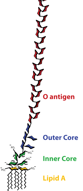
Lipopolysaccharides (LPS) are large molecules consisting of a lipid and a polysaccharide that are bacterial toxins. They are composed of an O-antigen, an outer core, and an inner core all joined by covalent bonds, and are found in the outer membrane of Gram-negative bacteria, such as E. coli and Salmonella. Today, the term endotoxin is often used synonymously with LPS, although there are a few endotoxins that are not related to LPS, such as the so-called delta endotoxin proteins produced by Bacillus thuringiensis.

Mycoplasma pneumoniae is a very small bacterium in the class Mollicutes. It is a human pathogen that causes the disease mycoplasma pneumonia, a form of atypical bacterial pneumonia related to cold agglutinin disease. M. pneumoniae is characterized by the absence of a peptidoglycan cell wall and resulting resistance to many antibacterial agents. The persistence of M. pneumoniae infections even after treatment is associated with its ability to mimic host cell surface composition.

Pathogenicity islands (PAIs), as termed in 1990, are a distinct class of genomic islands acquired by microorganisms through horizontal gene transfer. Pathogenicity islands are found in both animal and plant pathogens. Additionally, PAIs are found in both gram-positive and gram-negative bacteria. They are transferred through horizontal gene transfer events such as transfer by a plasmid, phage, or conjugative transposon. Therefore, PAIs contribute to microorganisms' ability to evolve.

<i>Neisseria meningitidis</i> Species of bacterium that can cause meningitis

Neisseria meningitidis, often referred to as the meningococcus, is a Gram-negative bacterium that can cause meningitis and other forms of meningococcal disease such as meningococcemia, a life-threatening sepsis. The bacterium is referred to as a coccus because it is round, and more specifically a diplococcus because of its tendency to form pairs.

Virulence factors are cellular structures, molecules and regulatory systems that enable microbial pathogens to achieve the following:

Porphyromonas gingivalis belongs to the phylum Bacteroidota and is a nonmotile, Gram-negative, rod-shaped, anaerobic, pathogenic bacterium. It forms black colonies on blood agar.

<span class="mw-page-title-main">Autotransporter family</span>

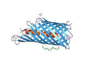
In molecular biology, an autotransporter domain is a structural domain found in some bacterial outer membrane proteins. The domain is always located at the C-terminal end of the protein and forms a beta-barrel structure. The barrel is oriented in the membrane such that the N-terminal portion of the protein, termed the passenger domain, is presented on the cell surface. These proteins are typically virulence factors, associated with infection or virulence in pathogenic bacteria.

In biology, phase variation is a method for dealing with rapidly varying environments without requiring random mutation. It involves the variation of protein expression, frequently in an on-off fashion, within different parts of a bacterial population. As such the phenotype can switch at frequencies that are much higher than classical mutation rates. Phase variation contributes to virulence by generating heterogeneity. Although it has been most commonly studied in the context of immune evasion, it is observed in many other areas as well and is employed by various types of bacteria, including Salmonella species.

Pathogenic <i>Escherichia coli</i> Strains of E. coli that can cause disease

Escherichia coli is a gram-negative, rod-shaped bacterium that is commonly found in the lower intestine of warm-blooded organisms (endotherms). Most E. coli strains are harmless, but pathogenic varieties cause serious food poisoning, septic shock, meningitis, or urinary tract infections in humans. Unlike normal flora E. coli, the pathogenic varieties produce toxins and other virulence factors that enable them to reside in parts of the body normally not inhabited by E. coli, and to damage host cells. These pathogenic traits are encoded by virulence genes carried only by the pathogens.

<span class="mw-page-title-main">Trimeric autotransporter adhesin</span> Proteins found on the outer membrane of Gram-negative bacteria

In molecular biology, trimeric autotransporter adhesins (TAAs), are proteins found on the outer membrane of Gram-negative bacteria. Bacteria use TAAs in order to infect their host cells via a process called cell adhesion. TAAs also go by another name, oligomeric coiled-coil adhesins, which is shortened to OCAs. In essence, they are virulence factors, factors that make the bacteria harmful and infective to the host organism.

<span class="mw-page-title-main">YadA bacterial adhesin protein domain</span>

In molecular biology, YadA is a protein domain which is short for Yersinia adhesin A. These proteins have strong sequence and structural homology, particularly at their C-terminal end. The function is to promote their pathogenicity and virulence in host cells, though cell adhesion. YadA is found in three pathogenic species of Yersinia, Y. pestis,Y. pseudotuberculosis, and Y. enterocolitica. The YadA domain is encoded for by a virulence plasmid in Yersinia, which encodes a type-III secretion (T3S) system consisting of the Ysc injectisome and the Yop effectors.

Enteroaggregative Escherichia coli are a pathotype of Escherichia coli which cause acute and chronic diarrhea in both the developed and developing world. They may also cause urinary tract infections. EAEC are defined by their "stacked-brick" pattern of adhesion to the human laryngeal epithelial cell line HEp-2. The pathogenesis of EAEC involves the aggregation of and adherence of the bacteria to the intestinal mucosa, where they elaborate enterotoxins and cytotoxins that damage host cells and induce inflammation that results in diarrhea.

Chaperone-usher fimbriae (CU) are linear, unbranching, outer-membrane pili secreted by gram-negative bacteria through the chaperone-usher system rather than through type IV secretion or extracellular nucleation systems. These fimbriae are built up out of modular pilus subunits, which are transported into the periplasm in a Sec dependent manner. Chaperone-usher secreted fimbriae are important pathogenicity factors facilitating host colonisation, localisation and biofilm formation in clinically important species such as uropathogenic Escherichia coli and Pseudomonas aeruginosa.

The fim switch in Escherichia coli is the mechanism by which the fim gene cluster, encoding Type I Pili, is transcriptionally controlled.

Antivirulence is the concept of blocking virulence factors. In regards to bacteria, the idea is to design agents that block virulence rather than kill bacteria en masse, as the current regime results in much more selective pressure.

P fimbriae or P pili or Pap are chaperone-usher type fimbrial appendages found on the surface of many Escherichia coli bacteria. The P fimbriae is considered to be one of the most important virulence factor in uropathogenic E. coli and plays an important role in upper urinary tract infections. P fimbriae mediate adherence to host cells, a key event in the pathogenesis of urinary tract infections.

References

  1. Coutte L, Alonso S, Reveneau N, Willery E, Quatannens B, Locht C, Jacob-Dubuisson F (2003). "Role of adhesin release for mucosal colonization by a bacterial pathogen". J Exp Med. 197 (6): 735–42. doi:10.1084/jem.20021153. PMC   2193847 . PMID   12629063.
  2. Krachler, AM; Orth, K (2014). "Targeting the bacteria-host interface: strategies in anti-adhesion therapy". Virulence. 4 (4): 284–94. doi:10.4161/viru.24606. PMC   3710331 . PMID   23799663.
  3. 1 2 3 4 5 6 7 8 9 10 11 12 Klemm P, Schembri MA (March 2000). "Bacterial adhesins: function and structure". Int. J. Med. Microbiol. 290 (1): 27–35. doi:10.1016/S1438-4221(00)80102-2. PMID   11043979.
  4. Kline, Kimberly A.; Fälker, Stefan; Dahlberg, Sofia; Normark, Staffan; Henriques-Normark, Birgitta (2009). "Bacterial Adhesins in Host-Microbe Interactions". Cell Host & Microbe. 5 (6): 580–592. doi: 10.1016/j.chom.2009.05.011 . PMID   19527885.
  5. 1 2 Choudhury D, Thompson A, Stojanoff V, et al. (August 1999). "X-ray structure of the FimC-FimH chaperone-adhesin complex from uropathogenic Escherichia coli". Science. 285 (5430): 1061–6. doi:10.1126/science.285.5430.1061. PMID   10446051.
  6. Schembri MA, Klemm P (May 1998). "Heterobinary adhesins based on the Escherichia coli FimH fimbrial protein". Appl. Environ. Microbiol. 64 (5): 1628–33. Bibcode:1998ApEnM..64.1628S. doi:10.1128/AEM.64.5.1628-1633.1998. PMC   106206 . PMID   9572927.
  7. Levine, M. M.; Giron, J. A.; Noriega, E. R. (1994). "Fimbrial vaccines". In Klemm, Per (ed.). Fimbriae : adhesion, genetics, biogenesis, and vaccines. Boca Raton: CRC Press. pp. 255–270. ISBN   978-0849348945.
  8. Tao Y, Pinzón-Arango PA, Howell AB, Camesano TA (2011). "Oral consumption of cranberry juice cocktail inhibits molecular-scale adhesion of clinical uropathogenic Escherichia coli". J Med Food. 14 (7–8): 739–45. doi:10.1089/jmf.2010.0154. PMC   3133681 . PMID   21480803.
  9. Davies, J. K.; Koomey, J. M.; Seifert, H. S. (1994). "Pili (fimbriae) of Neisseria gonorrhoeae". In Klemm, Per (ed.). Fimbriae : adhesion, genetics, biogenesis, and vaccines. Boca Raton: CRC Press. pp. 147–155. ISBN   978-0849348945.
  10. Langermann S, Möllby R, Burlein J, Palaszynski S, Auguste C, DeFusco A, Strouse R, Schenerman M, Hultgren S, Pinkner J, Winberg J, Guldevall L, Söderhäll M, Ishikawa K, Normark S, Koenig S (2000). "Vaccination with FimH adhesin protects cynomolgus monkeys from colonization and infection by uropathogenic Escherichia coli". J Infect Dis. 181 (2): 774–8. doi:10.1086/315258. PMID   10669375.
  11. Langermann S, Palaszynski S, Barnhart M, et al. (April 1997). "Prevention of mucosal Escherichia coli infection by FimH-adhesin-based systemic vaccination". Science. 276 (5312): 607–11. doi:10.1126/science.276.5312.607. PMID   9110982.
  12. 1 2 3 4 Identified Virulence Factors of UPEC : Adherence, State Key Laboratory for Moleclular Virology and Genetic Engineering, Beijing. Retrieved July 2011
  13. Zhang L, Foxman B, Tallman P, Cladera E, Le Bouguenec C, Marrs CF (June 1997). "Distribution of drb genes coding for Dr binding adhesins among uropathogenic and fecal Escherichia coli isolates and identification of new subtypes". Infection and Immunity. 65 (6): 2011–8. doi:10.1128/iai.65.6.2011-2018.1997. PMC   175278 . PMID   9169726.
  14. Swanson TN, Bilge SS, Nowicki B, Moseley SL (January 1991). "Molecular structure of the Dr adhesin: nucleotide sequence and mapping of receptor-binding domain by use of fusion constructs". Infection and Immunity. 59 (1): 261–8. doi:10.1128/iai.59.1.261-268.1991. PMC   257736 . PMID   1670929.
  15. Krachler, Anne Marie; Ham, Hyeilin; Orth, Kim (2011-07-12). "Outer membrane adhesion factor multivalent adhesion molecule 7 initiates host cell binding during infection by gram-negative pathogens". Proceedings of the National Academy of Sciences of the United States of America. 108 (28): 11614–11619. Bibcode:2011PNAS..10811614K. doi: 10.1073/pnas.1102360108 . ISSN   1091-6490. PMC   3136308 . PMID   21709226.
  16. Huebinger, Ryan M.; Stones, Daniel H.; de Souza Santos, Marcela; Carlson, Deborah L.; Song, Juquan; Vaz, Diana Pereira; Keen, Emma; Wolf, Steven E.; Orth, Kim (2016-12-20). "Targeting bacterial adherence inhibits multidrug-resistant Pseudomonas aeruginosa infection following burn injury". Scientific Reports. 6: 39341. Bibcode:2016NatSR...639341H. doi:10.1038/srep39341. ISSN   2045-2322. PMC   5171828 . PMID   27996032.
  17. Söderholm N, Vielfort K, Hultenby K, Aro H (2011). "Pathogenic Neisseria hitchhike on the uropod of human neutrophils". PLOS ONE. 6 (9): e24353. Bibcode:2011PLoSO...624353S. doi: 10.1371/journal.pone.0024353 . PMC   3174955 . PMID   21949708.
  18. Gaastra W, de Graaf FK (June 1982). "Host-specific fimbrial adhesins of noninvasive enterotoxigenic Escherichia coli strains". Microbiol. Rev. 46 (2): 129–61. doi:10.1128/mr.46.2.129-161.1982. PMC   281536 . PMID   6126799.
  19. 1 2 3 Tchesnokova V, Aprikian P, Kisiela D, et al. (October 2011). "Type 1 fimbrial adhesin FimH elicits an immune response that enhances cell adhesion of Escherichia coli". Infect. Immun. 79 (10): 3895–904. doi:10.1128/IAI.05169-11. PMC   3187269 . PMID   21768279.

Adhesins are also used in cell communication, and bind to surface communicators. Can also be used to bind to other bacteria.