The larynx, commonly called the voice box, is an organ in the top of the neck involved in breathing, producing sound and protecting the trachea against food aspiration. The opening of larynx into pharynx known as the laryngeal inlet is about 4–5 centimeters in diameter. The larynx houses the vocal cords, and manipulates pitch and volume, which is essential for phonation. It is situated just below where the tract of the pharynx splits into the trachea and the esophagus. The word 'larynx' comes from the Ancient Greek word lárunx ʻlarynx, gullet, throat.ʼ

The neck is the part of the body on many vertebrates that connects the head with the torso. The neck supports the weight of the head and protects the nerves that carry sensory and motor information from the brain down to the rest of the body. In addition, the neck is highly flexible and allows the head to turn and flex in all directions. The structures of the human neck are anatomically grouped into four compartments: vertebral, visceral and two vascular compartments. Within these compartments, the neck houses the cervical vertebrae and cervical part of the spinal cord, upper parts of the respiratory and digestive tracts, endocrine glands, nerves, arteries and veins. Muscles of the neck are described separately from the compartments. They bound the neck triangles.

The tongue is a muscular organ in the mouth of a typical tetrapod. It manipulates food for chewing and swallowing as part of the digestive process, and is the primary organ of taste. The tongue's upper surface (dorsum) is covered by taste buds housed in numerous lingual papillae. It is sensitive and kept moist by saliva and is richly supplied with nerves and blood vessels. The tongue also serves as a natural means of cleaning the teeth. A major function of the tongue is the enabling of speech in humans and vocalization in other animals.
Swallowing, also called deglutition or inglutition in scientific contexts, is the process in the body of a human or other animal that allows for a substance to pass from the mouth, to the pharynx, and into the esophagus, while shutting the epiglottis. Swallowing is an important part of eating and drinking. If the process fails and the material goes through the trachea, then choking or pulmonary aspiration can occur. In the human body the automatic temporary closing of the epiglottis is controlled by the swallowing reflex.

The facial nerve, also known as the seventh cranial nerve, cranial nerve VII, or simply CN VII, is a cranial nerve that emerges from the pons of the brainstem, controls the muscles of facial expression, and functions in the conveyance of taste sensations from the anterior two-thirds of the tongue. The nerve typically travels from the pons through the facial canal in the temporal bone and exits the skull at the stylomastoid foramen. It arises from the brainstem from an area posterior to the cranial nerve VI and anterior to cranial nerve VIII.

The hyoid bone is a horseshoe-shaped bone situated in the anterior midline of the neck between the chin and the thyroid cartilage. At rest, it lies between the base of the mandible and the third cervical vertebra.

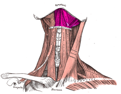
The omohyoid muscle is a muscle in the neck. It is one of the infrahyoid muscles. It consists of two bellies separated by an intermediate tendon. Its inferior belly is attached to the scapula; its superior belly is attached to the hyoid bone. Its intermediate tendon is anchored to the clavicle and first rib by a fascial sling. The omohyoid is innervated by the ansa cervicalis of the cervical plexus. It acts to depress the hyoid bone.

The digastric muscle is a bilaterally paired suprahyoid muscle located under the jaw. Its posterior belly is attached to the mastoid notch of temporal bone, and its anterior belly is attached to the digastric fossa of mandible; the two bellies are united by an intermediate tendon which is held in a loop that attaches to the hyoid bone. The anterior belly is innervated via the mandibular nerve, and the posterior belly is innervated via the facial nerve. It may act to depress the mandible or elevate the hyoid bone.

The geniohyoid muscle is a narrow paired muscle situated superior to the medial border of the mylohyoid muscle. It is named for its passage from the chin to the hyoid bone.

The mylohyoid muscle or diaphragma oris is a paired muscle of the neck. It runs from the mandible to the hyoid bone, forming the floor of the oral cavity of the mouth. It is named after its two attachments near the molar teeth. It forms the floor of the submental triangle. It elevates the hyoid bone and the tongue, important during swallowing and speaking.

The stylohyoid muscle is one of the suprahyoid muscles. Its originates from the styloid process of the temporal bone; it inserts onto hyoid bone. It is innervated by a branch of the facial nerve. It acts draw the hyoid bone upwards and backwards.

The thyrohyoid muscle is a small skeletal muscle of the neck. Above, it attaches onto the greater cornu of the hyoid bone; below, it attaches onto the oblique line of the thyroid cartilage. It is innervated by fibres derived from the cervical spinal nerve 1 that run with the hypoglossal nerve to reach this muscle. The thyrohyoid muscle depresses the hyoid bone and elevates the larynx during swallowing. By controlling the position and shape of the larynx, it aids in making sound.

The hyoglossus is a thin and quadrilateral extrinsic muscle of the tongue. It originates from the hyoid bone; it inserts onto the side of the tongue. It is innervated by the hypoglossal nerve. It acts to depress and retract the tongue.

The middle pharyngeal constrictor is a fan-shaped muscle located in the neck. It is one of three pharyngeal constrictor muscles. It is smaller than the inferior pharyngeal constrictor muscle.

The pharyngeal arches, also known as visceral arches, are structures seen in the embryonic development of vertebrates that are recognisable precursors for many structures. In fish, the arches are known as the branchial arches, or gill arches.
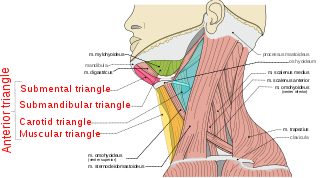
The anterior triangle is a region of the neck.

The deep cervical fascia lies under cover of the platysma, and invests the muscles of the neck; it also forms sheaths for the carotid vessels, and for the structures situated in front of the vertebral column. Its attachment to the hyoid bone prevents the formation of a dewlap.

Cervical lymph nodes are lymph nodes found in the neck. Of the 800 lymph nodes in the human body, 300 are in the neck. Cervical lymph nodes are subject to a number of different pathological conditions including tumours, infection and inflammation.

The submandibular space is a fascial space of the head and neck. It is a potential space, and is paired on either side, located on the superficial surface of the mylohyoid muscle between the anterior and posterior bellies of the digastric muscle. The space corresponds to the anatomic region termed the submandibular triangle, part of the anterior triangle of the neck.

The submental space is a fascial space of the head and neck. It is a potential space located between the mylohyoid muscle superiorly, the platysma muscle inferiorly, under the chin in the midline. The space coincides with the anatomic region termed the submental triangle, part of the anterior triangle of the neck.