Swallowing

Last updated

Swallowing, also called deglutition or inglutition [1] in scientific contexts, is the process in the body of a human or other animal that allows for a substance to pass from the mouth, to the pharynx, and into the esophagus, while shutting the epiglottis. Swallowing is an important part of eating and drinking. If the process fails and the material (such as food, drink, or medicine) goes through the trachea, then choking or pulmonary aspiration can occur. In the human body the automatic temporary closing of the epiglottis is controlled by the swallowing reflex.

Contents

The portion of food, drink, or other material that will move through the neck in one swallow is called a bolus.

In colloquial English, the term "swallowing" is also used to describe the action of taking in a large mouthful of food without any biting, where words like gulping or devouring are more adequate.

In humans

Swallowing comes so easily to most people that the process rarely prompts much thought. However, from the viewpoints of physiology, of speech–language pathology, and of health care for people with difficulty in swallowing (dysphagia), it is an interesting topic with extensive scientific literature.

Coordination and control

Eating and swallowing are complex neuromuscular activities consisting essentially of three phases, an oral, pharyngeal and esophageal phase. Each phase is controlled by a different neurological mechanism. The oral phase, which is entirely voluntary, is mainly controlled by the medial temporal lobes and limbic system of the cerebral cortex with contributions from the motor cortex and other cortical areas. The pharyngeal swallow is started by the oral phase and subsequently is coordinated by the swallowing center on the medulla oblongata and pons. The reflex is initiated by touch receptors in the pharynx as a bolus of food is pushed to the back of the mouth by the tongue, or by stimulation of the palate (palatal reflex).

Swallowing is a complex mechanism using both skeletal muscle (tongue) and smooth muscles of the pharynx and esophagus. The autonomic nervous system (ANS) coordinates this process in the pharyngeal and esophageal phases.

Real-time MRI – swallowing
Sagittal view of mouth and pharynx Sagittalmouth.png
Sagittal view of mouth and pharynx

Phases

Oral phase

Prior to the following stages of the oral phase, the mandible depresses and the lips abduct to allow food or liquid to enter the oral cavity. Upon entering the oral cavity, the mandible elevates and the lips adduct to assist in oral containment of the food and liquid. The following stages describe the normal and necessary actions to form the bolus, which is defined as the state of the food in which it is ready to be swallowed.

1) Moistening

Food is moistened by saliva from the salivary glands (parasympathetic).

2) Mastication

Food is mechanically broken down by the action of the teeth controlled by the muscles of mastication (V3) acting on the temporomandibular joint. This results in a bolus which is moved from one side of the oral cavity to the other by the tongue. Buccinator (VII) helps to contain the food against the occlusal surfaces of the teeth. The bolus is ready for swallowing when it is held together by saliva (largely mucus), sensed by the lingual nerve of the tongue (VII—chorda tympani and IX—lesser petrosal) (V3). Any food that is too dry to form a bolus will not be swallowed.

3) Trough formation

A trough is then formed at the back of the tongue by the intrinsic muscles (XII). The trough obliterates against the hard palate from front to back, forcing the bolus to the back of the tongue. The intrinsic muscles of the tongue (XII) contract to make a trough (a longitudinal concave fold) at the back of the tongue. The tongue is then elevated to the roof of the mouth (by the mylohyoid (mylohyoid nerve—V3), genioglossus, styloglossus and hyoglossus (the rest XII)) such that the tongue slopes downwards posteriorly. The contraction of the genioglossus and styloglossus (both XII) also contributes to the formation of the central trough.

4) Movement of the bolus posteriorly

At the end of the oral preparatory phase, the food bolus has been formed and is ready to be propelled posteriorly into the pharynx. In order for anterior to posterior transit of the bolus to occur, orbicularis oris contracts and adducts the lips to form a tight seal of the oral cavity. Next, the superior longitudinal muscle elevates the apex of the tongue to make contact with the hard palate and the bolus is propelled to the posterior portion of the oral cavity. Once the bolus reaches the palatoglossal arch of the oropharynx, the pharyngeal phase, which is reflex and involuntary, then begins. Receptors initiating this reflex are proprioceptive (afferent limb of reflex is IX and efferent limb is the pharyngeal plexus- IX and X). They are scattered over the base of the tongue, the palatoglossal and palatopharyngeal arches, the tonsillar fossa, uvula and posterior pharyngeal wall. Stimuli from the receptors of this phase then provoke the pharyngeal phase. In fact, it has been shown that the swallowing reflex can be initiated entirely by peripheral stimulation of the internal branch of the superior laryngeal nerve. This phase is voluntary and involves important cranial nerves: V (trigeminal), VII (facial) and XII (hypoglossal).

Pharyngeal phase

For the pharyngeal phase to work properly all other egress from the pharynx must be occluded—this includes the nasopharynx and the larynx. When the pharyngeal phase begins, other activities such as chewing, breathing, coughing and vomiting are concomitantly inhibited.

5) Closure of the nasopharynx

The soft palate is tensed by tensor palatini (Vc), and then elevated by levator palatini (pharyngeal plexus—IX, X) to close the nasopharynx. There is also the simultaneous approximation of the walls of the pharynx to the posterior free border of the soft palate, which is carried out by the palatopharyngeus (pharyngeal plexus—IX, X) and the upper part of the superior constrictor (pharyngeal plexus—IX, X).

6) The pharynx prepares to receive the bolus

The pharynx is pulled upwards and forwards by the suprahyoid and longitudinal pharyngeal muscles – stylopharyngeus (IX), salpingopharyngeus (pharyngeal plexus—IX, X) and palatopharyngeus (pharyngeal plexus—IX, X) to receive the bolus. The palatopharyngeal folds on each side of the pharynx are brought close together through the superior constrictor muscles, so that only a small bolus can pass.

7) Opening of the auditory tube

The actions of the levator palatini (pharyngeal plexus—IX, X), tensor palatini (Vc) and salpingopharyngeus (pharyngeal plexus—IX, X) in the closure of the nasopharynx and elevation of the pharynx opens the auditory tube, which equalises the pressure between the nasopharynx and the middle ear. This does not contribute to swallowing, but happens as a consequence of it.

8) Closure of the oropharynx

The oropharynx is kept closed by palatoglossus (pharyngeal plexus—IX, X), the intrinsic muscles of tongue (XII) and styloglossus (XII).

9) Laryngeal closure

The primary laryngopharyngeal protective mechanism to prevent aspiration during swallowing is via the closure of the true vocal folds. The adduction of the vocal cords is affected by the contraction of the lateral cricoarytenoids and the oblique and transverse arytenoids (all recurrent laryngeal nerve of vagus). Since the true vocal folds adduct during the swallow, a finite period of apnea (swallowing apnea) must necessarily take place with each swallow. When relating swallowing to respiration, it has been demonstrated that swallowing occurs most often during expiration, even at full expiration a fine air jet is expired probably to clear the upper larynx from food remnants or liquid. The clinical significance of this finding is that patients with a baseline of compromised lung function will, over a period of time, develop respiratory distress as a meal progresses. Subsequently, false vocal fold adduction, adduction of the aryepiglottic folds and retroversion of the epiglottis take place. The aryepiglotticus (recurrent laryngeal nerve of vagus) contracts, causing the arytenoids to appose each other (closes the laryngeal aditus by bringing the aryepiglottic folds together), and draws the epiglottis down to bring its lower half into contact with arytenoids, thus closing the aditus. Retroversion of the epiglottis, while not the primary mechanism of protecting the airway from laryngeal penetration and aspiration, acts to anatomically direct the food bolus laterally towards the piriform fossa. Additionally, the larynx is pulled up with the pharynx under the tongue by stylopharyngeus (IX), salpingopharyngeus (pharyngeal plexus—IX, X), palatopharyngeus (pharyngeal plexus—IX, X) and inferior constrictor (pharyngeal plexus—IX, X). This phase is passively controlled reflexively and involves cranial nerves V, X (vagus), XI (accessory) and XII (hypoglossal). The respiratory center of the medulla is directly inhibited by the swallowing center for the very brief time that it takes to swallow. This means that it is briefly impossible to breathe during this phase of swallowing and the moment where breathing is prevented is known as deglutition apnea .

10) Hyoid elevation

The hyoid is elevated by digastric (V & VII) and stylohyoid (VII), lifting the pharynx and larynx up even further.

11) Bolus transits pharynx

The bolus moves down towards the esophagus by pharyngeal peristalsis which takes place by sequential contraction of the superior, middle and inferior pharyngeal constrictor muscles (pharyngeal plexus—IX, X). The lower part of the inferior constrictor (cricopharyngeus) is normally closed and only opens for the advancing bolus. Gravity plays only a small part in the upright position—in fact, it is possible to swallow solid food even when standing on one's head. The velocity through the pharynx depends on a number of factors such as viscosity and volume of the bolus. In one study, bolus velocity in healthy adults was measured to be approximately 30–40 cm/s. [2]

Esophageal phase

12) Esophageal peristalsis

Like the pharyngeal phase of swallowing, the esophageal phase of swallowing is under involuntary neuromuscular control. However, propagation of the food bolus is significantly slower than in the pharynx. The bolus enters the esophagus and is propelled downwards first by striated muscle (recurrent laryngeal, X) then by the smooth muscle (X) at a rate of 3–5 cm/s. The upper esophageal sphincter relaxes to let food pass, after which various striated constrictor muscles of the pharynx as well as peristalsis and relaxation of the lower esophageal sphincter sequentially push the bolus of food through the esophagus into the stomach.

13) Relaxation phase

Finally the larynx and pharynx move down with the hyoid mostly by elastic recoil. Then the larynx and pharynx move down from the hyoid to their relaxed positions by elastic recoil. Swallowing therefore depends on coordinated interplay between many various muscles, and although the initial part of swallowing is under voluntary control, once the deglutition process is started, it is quite hard to stop it.

Clinical significance

Swallowing becomes a great concern for the elderly since strokes and Alzheimer's disease can interfere with the autonomic nervous system. Speech pathologists commonly diagnose and treat this condition since the speech process uses the same neuromuscular structures as swallowing. Diagnostic procedures commonly performed by a speech pathologist to evaluate dysphagia include Fiberoptic Endoscopic Evaluation of Swallowing and Modified Barium Swallow Study. Occupational Therapists may also offer swallowing rehabilitation services as well as prescribing modified feeding techniques and utensils. Consultation with a dietician is essential, in order to ensure that the individual with dysphagia is able to consume sufficient calories and nutrients to maintain health. In terminally ill patients, a failure of the reflex to swallow leads to a build-up of mucus or saliva in the throat and airways, producing a noise known as a death rattle (not to be confused with agonal respiration, which is an abnormal pattern of breathing due to cerebral ischemia or hypoxia).

Abnormalities of the pharynx and/or oral cavity may lead to oropharyngeal dysphagia. Abnormalities of the esophagus may lead to esophageal dysphagia. The failure of the lower esophagus sphincter to respond properly to swallowing is called achalasia.

M-Type Swallowing

With practice, people can learn to swallow fluidly without closing the mouth by merely manipulating the tongue and jaw to drive fluids or foods down the esophagus. With a continuous motion, an individual forges breathing and priorities the swallowed matter. This intermediate level of muscle manipulation is similar to the techniques used by sword swallowers.

In non-mammal animals

A pelican swallowing a fish Pelican swallowing a fish part 4.JPG
A pelican swallowing a fish

In many birds, the esophagus is largely a mere gravity chute, and in such events as a seagull swallowing a fish or a stork swallowing a frog, swallowing consists largely of the bird lifting its head with its beak pointing up and guiding the prey with tongue and jaws so that the prey slides inside and down.

In fish, the tongue is largely bony and much less mobile and getting the food to the back of the pharynx is helped by pumping water in its mouth and out of its gills.

In snakes, the work of swallowing is done by raking with the lower jaw until the prey is far enough back to be helped down by body undulations.

See also

Related Research Articles

<span class="mw-page-title-main">Larynx</span> Voice box, an organ in the neck of amphibians, reptiles, and mammals

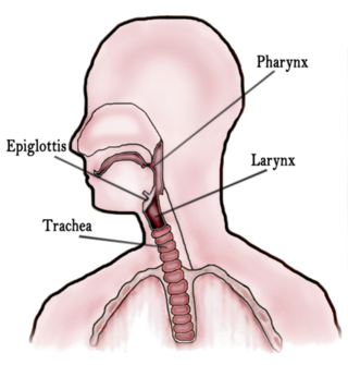
The larynx, commonly called the voice box, is an organ in the top of the neck involved in breathing, producing sound and protecting the trachea against food aspiration. The opening of larynx into pharynx known as the laryngeal inlet is about 4–5 centimeters in diameter. The larynx houses the vocal cords, and manipulates pitch and volume, which is essential for phonation. It is situated just below where the tract of the pharynx splits into the trachea and the esophagus. The word 'larynx' comes from the Ancient Greek word lárunx ʻlarynx, gullet, throat.ʼ

<span class="mw-page-title-main">Throat</span> Anterior part of the neck, in front of the vertebra

In vertebrate anatomy, the throat is the front part of the neck, internally positioned in front of the vertebrae. It contains the pharynx and larynx. An important section of it is the epiglottis, separating the esophagus from the trachea (windpipe), preventing food and drinks being inhaled into the lungs. The throat contains various blood vessels, pharyngeal muscles, the nasopharyngeal tonsil, the tonsils, the palatine uvula, the trachea, the esophagus, and the vocal cords. Mammal throats consist of two bones, the hyoid bone and the clavicle. The "throat" is sometimes thought to be synonymous for the fauces.

<span class="mw-page-title-main">Esophagus</span> Vertebrate organ through which food passes to the stomach

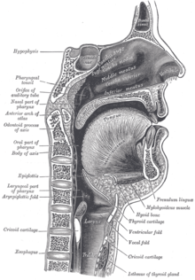
The esophagus or oesophagus, colloquially known also as the food pipe, food tube, or gullet, is an organ in vertebrates through which food passes, aided by peristaltic contractions, from the pharynx to the stomach. The esophagus is a fibromuscular tube, about 25 cm (10 in) long in adults, that travels behind the trachea and heart, passes through the diaphragm, and empties into the uppermost region of the stomach. During swallowing, the epiglottis tilts backwards to prevent food from going down the larynx and lungs. The word oesophagus is from Ancient Greek οἰσοφάγος (oisophágos), from οἴσω (oísō), future form of φέρω + ἔφαγον.

<span class="mw-page-title-main">Glossopharyngeal nerve</span> Cranial nerve IX, for the tongue and pharynx

The glossopharyngeal nerve, also known as the ninth cranial nerve, cranial nerve IX, or simply CN IX, is a cranial nerve that exits the brainstem from the sides of the upper medulla, just anterior to the vagus nerve. Being a mixed nerve (sensorimotor), it carries afferent sensory and efferent motor information. The motor division of the glossopharyngeal nerve is derived from the basal plate of the embryonic medulla oblongata, whereas the sensory division originates from the cranial neural crest.

<span class="mw-page-title-main">Epiglottis</span> Leaf-shaped flap in the throat that prevents food from entering the windpipe and the lungs

The epiglottis is a leaf-shaped flap in the throat that prevents food and water from entering the trachea and the lungs. It stays open during breathing, allowing air into the larynx. During swallowing, it closes to prevent aspiration of food into the lungs, forcing the swallowed liquids or food to go along the esophagus toward the stomach instead. It is thus the valve that diverts passage to either the trachea or the esophagus.

The pharyngeal reflex or gag reflex is a reflex muscular contraction of the back of the throat, evoked by touching the roof of the mouth, back of the tongue, area around the tonsils, uvula, and back of the throat. It, along with other aerodigestive reflexes such as reflexive pharyngeal swallowing, prevents objects in the oral cavity from entering the throat except as part of normal swallowing and helps prevent choking, and is a form of coughing. The pharyngeal reflex is different from the laryngeal spasm, which is a reflex muscular contraction of the vocal cords.

<span class="mw-page-title-main">Nucleus ambiguus</span>

The nucleus ambiguus is a group of large motor neurons, situated deep in the medullary reticular formation named by Jacob Clarke. The nucleus ambiguus contains the cell bodies of neurons that innervate the muscles of the soft palate, pharynx, and larynx which are associated with speech and swallowing. As well as motor neurons, the nucleus ambiguus contains preganglionic parasympathetic neurons which innervate postganglionic parasympathetic neurons in the heart.

<span class="mw-page-title-main">Levator veli palatini</span> Muscle of the soft palate

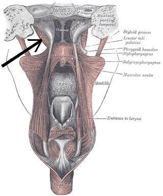
The levator veli palatini is a muscle of the soft palate and pharynx. It is innervated by the vagus nerve via its pharyngeal plexus. During swallowing, it contracts, elevating the soft palate to help prevent food from entering the nasopharynx.

<span class="mw-page-title-main">Middle pharyngeal constrictor muscle</span> Muscle in the neck

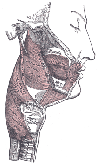
The middle pharyngeal constrictor is a fan-shaped muscle located in the neck. It is one of three pharyngeal constrictor muscles. It is smaller than the inferior pharyngeal constrictor muscle.

<span class="mw-page-title-main">Inferior pharyngeal constrictor muscle</span> Skeletal muscle of the pharynx

The inferior pharyngeal constrictor muscle is a skeletal muscle of the neck. It is the thickest of the three outer pharyngeal muscles. It arises from the sides of the cricoid cartilage and the thyroid cartilage. It is supplied by the vagus nerve. It is active during swallowing, and partially during breathing and speech. It may be affected by Zenker's diverticulum.

<span class="mw-page-title-main">Superior pharyngeal constrictor muscle</span> Muscle

The superior pharyngeal constrictor muscle is a quadrilateral muscle of the pharynx. It is the uppermost and thinnest of the three pharyngeal constrictors.

<span class="mw-page-title-main">Stylopharyngeus muscle</span> Muscle

The stylopharyngeus muscle is a muscle in the head. It originates from the temporal styloid process. Some of its fibres insert onto the thyroid cartilage, while others end by intermingling with proximal structures. It is innervated by the glossopharyngeal nerve. It acts to elevate the larynx and pharynx, and dilate the pharynx, thus facilitating swallowing.

<span class="mw-page-title-main">Inferior thyroid artery</span> Artery of the neck

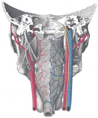
The inferior thyroid artery is an artery in the neck. It arises from the thyrocervical trunk and passes upward, in front of the vertebral artery and longus colli muscle. It then turns medially behind the carotid sheath and its contents, and also behind the sympathetic trunk, the middle cervical ganglion resting upon the vessel.

<span class="mw-page-title-main">Superior laryngeal nerve</span> Branch of the vagus nerve

The superior laryngeal nerve is a branch of the vagus nerve. It arises from the middle of the inferior ganglion of vagus nerve and additionally also receives a sympathetic branch from the superior cervical ganglion.

<span class="mw-page-title-main">Oropharyngeal dysphagia</span> Difficulty controlling the mouth or throat for swallowing

Oropharyngeal dysphagia is the inability to empty material from the oropharynx into the esophagus as a result of malfunction near the esophagus. Oropharyngeal dysphagia manifests differently depending on the underlying pathology and the nature of the symptoms. Patients with dysphagia can experience feelings of food sticking to their throats, coughing and choking, weight loss, recurring chest infections, or regurgitation. Depending on the underlying cause, age, and environment, dysphagia prevalence varies. In research including the general population, the estimated frequency of oropharyngeal dysphagia has ranged from 2 to 16 percent.

<span class="mw-page-title-main">Pharyngeal muscles</span>

The pharyngeal muscles are a group of muscles that form the pharynx, which is posterior to the oral cavity, determining the shape of its lumen, and affecting its sound properties as the primary resonating cavity.

<span class="mw-page-title-main">Pharyngeal plexus of vagus nerve</span> Nerve fibers innervating most of the palate and pharynx

The pharyngeal plexus is a nerve plexus located upon the outer surface of the pharynx. It contains a motor component, a sensory component, and sympathetic component.

Presbyphagia refers to characteristic changes in the swallowing mechanism of otherwise healthy older adults. Although age-related changes place older adults at risk swallowing disorders, an older adult's swallow is not necessarily an impaired swallow. Clinicians are becoming more aware of the need to distinguish among swallowing disorders, presbyphagia and other related diagnoses in order to avoid over diagnosing and over treating presbyphagia. Older adults are more vulnerable and with the increased threat of acute illnesses, medications and any number of age-related conditions, they can cross the line from having a healthy older swallow to being dysphagic.

<span class="mw-page-title-main">Pharynx</span> Part of the throat that is behind the mouth and nasal cavity

The pharynx is the part of the throat behind the mouth and nasal cavity, and above the esophagus and trachea. It is found in vertebrates and invertebrates, though its structure varies across species. The pharynx carries food to the esophagus and air to the larynx. The flap of cartilage called the epiglottis stops food from entering the larynx.

<span class="mw-page-title-main">Human digestive system</span> Digestive system in humans

The human digestive system consists of the gastrointestinal tract plus the accessory organs of digestion. Digestion involves the breakdown of food into smaller and smaller components, until they can be absorbed and assimilated into the body. The process of digestion has three stages: the cephalic phase, the gastric phase, and the intestinal phase.

References

  1. "inglutition". Oxford English Dictionary .
  2. Clave, P.; De Kraa, M.; Arreola, V.; Girvent, M.; Farre, R.; Palomera, E.; Serra-Prat, M. (2006). "The effect of bolus viscosity on swallowing function in neurogenic dysphagia". Alimentary Pharmacology & Therapeutics. Wiley. 24 (9): 1385–1394. doi:10.1111/j.1365-2036.2006.03118.x. PMID   17059520. S2CID   22881225.