Abducens nerve

Last updated
Abducens nerve
Abducens nerve1.png
The path of the abducens nerve
Brain human normal inferior view with labels en.svg
Inferior view of the human brain, with the cranial nerves labelled.
Details
From Abducens nucleus
Innervates Lateral rectus muscle
Identifiers
Latin nervus abducens
MeSH D000010
NeuroNames 550
TA98 A14.2.01.098
TA2 6283
FMA 50867
Anatomical terms of neuroanatomy

The abducens nerve or abducent nerve, also known as the sixth cranial nerve, cranial nerve VI, or simply CN VI, is a cranial nerve in humans and various other animals that controls the movement of the lateral rectus muscle, one of the extraocular muscles responsible for outward gaze. It is a somatic efferent nerve.

Contents

Structure

Nucleus

Axial section of the Brainstem (Pons) at the level of the Facial Colliculus Pons section at facial colliculus.png
Axial section of the Brainstem (Pons) at the level of the Facial Colliculus

The abducens nucleus is located in the pons, on the floor of the fourth ventricle, at the level of the facial colliculus. Axons from the facial nerve loop around the abducens nucleus, creating a slight bulge (the facial colliculus) that is visible on the dorsal surface of the floor of the fourth ventricle. The abducens nucleus is close to the midline, like the other motor nuclei that control eye movements (the oculomotor and trochlear nuclei).[ citation needed ]

Motor axons leaving the abducens nucleus run ventrally and caudally through the pons. They pass lateral to the corticospinal tract (which runs longitudinally through the pons at this level) before exiting the brainstem at the pontomedullary junction.[ citation needed ]

Course

The Clivus Clivus.png
The Clivus

The abducens nerve emerges from the brainstem at the junction of the pons and the medulla, [1] superior to the medullary pyramid, [2] and medial to the facial nerve. It runs upwards and forwards from this position to reach the eye.

The nerve enters the subarachnoid space (more precisely, the pontine cistern [2] ) when it emerges from the brainstem. It runs upward between the pons and the clivus, and then pierces the dura mater to run between the dura and the skull through Dorello's canal. [3] [4] At the apex of the petrous part of the temporal bone, it makes a sharp turn forward to enter the cavernous sinus. [1] In the cavernous sinus, it runs anterior-ward alongside (inferolateral to) the internal carotid artery. It enters the orbit through (medial end of) the superior orbital fissure, passing through the common tendinous ring to reach and innervate the lateral rectus muscle of the eye. [2]

Development

The human abducens nerve is derived from the basal plate of the embryonic pons.

Function

The abducens nerve supplies the lateral rectus muscle of the human eye. This muscle is responsible for outward gaze. [1] The abducens nerve carries axons of type GSE, general somatic efferent.

Clinical significance

Damage

Limitation of abduction of the right eye. This individual tries to look to his right, but the right eye fails to turn to the side. Abducens palsy.jpg
Limitation of abduction of the right eye. This individual tries to look to his right, but the right eye fails to turn to the side.

Damage to the peripheral part of the abducens nerve will cause double vision (diplopia), due to the unopposed muscle tone of the medial rectus muscle. The affected eye is pulled to look towards the midline. In order to see without double vision, patients will rotate their heads so that both eyes are toward the temple.[ citation needed ] Partial damage to the abducens nerve causes weak or incomplete abduction of the affected eye. The diplopia is worse on attempts at looking laterally.

The long course of the abducens nerve between the brainstem and the eye makes it vulnerable to injury at many levels. For example, fractures of the petrous temporal bone can selectively damage the nerve, as can aneurysms of the intracavernous carotid artery. Mass lesions that push the brainstem downward can damage the nerve by stretching it between the point where it emerges from the pons and the point where it hooks over the petrous temporal bone.

The central anatomy of the sixth nerve predicts (correctly) that infarcts affecting the dorsal pons at the level of the abducens nucleus can also affect the facial nerve, producing an ipsilateral facial palsy together with a lateral rectus palsy. The anatomy also predicts (correctly) that infarcts involving the ventral pons can affect the sixth nerve and the corticospinal tract simultaneously, producing a lateral rectus palsy associated with a contralateral hemiparesis. These rare syndromes are of interest primarily as useful summaries of the anatomy of the brainstem.

Peripheral lesions

Complete interruption of the peripheral sixth nerve causes diplopia (double vision), due to the unopposed action of the medial rectus muscle. The affected eye is pulled medially. In order to see without double vision, patients will turn their heads sideways so that both eyes are looking sideways. On formal testing, the affected eye cannot abduct past the midline – it cannot look sideways, toward the temple. Partial damage to the sixth nerve causes weak or incomplete abduction of the affected eye. The diplopia is worse on attempted lateral gaze, as would be expected (since the lateral gaze muscle is impaired).

Peripheral sixth nerve damage can be caused by tumors, aneurysms, or fractures – anything that directly compresses or stretches the nerve. Other processes that can damage the sixth nerve include strokes (infarctions), demyelination, infections (e.g. meningitis), cavernous sinus diseases and various neuropathies. Perhaps the most common overall cause of sixth nerve impairment is diabetic neuropathy. Iatrogenic injury is also known to occur, with the abducens nerve being the most commonly injured cranial nerve in halo orthosis placement. [5] The resultant palsy is identified through loss of lateral gaze after application of the orthosis.

Rare causes of isolated sixth nerve damage include Wernicke–Korsakoff syndrome and Tolosa–Hunt syndrome. Wernicke–Korsakoff syndrome is caused by thiamine deficiency, classically due to alcoholism. The characteristic ocular abnormalities are nystagmus and lateral rectus weakness. Tolosa-Hunt syndrome is an idiopathic granulomatous disease that causes painful oculomotor (especially sixth nerve) palsies.

Indirect damage to the sixth nerve can be caused by any process (brain tumor, hydrocephalus, pseudotumor cerebri, hemorrhage, edema) that exerts downward pressure on the brainstem, causing the nerve to stretch along the clivus. This type of traction injury can affect either side first. A right-sided brain tumor can produce either a right-sided or a left-sided sixth nerve palsy as an initial sign. Thus a right-sided sixth nerve palsy does not necessarily imply a right-sided cause. Sixth nerve palsies are infamous as "false localizing signs." Neurological signs are described as "false localizing" if they reflect dysfunction distant or remote from the expected anatomical location of pathology. Isolated sixth nerve palsies in children are assumed to be due to brain tumors until proven otherwise.

Nuclear lesions

Damage to the abducens nucleus does not produce an isolated sixth nerve palsy, but rather a horizontal gaze palsy that affects both eyes simultaneously. The abducens nucleus contains two types of cells: motor neurons that control the lateral rectus muscle on the same side, and interneurons that cross the midline and connect to the contralateral oculomotor nucleus (which controls the medial rectus muscle of the opposite eye). In normal vision, lateral movement of one eye (lateral rectus muscle) is precisely coupled to medial movement of the other eye (medial rectus muscle), so that both eyes remain fixed on the same object.

The control of conjugate gaze is mediated in the brainstem by the medial longitudinal fasciculus (MLF), a nerve tract that connects the three extraocular motor nuclei (abducens, trochlear and oculomotor) into a single functional unit. Lesions of the abducens nucleus and the MLF produce observable sixth nerve problems, most notably internuclear ophthalmoplegia (INO).

Supranuclear lesions

The sixth nerve is one of the final common pathways for numerous cortical systems that control eye movement in general. Cortical control of eye movement (saccades, smooth pursuit, accommodation) involves conjugate gaze, not unilateral eye movement.

Tuberculosis

15–40% of people with tuberculosis have some resulting cranial nerve deficit. The sixth nerve is the most commonly affected cranial nerve in immunocompetent people with tuberculosis. [6]

History

Etymology

The Latin name for the sixth cranial nerve is "nervus abducens". The Terminologia Anatomica officially recognizes two different English translations: "abducent nerve" and "abducens nerve". [7]

"Abducens" is more common in recent literature, while "abducent" predominates in the older literature. The United States National Library of Medicine uses "abducens nerve" in its Medical Subject Heading (MeSH) vocabulary to index the vast MEDLINE and PubMed biomedical databases. The 39th edition of Gray's Anatomy (2005) also prefers "abducens nerve." [1]

Other animals

The abducens nerve controls the movement of a single muscle, the lateral rectus muscle of the eye. In most other mammals it also innervates the musculus retractor bulbi, which can retract the eye for protection. Homologous abducens nerves are found in all vertebrates except lampreys and hagfishes.

See also

Related Research Articles

<span class="mw-page-title-main">Cranial nerves</span> Nerves that emerge directly from the brain and the brainstem

Cranial nerves are the nerves that emerge directly from the brain, of which there are conventionally considered twelve pairs. Cranial nerves relay information between the brain and parts of the body, primarily to and from regions of the head and neck, including the special senses of vision, taste, smell, and hearing.

<span class="mw-page-title-main">Facial nerve</span> Cranial nerve VII, for the face and tasting

The facial nerve, also known as the seventh cranial nerve, cranial nerve VII, or simply CN VII, is a cranial nerve that emerges from the pons of the brainstem, controls the muscles of facial expression, and functions in the conveyance of taste sensations from the anterior two-thirds of the tongue. The nerve typically travels from the pons through the facial canal in the temporal bone and exits the skull at the stylomastoid foramen. It arises from the brainstem from an area posterior to the cranial nerve VI and anterior to cranial nerve VIII.

<span class="mw-page-title-main">Pons</span> Part of the brainstem in humans and other bipeds

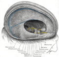
The pons is part of the brainstem that in humans and other mammals, lies inferior to the midbrain, superior to the medulla oblongata and anterior to the cerebellum.

<span class="mw-page-title-main">Brainstem</span> Posterior part of the brain, adjoining and structurally continuous

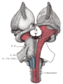
The brainstem is the stalk-like part of the brain that interconnects the cerebrum and diencephalon with the spinal cord. In the human brain, the brainstem is composed of the midbrain, the pons, and the medulla oblongata. The midbrain is continuous with the thalamus of the diencephalon through the tentorial notch.

<span class="mw-page-title-main">Oculomotor nerve</span> Cranial nerve III, for eye movements

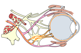
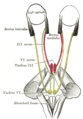
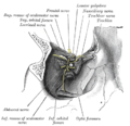
The oculomotor nerve, also known as the third cranial nerve, cranial nerve III, or simply CN III, is a cranial nerve that enters the orbit through the superior orbital fissure and innervates extraocular muscles that enable most movements of the eye and that raise the eyelid. The nerve also contains fibers that innervate the intrinsic eye muscles that enable pupillary constriction and accommodation. The oculomotor nerve is derived from the basal plate of the embryonic midbrain. Cranial nerves IV and VI also participate in control of eye movement.

<span class="mw-page-title-main">Trochlear nerve</span> Cranial nerve IV, for eye movements

The trochlear nerve, also known as the fourth cranial nerve, cranial nerve IV, or CN IV, is a cranial nerve that innervates a single muscle - the superior oblique muscle of the eye. Unlike most other cranial nerves, the trochlear nerve is exclusively a motor nerve.

<span class="mw-page-title-main">Medial longitudinal fasciculus</span> Nerve tracts in the brainstem

The medial longitudinal fasciculus (MLF) is an area of crossed over tracts, on each side of the brainstem. These bundles of axons are situated near the midline of the brainstem. They are made up of both ascending and descending fibers that arise from a number of sources and terminate in different areas, including the superior colliculus, the vestibular nuclei, and the cerebellum. It contains the interstitial nucleus of Cajal, responsible for oculomotor control, head posture, and vertical eye movement.

<span class="mw-page-title-main">Eye movement</span> Movement of the eyes

Eye movement includes the voluntary or involuntary movement of the eyes. Eye movements are used by a number of organisms to fixate, inspect and track visual objects of interests. A special type of eye movement, rapid eye movement, occurs during REM sleep.

<span class="mw-page-title-main">Lateral rectus muscle</span> Muscle on lateral side of the eye

The lateral rectus muscle is a muscle on the lateral side of the eye in the orbit. It is one of six extraocular muscles that control the movements of the eye. The lateral rectus muscle is responsible for lateral movement of the eyeball, specifically abduction. Abduction describes the movement of the eye away from the midline, allowing the eyeball to move horizontally in the lateral direction, bringing the pupil away from the midline of the body.

<span class="mw-page-title-main">Inferior oblique muscle</span> Part of the eye

The inferior oblique muscle or obliquus oculi inferior is a thin, narrow muscle placed near the anterior margin of the floor of the orbit. The inferior oblique is one of the extraocular muscles, and is attached to the maxillary bone (origin) and the posterior, inferior, lateral surface of the eye (insertion). The inferior oblique is innervated by the inferior branch of the oculomotor nerve.

<span class="mw-page-title-main">Extraocular muscles</span> Seven extrinsic muscles of the eye

The extraocular muscles, or extrinsic ocular muscles, are the seven extrinsic muscles of the eye in humans and other animals. Six of the extraocular muscles, the four recti muscles, and the superior and inferior oblique muscles, control movement of the eye. The other muscle, the levator palpebrae superioris, controls eyelid elevation. The actions of the six muscles responsible for eye movement depend on the position of the eye at the time of muscle contraction.

<span class="mw-page-title-main">Abducens nucleus</span>

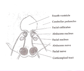
The abducens nucleus is the originating nucleus from which the abducens nerve (VI) emerges—a cranial nerve nucleus. This nucleus is located beneath the fourth ventricle in the caudal portion of the pons near the midline, medial to the sulcus limitans.

<span class="mw-page-title-main">Sixth nerve palsy</span> Medical condition

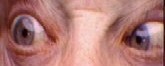
Sixth nerve palsy, or abducens nerve palsy, is a disorder associated with dysfunction of cranial nerve VI, which is responsible for causing contraction of the lateral rectus muscle to abduct the eye. The inability of an eye to turn outward, results in a convergent strabismus or esotropia of which the primary symptom is diplopia in which the two images appear side-by-side. Thus, the diplopia is horizontal and worse in the distance. Diplopia is also increased on looking to the affected side and is partly caused by overaction of the medial rectus on the unaffected side as it tries to provide the extra innervation to the affected lateral rectus. These two muscles are synergists or "yoke muscles" as both attempt to move the eye over to the left or right. The condition is commonly unilateral but can also occur bilaterally.

<span class="mw-page-title-main">Facial colliculus</span>

The facial colliculus is an elevated area located in the pontine tegmentum, within the floor of the fourth ventricle. It is formed by fibres from the facial motor nucleus looping over the abducens nucleus. The facial colliculus is an essential landmark of the rhomboid fossa.

Conjugate gaze palsies are neurological disorders affecting the ability to move both eyes in the same direction. These palsies can affect gaze in a horizontal, upward, or downward direction. These entities overlap with ophthalmoparesis and ophthalmoplegia.

<span class="mw-page-title-main">Oculomotor nerve palsy</span> Medical condition

Oculomotor nerve palsy or oculomotor neuropathy is an eye condition resulting from damage to the third cranial nerve or a branch thereof. As the name suggests, the oculomotor nerve supplies the majority of the muscles controlling eye movements. Damage to this nerve will result in an inability to move the eye normally. The nerve also supplies the upper eyelid muscle and is accompanied by parasympathetic fibers innervating the muscles responsible for pupil constriction. The limitations of eye movement resulting from the condition are generally so severe that patients are often unable to maintain normal eye alignment when gazing straight ahead, leading to strabismus and, as a consequence, double vision (diplopia).

A horizontal gaze palsy is a subtype of gaze palsy in which conjugate, horizontal eye movements are limited by neurologic deficits. Horizontal gaze palsies typically result from an ipsilateral pontine lesion or a contralateral frontal lobe lesion.

The term gaze is frequently used in physiology to describe coordinated motion of the eyes and neck. The lateral gaze is controlled by the paramedian pontine reticular formation (PPRF). The vertical gaze is controlled by the rostral interstitial nucleus of medial longitudinal fasciculus and the interstitial nucleus of Cajal.

Alternating hemiplegia is a form of hemiplegia that has an ipsilateral cranial nerve palsies and contralateral hemiplegia or hemiparesis of extremities of the body. The disorder is characterized by recurrent episodes of paralysis on one side of the body. There are multiple forms of alternating hemiplegia, Weber's syndrome, middle alternating hemiplegia, and inferior alternating hemiplegia. This type of syndrome can result from a unilateral lesion in the brainstem affecting both upper motor neurons and lower motor neurons. The muscles that would receive signals from these damaged upper motor neurons result in spastic paralysis. With a lesion in the brainstem, this affects the majority of limb and trunk muscles on the contralateral side due to the upper motor neurons decussation after the brainstem. The cranial nerves and cranial nerve nuclei are also located in the brainstem making them susceptible to damage from a brainstem lesion. Cranial nerves III (Oculomotor), VI (Abducens), and XII (Hypoglossal) are most often associated with this syndrome given their close proximity with the pyramidal tract, the location which upper motor neurons are in on their way to the spinal cord. Damages to these structures produce the ipsilateral presentation of paralysis or palsy due to the lack of cranial nerve decussation before innervating their target muscles. The paralysis may be brief or it may last for several days, many times the episodes will resolve after sleep. Some common symptoms of alternating hemiplegia are mental impairment, gait and balance difficulties, excessive sweating and changes in body temperature.

<span class="mw-page-title-main">Corticomesencephalic tract</span>

In neuroanatomy, corticomesencephalic tract is a descending nerve tract that originates in the frontal eye field and terminate in the midbrain. Its fibers mediate conjugate eye movement.

References

  1. 1 2 3 4 Gray's Anatomy 2008, pp. 666–7.
  2. 1 2 3 Chummy S. Sinnatamby (2011). Last's anatomy: regional and applied (12th ed.). Edinburgh. ISBN   978-0-7020-4839-5. OCLC   764565702.{{cite book}}: CS1 maint: location missing publisher (link)
  3. Umansky, Felix; Elidan, Josef; Valarezo, Alberto (1 August 1991). "Dorello's canal: a microanatomical study". Journal of Neurosurgery . 75 (2): 294–298. doi:10.3171/jns.1991.75.2.0294. PMID   2072168.
  4. Özveren, Mehmet Faik; Erol, Fatih Serhat; Alkan, Alpay; Kocak, Ayhan; Önal, Cagatay; Türe, Uğur (1 February 2007). "Microanatomical Architecture of Dorello's Canal and Its Clinical Implications". Operative Neurosurgery . 60 (suppl_2): ONS1-7, discussion ONS7-8. doi:10.1227/01.neu.0000249229.89988.4d. ISSN   2332-4252. PMID   17297359. S2CID   36731926.
  5. "Halo Orthosis Immobilization – Spine – Orthobullets". www.orthobullets.com.
  6. Smith, Daniel E.; Blasi, Ashley (August 2009). "Acquired abducens nerve palsy secondary to tuberculosis". Optometry (Journal of the American Optometric Association). 80 (10): 567–571. doi:10.1016/j.optm.2009.02.012. PMID   19801340.
  7. Federative Committee on Anatomical Terminology. Terminologia Anatomica. Thieme, 1998
Books

Additional images