Anatomical terms of neuroanatomy

Last updated

This article describes anatomical terminology that is used to describe the central and peripheral nervous systems - including the brain, brainstem, spinal cord, and nerves.

Contents

Anatomical terminology in neuroanatomy

Neuroanatomy, like other aspects of anatomy, uses specific terminology to describe anatomical structures. This terminology helps ensure that a structure is described accurately, with minimal ambiguity. Terms also help ensure that structures are described consistently, depending on their structure or function. Terms are often derived from Latin and Greek, and like other areas of anatomy are generally standardised based on internationally accepted lexicons such as Terminologia Anatomica.

To help with consistency, humans and other species are assumed when described to be in standard anatomical position , with the body standing erect and facing observer, arms at sides, palms forward.

Location

Anatomical terms of location depend on the location and species that is being described.

To understand the terms used for anatomical localisation, consider an animal with a straight CNS, such as a fish or lizard. In such animals the terms "rostral", "caudal", "ventral" and "dorsal" mean respectively towards the rostrum, towards the tail, towards the belly and towards the back. For a full discussion of those terms, see anatomical terms of location.

For many purposes of anatomical description, positions and directions are relative to the standard anatomical planes and axes. Such reference to the anatomical planes and axes is called the stereotactic approach.

Standard terms used throughout anatomy include anterior / posterior for the front and back of a structure, superior / inferior for above and below, medial / lateral for structures close to and away from the midline respectively, and proximal / distal for structures close to and far away from a set point.

Some terms are used more commonly in neuroanatomy, particularly:

Planes and axes

The anatomical axes of orientation of the human brain are at odds with the anatomical axes of the human body in the standard anatomical position.
Red axis shows how the head bent forward as the back pointed upwards:
c: Caudal
r: Rostral
Yellow axes show the conventions for naming directions in the brain itself:
c: Caudal (though not tail direction),
d: Dorsal
r: Rostral (effectively unchanged)
v: Ventral (though not belly direction) Human brain anatomical axes alterations.jpg
The anatomical axes of orientation of the human brain are at odds with the anatomical axes of the human body in the standard anatomical position.
Red axis shows how the head bent forward as the back pointed upwards:
c: Caudal
r: Rostral
Yellow axes show the conventions for naming directions in the brain itself:
c: Caudal (though not tail direction),
d: Dorsal
r: Rostral (effectively unchanged)
v: Ventral (though not belly direction)
Anatomical axes, planes and localisations in the human brain. Three axes:
.mw-parser-output .plainlist ol,.mw-parser-output .plainlist ul{line-height:inherit;list-style:none;margin:0;padding:0}.mw-parser-output .plainlist ol li,.mw-parser-output .plainlist ul li{margin-bottom:0}
d: Axial, Superior-inferior or Dorsoventral axis (yellow)
l: Left-right, or Lateral axis (cyan, blue-green)
m: Median, Antero-posterior or Rostral-to-caudal axis (magenta)
Three major planes:
A: Axial (blue), containing the lateral axis and also the Medial axis
C: Coronal plane (green), containing the axial axis and the lateral axis
S: Sagittal plane (red), containing the axial axis and the medial axis
Also:
e: The eye at the anterior end of the brain
P: A Parasagittal plane (yellow) through one eye; parasagittal planes comprise the class of planes parallel to (and therefore lateral to) the sagittal plane. Human brain anatomical planes letter annotations.jpg
Anatomical axes, planes and localisations in the human brain. Three axes:
  • d: Axial, Superior-inferior or Dorsoventral axis (yellow)
  • l: Left-right, or Lateral axis (cyan, blue-green)
  • m: Median, Antero-posterior or Rostral-to-caudal axis (magenta)
Three major planes:
  • A: Axial (blue), containing the lateral axis and also the Medial axis
  • C: Coronal plane (green), containing the axial axis and the lateral axis
  • S: Sagittal plane (red), containing the axial axis and the medial axis
Also:
  • e: The eye at the anterior end of the brain
  • P: A Parasagittal plane (yellow) through one eye; parasagittal planes comprise the class of planes parallel to (and therefore lateral to) the sagittal plane.

Standard anatomical planes and anatomical axes are used to describe structures in animals. In humans and many other primates the axis of the central nervous system is not straight, but bent to allow for forward vision when the body is vertical. This means that differences in terminology are needed to reflect the differences between the brains of primates and the brains of nearly all other vertebrates. For example, to describe the human brain, "rostral" still means "towards the beak or snout (Latin rostrum)", or at any rate, the interior of the cranial cavity just behind the face. "Caudal" means "towards the tail (Latin cauda"), but not "towards the back of the cranial cavity", which is "posterior" (behind, in ordinary motion). The rostro-caudal axis of the human central nervous system (magenta in the diagram) makes a near 90° bend at the level of the midbrain and continues through the brain-stem and spinal cord. In human anatomy, the occipital lobes and the back of the head are posterior but not caudal to the frontal lobes and the face.

"Superior" and "inferior" are adjectives from human anatomy, respectively meaning towards to top of the head or the soles of the feet when standing. The brain is superior to the spinal cord in people, but in quadrupeds the brain is anterior (forward in motion) to the spinal cord.

"Dorsal" means "in the direction away from the ridge of the human back or its equivalent in other animals. In human neuroanatomy the word is somewhat distorted, becoming synonymous with "superior" in the forebrain, i.e. in the direction of the roof of the cranial cavity"cranial cavity and thence to the body. "Ventral" in the central nervous system also refers to the rostro-caudal axis, which changes within the head.

These three axes of the human brain match the three planes within which they lie, even though the terms for the planes have not been changed from the terms for the bodily planes. The most commonly used reference planes are:

Nerves

Function

Specific terms are used for peripheral nerves that originate from, or arrive at, a specific point.

An afferent nerve fiber is a fibre originating at the present point. For example, a striatal afferent is an afferent originating at the striatum.

An efferent nerve fiber is one that arrives at the present point. For example, a cortical efferent is a fibre coming from elsewhere, and arriving to the cortex. That is the opposite of the direction in which the nerve fibre conducts signals.

Nerve fibre crossings

Specific terms are also used to describe the route of a nerve or nerve fibre:

A chiasm (from Greek Chi ) is used to describe different types of crossings of or within peripheral nerve fibres between the cerebral hemispheres. The major example in the human brain is the Optic chiasm.

A decussation (from Latin decussis 'ten', written as a capital X) refers to nerve fibers that cross the sagittal plane from one side of the central nervous system to the other, and connect different brain regions. There are two kinds:

The first type is known also for invertebrates, whereas the second type only occurs in vertebrates. The second type is thought to be due to an axial twist, such that each hemisphere of the forebrain represents predominantly the contralateral side of the body.

A commissure is a bilateral connection of axons connecting the left and right side of the same brain region. For example, nerve fibre tracts that cross between the two cerebral hemispheres, are the anterior commissure, posterior commissure, corpus callosum, hippocampal commissure, and habenular commissure. The spinal cord contains a commissure as well: the anterior white commissure.

A ganglion can also have the form of crossing nerves, but a ganglion always contains synapses between neurons as well as their cell bodies. The other kinds of nerve crossings never contain synapses of cell bodies of neurons.

The difference between a chiasm and a decussation is that the first refers to peripheral nerves whereas the latter refers to crossings inside central nervous system. A commissure connects the same brain region of each side whereas a decussation connects different brain regions.

Brain

Image of the human brain showing sulci, gyri, and fundi shown in a Coronal section. Sulci Gyri Fundi in section of Human brain.jpg
Image of the human brain showing sulci, gyri, and fundi shown in a Coronal section.

Specific terms are used to represent the gross anatomy of the brain:

A gyrus is an outward folding of the brain, for example the precentral gyrus. A sulcus is an inward fold, or valley in the brain's surface - for example the central sulcus. Additional terms used to describe these may include:

A fissure is used to describe:

  1. A deep groove produced by opercularisation. An example is the Sylvian Fissure.
  2. A deep groove produced by the differentiation of the telencephalic vesicles. An example is the longitudinal fissure, also known as the interhemispheric fissure.

Imaging

Specific acronyms are used to represent imaging. Some common acronyms include MRI (Magnetic Resonance Imaging) and CT (Computed tomography).

See also

Related Research Articles

<span class="mw-page-title-main">Anatomical terms of location</span> Standard terms for unambiguous description of relative placement of body parts

Standard anatomical terms of location are used to unambiguously describe the anatomy of animals, including humans. The terms, typically derived from Latin or Greek roots, describe something in its standard anatomical position. This position provides a definition of what is at the front ("anterior"), behind ("posterior") and so on. As part of defining and describing terms, the body is described through the use of anatomical planes and anatomical axes.

Articles related to anatomy include:

<span class="mw-page-title-main">Brainstem</span> Posterior part of the brain, adjoining and structurally continuous

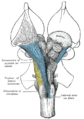
The brainstem is the stalk-like part of the brain that interconnects the cerebrum and diencephalon with the spinal cord. In the human brain, the brainstem is composed of the midbrain, the pons, and the medulla oblongata. The midbrain is continuous with the thalamus of the diencephalon through the tentorial notch.

<span class="mw-page-title-main">Oculomotor nerve</span> Cranial nerve III, for eye movements

The oculomotor nerve, also known as the third cranial nerve, cranial nerve III, or simply CN III, is a cranial nerve that enters the orbit through the superior orbital fissure and innervates extraocular muscles that enable most movements of the eye and that raise the eyelid. The nerve also contains fibers that innervate the intrinsic eye muscles that enable pupillary constriction and accommodation. The oculomotor nerve is derived from the basal plate of the embryonic midbrain. Cranial nerves IV and VI also participate in control of eye movement.

<span class="mw-page-title-main">Third ventricle</span> Ventricle of the brain located between the two thalami

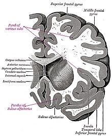
The third ventricle is one of the four connected ventricles of the ventricular system within the mammalian brain. It is a slit-like cavity formed in the diencephalon between the two thalami, in the midline between the right and left lateral ventricles, and is filled with cerebrospinal fluid (CSF).

<span class="mw-page-title-main">Midbrain</span> Forward-most portion of the brainstem

The midbrain or mesencephalon is the rostral-most portion of the brainstem connecting the diencephalon and cerebrum with the pons. It consists of the cerebral peduncles, tegmentum, and tectum.

<span class="mw-page-title-main">Spinothalamic tract</span> Sensory pathway from the skin to the thalamus

The spinothalamic tract is a part of the anterolateral system or the ventrolateral system, a sensory pathway to the thalamus. From the ventral posterolateral nucleus in the thalamus, sensory information is relayed upward to the somatosensory cortex of the postcentral gyrus.

<span class="mw-page-title-main">Spinocerebellar tract</span> Nerve tract in humans

The spinocerebellar tract is a nerve tract originating in the spinal cord and terminating in the same side (ipsilateral) of the cerebellum.

<span class="mw-page-title-main">Subarachnoid cisterns</span> Spaces around the brain filled with cerebrospinal fluid

The subarachnoid cisterns are spaces formed by openings in the subarachnoid space, an anatomic space in the meninges of the brain. The space is situated between the two meninges, the arachnoid mater and the pia mater. These cisterns are filled with cerebrospinal fluid (CSF).

<span class="mw-page-title-main">Trochlear nucleus</span> Motor nucleus of cranial nerve IV

The nucleus of the trochlear nerve is a motor nucleus in the medial midbrain giving rise to the trochlear nerve.

<span class="mw-page-title-main">Superior cerebellar peduncle</span>

In the human brain, the superior cerebellar peduncle is a paired structure of white matter that connects the cerebellum to the midbrain. It consists mainly of efferent fibers, the cerebellothalamic tract that runs from a cerebellar hemisphere to the contralateral thalamus, and the cerebellorubral tract that runs from a cerebellar hemisphere to the red nucleus. It also contains afferent tracts, most prominent of which is the ventral spinocerebellar tract. Other afferent tracts are the trigeminothalamic fibers, tectocerebellar fibers, and noradrenergic fibers from the locus coeruleus. The superior peduncle emerges from the upper and medial parts of the white matter of each hemisphere and is placed under cover of the upper part of the cerebellum.

<span class="mw-page-title-main">Decussation</span> Biological term to describe a crossing

Decussation is used in biological contexts to describe a crossing. In Latin anatomical terms, the form decussatio is used, e.g. decussatio pyramidum.

<span class="mw-page-title-main">Anterior white commissure</span>

The anterior white commissure is a bundle of nerve fibers which cross the midline of the spinal cord just anterior to the gray commissure. A delta fibers and C fibers carrying pain sensation in the spinothalamic tract contribute to this commissure, as do fibers of the anterior corticospinal tract, which carry motor signals from the primary motor cortex.

<span class="mw-page-title-main">Spinal cord</span> Long, tubular central nervous system structure in the vertebral column

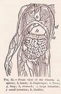
The spinal cord is a long, thin, tubular structure made up of nervous tissue that extends from the medulla oblongata in the brainstem to the lumbar region of the vertebral column (backbone) of vertebrate animals. The center of the spinal cord is hollow and contains a structure called central canal, which contains cerebrospinal fluid. The spinal cord is also covered by meninges and enclosed by the neural arches. Together, the brain and spinal cord make up the central nervous system (CNS).

<span class="mw-page-title-main">Nerve tract</span> Bundle of nerve fibers (axons) connecting nuclei of the central nervous system

A nerve tract is a bundle of nerve fibers (axons) connecting nuclei of the central nervous system. In the peripheral nervous system this is known as a nerve fascicle, and has associated connective tissue. The main nerve tracts in the central nervous system are of three types: association fibers, commissural fibers, and projection fibers. A nerve tract may also be referred to as a commissure, decussation, or neural pathway. A commissure connects the two cerebral hemispheres at the same levels, while a decussation connects at different levels.

<span class="mw-page-title-main">Anatomical plane</span> Anatomy method to describe locations

An anatomical plane is a hypothetical plane used to transect the body, in order to describe the location of structures or the direction of movements. In human and non-human anatomy, three principal planes are used:

<span class="mw-page-title-main">Anatomical terminology</span> Scientific terminology used by anatomists, zoologists, and health professionals

Anatomical terminology is a form of scientific terminology used by anatomists, zoologists, and health professionals such as doctors, physicians, and pharmacists.

<span class="mw-page-title-main">Contralateral brain</span> Each side of the forebrain represents the opposite side of the body

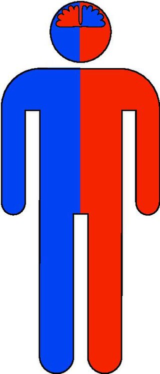
The contralateral organization of the forebrain is the property that the hemispheres of the cerebrum and the thalamus represent mainly the contralateral side of the body. Consequently, the left side of the forebrain mostly represents the right side of the body, and the right side of the brain primarily represents the left side of the body. The contralateral organization involves both executive and sensory functions. The contralateral organization is only present in vertebrates.

<span class="mw-page-title-main">Chiasm (anatomy)</span> Nerve crossings outside the central nervous system

In anatomy a chiasm is the spot where two structures cross, forming an X-shape. Examples of chiasms are:

References

  1. 1 2 Hal., Blumenfeld (2010). Neuroanatomy through clinical cases (2nd ed.). Sunderland, Mass.: Sinauer Associates. ISBN   9780878930586. OCLC   473478856.