Related Research Articles
Cranial nerves are the nerves that emerge directly from the brain, in contrast to spinal nerves. Ten of the cranial nerves originate in the brainstem. Cranial nerves relay information between the brain and parts of the body, primarily to and from regions of the head and neck.

The vagus nerve, historically cited as the pneumogastric nerve, is the tenth cranial nerve or CN X, and interfaces with the parasympathetic control of the heart, lungs, and digestive tract. The vagus nerves are normally referred to in the singular. It is the longest nerve of the autonomic nervous system in the human body. The ending part of vagus nerve is known as spinal accessory nucleus.

The facial nerve is the seventh cranial nerve, or simply CN VII. It emerges from the pons of the brainstem, controls the muscles of facial expression, and functions in the conveyance of taste sensations from the anterior two-thirds of the tongue. The nerves typically travels from the pons through the facial canal in the temporal bone and exits the skull at the stylomastoid foramen. It arises from the brainstem from an area posterior to the cranial nerve VI and anterior to cranial nerve VIII.
The glossopharyngeal nerve, known as the ninth cranial nerve, is a mixed nerve that carries afferent sensory and efferent motor information. It exits the brainstem out from the sides of the upper medulla, just anterior to the vagus nerve. The motor division of the glossopharyngeal nerve is derived from the basal plate of the embryonic medulla oblongata, while the sensory division originates from the cranial neural crest.

The mandibular nerve (V3) is the largest of the three divisions of the trigeminal nerve, the fifth cranial nerve (CN V).

The internal carotid artery is located in the inner side of the neck in contrast to the external carotid artery. In human anatomy, they arise from the common carotid arteries where these bifurcate into the internal and external carotid arteries at cervical vertebral level 3 or 4; the internal carotid artery supplies the brain including eyes, while the external carotid nourishes other portions of the head, such as face, scalp, skull, and meninges.

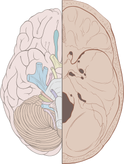
Dura mater is a thick membrane made of dense irregular connective tissue that surrounds the brain and spinal cord. It is the outermost of the three layers of membrane called the meninges that protect the central nervous system. The other two meningeal layers are the arachnoid mater and the pia mater. The dura surrounds the brain and the spinal cord and is responsible for keeping in the cerebrospinal fluid. It is derived primarily from the neural crest cell population, with postnatal contributions of the paraxial mesoderm.

Dura mater is the outermost meningeal layer that covers the brain and spinal cord. It consists of two layers; the inner meningeal layer and the outer periosteal layer. The potential space between the layers of the dura mater is called the epidural space. The epidural space exists around both the brain and the spinal cord.

The middle meningeal artery is typically the third branch of the first portion of the maxillary artery, one of the two terminal branches of the external carotid artery. After branching off the maxillary artery in the infratemporal fossa, it runs through the foramen spinosum to supply the dura mater the outer meningeal layer, and the calvaria. The middle meningeal artery is the largest of the three (paired) arteries that supply the meninges, the others being the anterior meningeal artery and the posterior meningeal artery.
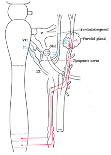
The auriculotemporal nerve is a branch of the mandibular nerve (V3) that runs with the superficial temporal artery and vein, and provides sensory innervation to various regions on the side of the head.
The auricular branch of the vagus nerve is often termed the Alderman's nerve or Arnold's nerve. The latter name is an eponym for Friedrich Arnold. The auricular branch of the vagus nerve supplies sensory innervation to the skin of the ear canal, tragus, and auricle.

The foramen spinosum is one of two foramina located in the base of the human skull, on the sphenoid bone. It is situated just anterior to the spine of the sphenoid bone, and just lateral to the foramen ovale. The middle meningeal artery, middle meningeal vein, and the meningeal branch of the mandibular nerve pass through the foramen.

The maxillary nerve (CN V2) is one of the three branches or divisions of the trigeminal nerve, the fifth (V) cranial nerve. It comprises the principal functions of sensation from the maxillary, nasal cavity, sinuses, the palate and subsequently that of the mid-face, and is intermediate, both in position and size, between the ophthalmic nerve and the mandibular nerve.

The ophthalmic nerve is the first branch of the trigeminal nerve. The ophthalmic nerve is a sensory nerve mostly carrying general somatic afferent fibers that transmit sensory information to the CNS from structures of the eyeball, the skin of the upper face and anterior scalp, the lining of the upper part of the nasal cavity and air cells, and the meninges of the anterior cranial fossa. Some of ophthalmic nerve branches also convey parasympathetic fibers.

The occipital artery arises from the external carotid artery opposite the facial artery. Its path is below the posterior belly of digastric to the occipital region. This artery supplies blood to the back of the scalp and sternocleidomastoid muscles, and deep muscles in the back and neck.

In human anatomy, the pterygopalatine fossa is a fossa in the skull. A human skull contains two pterygopalatine fossae—one on the left side, and another on the right side. Each fossa is a cone-shaped paired depression deep to the infratemporal fossa and posterior to the maxilla on each side of the skull, located between the pterygoid process and the maxillary tuberosity close to the apex of the orbit. It is the indented area medial to the pterygomaxillary fissure leading into the sphenopalatine foramen. It communicates with the nasal and oral cavities, infratemporal fossa, orbit, pharynx, and middle cranial fossa through eight foramina.

The petrous part of the temporal bone is pyramid-shaped and is wedged in at the base of the skull between the sphenoid and occipital bones. Directed medially, forward, and a little upward, it presents a base, an apex, three surfaces, and three angles, and houses in its interior, the components of the inner ear. The petrous portion is among the most basal elements of the skull and forms part of the endocranium. Petrous comes from the Latin word petrosus, meaning "stone-like, hard". It is one of the densest bones in the body.
The superior ganglion of the vagus nerve, (jugular ganglion) is a sensory ganglion of the peripheral nervous system. It is located within the jugular foramen, where the vagus nerve exits the skull. It is smaller than and above the inferior ganglion of the vagus nerve.

The middle cranial fossa, deeper than the anterior cranial fossa, is narrow medially and widens laterally to the sides of the skull. It is separated from the posterior fossa by the clivus and the petrous crest.

The infratemporal fossa is an irregularly shaped cavity, situated below and medial to the zygomatic arch. It is not fully enclosed by bone in all directions, and it contains superficial muscles that are visible during dissection after removing skin and fascia: namely, the lower part of the temporalis muscle, the lateral pterygoid, and the medial pterygoid.
References
- ↑ Kemp, William J.; Tubbs, R. Shane; Cohen-Gadol, Aaron A. (2012-11-01). "The Innervation of the Cranial Dura Mater: Neurosurgical Case Correlates and a Review of the Literature". World Neurosurgery. 78 (5): 505–510. doi:10.1016/j.wneu.2011.10.045. ISSN 1878-8750. PMID 22120554.
- ↑ Keller, Jeffrey T.; Saunders, Mary C.; Beduk, Altay; Jollis, James G. (1985-01-01). "Innervation of the posterior fossa dura of the cat". Brain Research Bulletin. 14 (1): 97–102. doi:10.1016/0361-9230(85)90181-9. ISSN 0361-9230. PMID 3872702.