Hypoglossal nucleus

Last updated
Hypoglossal nucleus
Gray694.png
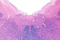
Section of the medulla oblongata. (Hypoglossal nucleus visible top left.)
Gray696.svg
The cranial nerve nuclei schematically represented; dorsal view. Motor nuclei in red; sensory in blue. (XII labeled at bottom left.)
Details
Identifiers
Latin nucleus nervi hypoglossi
NeuroNames 757
NeuroLex ID birnlex_2644
TA A14.1.04.227
FMA 54505
Anatomical terms of neuroanatomy

The hypoglossal nucleus is a cranial nerve nucleus, found within the medulla. Being a motor nucleus, it is close to the midline. In the open medulla, it is visible as what is known as the hypoglossal trigone , a raised area (medial to the vagal trigone) protruding slightly into the fourth ventricle.

Cranial nerve nucleus

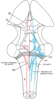
A cranial nerve nucleus is a collection of neurons in the brain stem that is associated with one or more cranial nerves. Axons carrying information to and from the cranial nerves form a synapse first at these nuclei. Lesions occurring at these nuclei can lead to effects resembling those seen by the severing of nerve(s) they are associated with. All the nuclei except that of the trochlear nerve supply nerves of the same side of the body.

Medulla oblongata structure of the brain stem

The medulla oblongata is a long stem-like structure which makes up part of the brainstem. It is anterior and partially inferior to the cerebellum. It is a cone-shaped neuronal mass responsible for autonomic (involuntary) functions ranging from vomiting to sneezing. The medulla contains the cardiac, respiratory, vomiting and vasomotor centers and therefore deals with the autonomic functions of breathing, heart rate and blood pressure as well as the sleep wake cycle.

Hypoglossal trigone

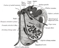
In the upper part of the medulla oblongata, the hypoglossal nucleus approaches the rhomboid fossa, where it lies close to the middle line, under an eminence named the hypoglossal trigone. It is a slight elevation in the floor of the inferior recess of the fourth ventricle, beneath which is the nucleus of origin of the twelfth cranial nerve.

Contents

The hypoglossal nucleus is located between the dorsal motor nucleus of the vagus and the midline of the medulla. Axons from the hypoglossal nucleus pass anteriorly through the medulla forming the hypoglossal nerve which exits between the pyramid and olive in a groove called the anterolateral sulcus.

Dorsal nucleus of vagus nerve

The dorsal nucleus of the vagus nerve is a cranial nerve nucleus for the vagus nerve in the medulla that lies ventral to the floor of the fourth ventricle. It mostly serves parasympathetic vagal functions in the gastrointestinal tract, lungs, and other thoracic and abdominal vagal innervations. The cell bodies for the preganglionic parasympathetic vagal neurons that innervate the heart reside in the nucleus ambiguus.

Hypoglossal nerve cranial nerve

The hypoglossal nerve is the twelfth cranial nerve, and innervates all the extrinsic and intrinsic muscles of the tongue, except for the palatoglossus which is innervated by the vagus nerve. It is a nerve with a solely motor function. The nerve arises from the hypoglossal nucleus in the medulla as a number of small rootlets, passes through the hypoglossal canal and down through the neck, and eventually passes up again over the tongue muscles it supplies into the tongue. There are two hypoglossal nerves in the body: one on the left, and one on the right.

Medullary pyramids (brainstem)

The medullary pyramids are paired white matter structures of the brainstem's medulla oblongata that contain motor fibers of the corticospinal and corticobulbar tracts – known together as the pyramidal tracts. The lower limit of the pyramids is marked when the fibers cross (decussate).

See also

Additional images

Micrograph process for producing pictures with a microscope

A micrograph or photomicrograph is a photograph or digital image taken through a microscope or similar device to show a magnified image of an object. This is opposed to a macrograph or photomacrograph, an image which is also taken on a microscope but is only slightly magnified, usually less than 10 times. Micrography is the practice or art of using microscopes to make photographs.

BrainMaps NIH-funded interactive zoomable high-resolution digital brain atlas and virtual microscope

BrainMaps is an NIH-funded interactive zoomable high-resolution digital brain atlas and virtual microscope that is based on more than 140 million megapixels of scanned images of serial sections of both primate and non-primate brains and that is integrated with a high-speed database for querying and retrieving data about brain structure and function over the internet.


Related Research Articles

Abducens nerve

The abducens nerve is the sixth cranial nerve (CNVI), in humans, that controls the movement of the lateral rectus muscle, responsible for outward gaze. It is a somatic efferent nerve.

Pons part of the brainstem

The pons is part of the brainstem, and in humans and other bipeds lies inferior to the midbrain, superior to the medulla oblongata and anterior to the cerebellum.

Brainstem posterior part of the brain, adjoining and structurally continuous with the spinal cord

The brainstem is the posterior part of the brain, continuous with the spinal cord. In the human brain the brainstem includes the midbrain, the pons and medulla oblongata of the hindbrain. The midbrain continues with the thalamus of the diencephalon through the tentorial notch, and sometimes the diencephalon is included in the brainstem.

Glossopharyngeal nerve

The glossopharyngeal nerve, known as the ninth cranial nerve, is a mixed nerve that carries afferent sensory and efferent motor information. It exits the brainstem out from the sides of the upper medulla, just rostral to the vagus nerve. The motor division of the glossopharyngeal nerve is derived from the basal plate of the embryonic medulla oblongata, while the sensory division originates from the cranial neural crest.

In neuroanatomy, a nucleus is a cluster of neurons in the central nervous system, located deep within the cerebral hemispheres and brainstem. The neurons in one nucleus usually have roughly similar connections and functions. Nuclei are connected to other nuclei by tracts, the bundles (fascicles) of axons extending from the cell bodies. A nucleus is one of the two most common forms of nerve cell organization, the other being layered structures such as the cerebral cortex or cerebellar cortex. In anatomical sections, a nucleus shows up as a region of gray matter, often bordered by white matter. The vertebrate brain contains hundreds of distinguishable nuclei, varying widely in shape and size. A nucleus may itself have a complex internal structure, with multiple types of neurons arranged in clumps (subnuclei) or layers.

Medial lemniscus

The medial lemniscus, also known as Reil's band or Reil's ribbon, is a large ascending bundle of heavily myelinated axons that decussate in the brainstem, specifically in the medulla oblongata. The medial lemniscus is formed by the crossings of the internal arcuate fibers. The internal arcuate fibers are composed of axons of nucleus gracilis and nucleus cuneatus. The axons of the nucleus gracilis and nucleus cuneatus in the medial lemniscus have cell bodies that lie contralaterally.

Oculomotor nucleus

The fibers of the oculomotor nerve arise from a nucleus in the midbrain, which lies in the gray substance of the floor of the cerebral aqueduct and extends in front of the aqueduct for a short distance into the floor of the third ventricle. From this nucleus the fibers pass forward through the tegmentum, the red nucleus, and the medial part of the substantia nigra, forming a series of curves with a lateral convexity, and emerge from the oculomotor sulcus on the medial side of the cerebral peduncle.

Solitary nucleus

In the human brainstem, the solitary nucleus(SN) is a series of purely sensory nuclei forming a vertical column of grey matter embedded in the medulla oblongata. Through the center of the SN runs the solitary tract, a white bundle of nerve fibers, including fibers from the facial, glossopharyngeal and vagus nerves, that innervate the SN. The SN projects to, among other regions, the reticular formation, parasympathetic preganglionic neurons, hypothalamus and thalamus, forming circuits that contribute to autonomic regulation. Cells along the length of the SN are arranged roughly in accordance with function; for instance, cells involved in taste are located In the rostrum part, while those receiving information from cardio-respiratory and gastrointestinal processes are found in the caudal part.

Nucleus ambiguus nucleus

The nucleus ambiguus is a group of large motor neurons, situated deep in the medullary reticular formation named by Jacob Clarke. The nucleus ambiguus contains the cell bodies of neurons that innervate the muscles of the soft palate, pharynx, and larynx which are associated with speech and swallowing. As well as motor neurons, the nucleus ambiguus contains preganglionic parasympathetic neurons which innervate postganglionic parasympathetic neurons in the heart.

Fourth ventricle

The fourth ventricle is one of the four connected fluid-filled cavities within the human brain. These cavities, known collectively as the ventricular system, consist of the left and right lateral ventricles, the third ventricle, and the fourth ventricle. The fourth ventricle extends from the cerebral aqueduct to the obex, and is filled with cerebrospinal fluid (CSF).

Olivary body

In anatomy, the olivary bodies or simply olives are a pair of prominent oval structures in the medulla oblongata, the lower portion of the brainstem. They contain the olivary nuclei.

Pontine tegmentum section of the mammalian brain

The pontine tegmentum, or dorsal pons, is located within the brainstem, and is one of two parts of the pons, the other being the ventral pons or basilar part of the pons. The pontine tegmentum can be defined in contrast to the basilar pons: basilar pons contains the corticospinal tract running craniocaudally and can be considered the rostral extension of the ventral medulla oblongata; however, basilar pons is distinguished from ventral medulla oblongata in that it contains additional transverse pontine fibres that continue laterally to become the middle cerebellar peduncle. The pontine tegmentum is all the material dorsal from the basilar pons to the fourth ventricle. Along with the dorsal surface of the medulla, it forms part of the rhomboid fossa – the floor of the fourth ventricle.

Trochlear nucleus

The nucleus of the trochlear nerve is located in the midbrain, at an intercollicular level between the superior colliculus and inferior colliculus. It is a motor nucleus, and so is located near the midline, embedded within the medial longitudinal fasciculus. The oculomotor nerve and trochlear nerve are the only two cranial nerves with nuclei in the midbrain, other than the trigeminal nerve, which has a midbrain nucleus called the mesencephalic nucleus of trigeminal nerve, which functions in preserving dentition.

Trigeminal nerve nuclei

The sensory trigeminal nerve nuclei are the largest of the cranial nerve nuclei, and extend through the whole of the midbrain, pons and medulla, and into the high cervical spinal cord.