
The autonomic nervous system (ANS), formerly referred to as the vegetative nervous system, is a division of the nervous system that operates internal organs, smooth muscle and glands. The autonomic nervous system is a control system that acts largely unconsciously and regulates bodily functions, such as the heart rate, its force of contraction, digestion, respiratory rate, pupillary response, urination, and sexual arousal. This system is the primary mechanism in control of the fight-or-flight response.
The parasympathetic nervous system (PSNS) is one of the three divisions of the autonomic nervous system, the others being the sympathetic nervous system and the enteric nervous system. The enteric nervous system is sometimes considered part of the autonomic nervous system, and sometimes considered an independent system.

The medulla oblongata or simply medulla is a long stem-like structure which makes up the lower part of the brainstem. It is anterior and partially inferior to the cerebellum. It is a cone-shaped neuronal mass responsible for autonomic (involuntary) functions, ranging from vomiting to sneezing. The medulla contains the cardiac, respiratory, vomiting and vasomotor centers, and therefore deals with the autonomic functions of breathing, heart rate and blood pressure as well as the sleep–wake cycle.

The pons is part of the brainstem that in humans and other mammals, lies inferior to the midbrain, superior to the medulla oblongata and anterior to the cerebellum.
The brainstem is the stalk-like part of the brain that interconnects the cerebrum and diencephalon with the spinal cord. In the human brain, the brainstem is composed of the midbrain, the pons, and the medulla oblongata. The midbrain is continuous with the thalamus of the diencephalon through the tentorial notch.

In neuroanatomy, the trigeminal nerve (lit. triplet nerve), also known as the fifth cranial nerve, cranial nerve V, or simply CN V, is a cranial nerve responsible for sensation in the face and motor functions such as biting and chewing; it is the most complex of the cranial nerves. Its name (trigeminal, from Latin tri- 'three', and -geminus 'twin') derives from each of the two nerves (one on each side of the pons) having three major branches: the ophthalmic nerve (V1), the maxillary nerve (V2), and the mandibular nerve (V3). The ophthalmic and maxillary nerves are purely sensory, whereas the mandibular nerve supplies motor as well as sensory (or "cutaneous") functions. Adding to the complexity of this nerve is that autonomic nerve fibers as well as special sensory fibers (taste) are contained within it.

The glossopharyngeal nerve, also known as the ninth cranial nerve, cranial nerve IX, or simply CN IX, is a cranial nerve that exits the brainstem from the sides of the upper medulla, just anterior to the vagus nerve. Being a mixed nerve (sensorimotor), it carries afferent sensory and efferent motor information. The motor division of the glossopharyngeal nerve is derived from the basal plate of the embryonic medulla oblongata, whereas the sensory division originates from the cranial neural crest.

The spinothalamic tract is a part of the anterolateral system or the ventrolateral system, a sensory pathway to the thalamus. From the ventral posterolateral nucleus in the thalamus, sensory information is relayed upward to the somatosensory cortex of the postcentral gyrus.

The solitary nucleus is a series of sensory nuclei forming a vertical column of grey matter in the medulla oblongata of the brainstem. It receives general visceral and/or special visceral inputs from the facial nerve, glossopharyngeal nerve and vagus nerve ; it receives and relays stimuli related to taste and visceral sensation. It sends outputs to various parts of the brain, such as the hypothalamus, thalamus, and reticular formation. Neuron cell bodies of the SN are roughly somatotopically arranged along its length according to function.

The spinocerebellar tract is a nerve tract originating in the spinal cord and terminating in the same side (ipsilateral) of the cerebellum.
The dorsal longitudinal fasciculus (DLF) is a longitudinal tract interconnecting the posterior hypothalamus, and the inferior medulla oblongata. It contains both ascending tracts and descending tracts, and serves to link the forebrain, and the visceral autonomic centres of the lower brainstem. It conveys both visceral motor signals, and sensory signals.
The inferior ganglion of the vagus nerve is one of the two sensory ganglia of each vagus nerve. It contains neuron cell bodies of general visceral afferent fibers and special visceral afferent fibers. It is situated within the jugular fossa just below the skull. It is situated just below the superior ganglion of vagus nerve.
The mammillothalamic tract is an efferent pathway of the mammillary body which projects to the anterior nuclei of thalamus. It consists of heavily myelinated fibres. It is part of a brain circuit involved in spatial memory.

The general visceral afferent (GVA) fibers conduct sensory impulses from the internal organs, glands, and blood vessels to the central nervous system. They are considered to be part of the visceral nervous system, which is closely related to the autonomic nervous system, but 'visceral nervous system' and 'autonomic nervous system' are not direct synonyms and care should be taken when using these terms. Unlike the efferent fibers of the autonomic nervous system, the afferent fibers are not classified as either sympathetic or parasympathetic.
Special visceral afferent fibers (SVA) are afferent fibers that develop in association with the gastrointestinal tract. They carry the special sense of taste (gustation). The cranial nerves containing SVA fibers are the facial nerve (VII), the glossopharyngeal nerve (IX), and the vagus nerve (X). The facial nerve receives taste from the anterior 2/3 of the tongue; the glossopharyngeal from the posterior 1/3, and the vagus nerve from the epiglottis. The sensory processes, using their primary cell bodies from the inferior ganglion, send projections to the medulla, from which they travel in the tractus solitarius, later terminating at the rostral nucleus solitarius.
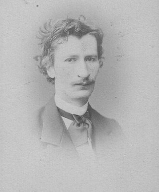
Johann Friedrich Wilhelm Krause was a German anatomist born in Hanover. He was the son of anatomist Karl Friedrich Theodor Krause (1797-1868).

The salivatory nuclei are two parasympathetic general visceral efferent cranial nerve nuclei - the superior salivatory nucleus and the inferior salivatory nucleus - that innervate the salivary glands. Both are located in the pontine tegmentum of the brainstem.

The gustatory nucleus is the rostral part of the solitary nucleus located in the medulla. The gustatory nucleus is associated with the sense of taste and has two sections, the rostral and lateral regions. A close association between the gustatory nucleus and visceral information exists for this function in the gustatory system, assisting in homeostasis - via the identification of food that might be possibly poisonous or harmful for the body. There are many gustatory nuclei in the brain stem. Each of these nuclei corresponds to three cranial nerves, the facial nerve (VII), the glossopharyngeal nerve (IX), and the vagus nerve (X) and GABA is the primary inhibitory neurotransmitter involved in its functionality. All visceral afferents in the vagus and glossopharyngeal nerves first arrive in the nucleus of the solitary tract and information from the gustatory system can then be relayed to the thalamus and cortex.
Hans Paul Bernhard Gierke was a German anatomist who was a native of Stettin.

