
The palate is the roof of the mouth in humans and other mammals. It separates the oral cavity from the nasal cavity. A similar structure is found in crocodilians, but in most other tetrapods, the oral and nasal cavities are not truly separate. The palate is divided into two parts, the anterior bony hard palate and the posterior fleshy soft palate.

The maxilla in animals is the upper fixed bone of the jaw formed from the fusion of two maxillary bones. The upper jaw includes the hard palate in the front of the mouth. The two maxillary bones are fused at the intermaxillary suture, forming the anterior nasal spine. This is similar to the mandible, which is also a fusion of two mandibular bones at the mandibular symphysis. The mandible is the movable part of the jaw.
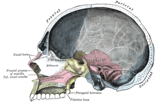
The palatine bones are two irregular bones of the facial skeleton in many animal species. Together with the maxillae they comprise the hard palate.

The vomer is one of the unpaired facial bones of the skull. It is located in the midsagittal line, and articulates with the sphenoid, the ethmoid, the left and right palatine bones, and the left and right maxillary bones. The vomer forms the inferior part of the nasal septum, with the superior part formed by the perpendicular plate of the ethmoid bone. The name is derived from the Latin word for a ploughshare and the shape of the bone.

The pterygopalatine ganglion is a parasympathetic ganglion found in the pterygopalatine fossa. It is largely innervated by the greater petrosal nerve ; and its axons project to the lacrimal glands and nasal mucosa. The flow of blood to the nasal mucosa, in particular the venous plexus of the conchae, is regulated by the pterygopalatine ganglion and heats or cools the air in the nose. It is one of four parasympathetic ganglia of the head and neck, the others being the submandibular ganglion, otic ganglion, and ciliary ganglion.

The palatopharyngeusmuscle is a small muscle in the roof of the mouth.
The maxillary nerve (CN V2) is one of the three branches or divisions of the trigeminal nerve, the fifth (V) cranial nerve. It comprises the principal functions of sensation from the maxillary, nasal cavity, sinuses, the palate and subsequently that of the mid-face, and is intermediate, both in position and size, between the ophthalmic nerve and the mandibular nerve.

The descending palatine artery is a branch of the third part of the maxillary artery supplying the hard and soft palate.

In the opening of the incisive foramen, the orifices of two lateral canals are visible; they are named the incisive canals or foramina of Stensen.
In the human mouth, the incisive foramen, also called anterior palatine foramen, or nasopalatine foramen is a funnel-shaped opening in the bone of the oral hard palate immediately behind the incisor teeth where blood vessels and nerves pass. The incisive foramen is continuous with the incisive canal, this foramen or group of foramina is located behind the central incisor teeth in the incisive fossa of the maxilla.

The sphenopalatine foramen is a foramen in the skull that connects the nasal cavity with the pterygopalatine fossa.
The greater palatine artery is a branch of the descending palatine artery and contributes to the blood supply of the hard palate and nasal septum.
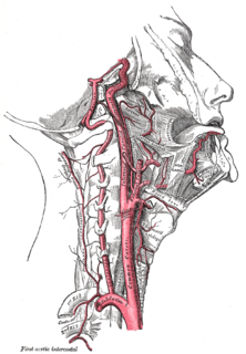
The ascending palatine artery is an artery in the head that branches off the facial artery and runs up the superior pharyngeal constrictor muscle.

At either posterior angle of the hard palate is the greater palatine foramen, for the transmission of the descending palatine vessels and greater palatine nerve; and running anteriorly (forward) and medially from it is a groove, for the same vessels and nerve.

Behind the greater palatine foramen is the pyramidal process of the palatine bone, perforated by one or more lesser palatine foramina which carry the lesser palatine nerve, and marked by the commencement of a transverse ridge, for the attachment of the tendinous expansion of the tensor veli palatini.
The pyramidal process of the palatine bone projects backward and lateralward from the junction of the horizontal and vertical parts, and is received into the angular interval between the lower extremities of the pterygoid plates.

In human anatomy of the mouth, the palatine process of maxilla, is a thick, horizontal process of the maxilla. It forms the anterior three quarters of the hard palate, the horizontal plate of the palatine bone making up the rest.

The greater palatine nerve is a branch of the pterygopalatine ganglion that carries both general sensory fibres from the maxillary nerve and parasympathetic fibers from the nerve of the pterygoid canal. It descends through the greater palatine canal, emerges upon the hard palate through the greater palatine foramen, and passes forward in a groove in the hard palate, nearly as far as the incisor teeth.

The greater palatine canal is a passage in the skull that transmits the descending palatine artery, vein, and greater and lesser palatine nerves between the pterygopalatine fossa and the oral cavity.
The lesser palatine nerve (posterior palatine nerve) is one of two palatine nerves that descends through the greater palatine canal, and emerges by the lesser palatine foramen. It is a branch of the maxillary nerve (CN V2) It also has nasal branches that innervate the nasal cavity.
The public domain consists of all the creative works to which no exclusive intellectual property rights apply. Those rights may have expired, been forfeited, expressly waived, or may be inapplicable.

Gray's Anatomy is an English language textbook of human anatomy originally written by Henry Gray and illustrated by Henry Vandyke Carter. Earlier editions were called Anatomy: Descriptive and Surgical and Gray's Anatomy: Descriptive and Applied, but the book's name is commonly shortened to, and later editions are titled, Gray's Anatomy. The book is widely regarded as an extremely influential work on the subject, and has continued to be revised and republished from its initial publication in 1858 to the present day. The latest edition of the book, the 41st, was published in September 2015.

