
The facial nerve, also known as the seventh cranial nerve, cranial nerve VII, or simply CN VII, is a cranial nerve that emerges from the pons of the brainstem, controls the muscles of facial expression, and functions in the conveyance of taste sensations from the anterior two-thirds of the tongue. The nerve typically travels from the pons through the facial canal in the temporal bone and exits the skull at the stylomastoid foramen. It arises from the brainstem from an area posterior to the cranial nerve VI and anterior to cranial nerve VIII.

The hypoglossal nerve, also known as the twelfth cranial nerve, cranial nerve XII, or simply CN XII, is a cranial nerve that innervates all the extrinsic and intrinsic muscles of the tongue except for the palatoglossus, which is innervated by the vagus nerve. CN XII is a nerve with a sole motor function. The nerve arises from the hypoglossal nucleus in the medulla as a number of small rootlets, pass through the hypoglossal canal and down through the neck, and eventually passes up again over the tongue muscles it supplies into the tongue.

The stylohyoid muscle is one of the suprahyoid muscles. Its originates from the styloid process of the temporal bone; it inserts onto hyoid bone. It is innervated by a branch of the facial nerve. It acts draw the hyoid bone upwards and backwards.

The facial artery is a branch of the external carotid artery that supplies structures of the superficial face.

The nasociliary nerve is a branch of the ophthalmic nerve (CN V1) (which is in turn a branch of the trigeminal nerve (CN V)). It is intermediate in size between the other two branches of the ophthalmic nerve, the frontal nerve and lacrimal nerve.

The pharyngeal arches, also known as visceral arches, are structures seen in the embryonic development of vertebrates that are recognisable precursors for many structures. In fish, the arches are known as the branchial arches, or gill arches.

The geniculate ganglion is a collection of pseudounipolar sensory neurons of the facial nerve located in the facial canal of the head. It receives fibers from the facial nerve. It sends fibers that supply the lacrimal glands, submandibular glands, sublingual glands, tongue, palate, pharynx, external auditory meatus, stapedius muscle, posterior belly of the digastric muscle, stylohyoid muscle, and muscles of facial expression.
The occipital artery is a branch of the external carotid artery that provides arterial supply to the back of the scalp, sternocleidomastoid muscles, and deep muscles of the back and neck.
The posterior auricular artery is a small artery that arises from the external carotid artery. It ascends along the side of the head. It supplies several muscles of the neck and several structures of the head.

The zygomaticotemporal nerve (zygomaticotemporal branch, temporal branch) is a cutaneous (sensory) nerve of the head. It is a branch of the zygomatic nerve (itself a branch of the maxillary nerve (CN V2)). It arises in the orbit and exits the orbit through the zygomaticotemporal foramen in the zygomatic bone to enter the temporal fossa. It is distributed to the skin of the side of the forehead. It also contains a parasympathetic secretomotor component for the lacrimal gland which it confers to the lacrimal nerve (which then delivers it to the gland).

The temporal styloid process is a slender bony process of the temporal bone extending downward and forward from the undersurface of the temporal bone just below the ear. The styloid process gives attachments to several muscles, and ligaments.
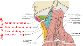
The anterior triangle is a region of the neck.

The masseteric nerve is a nerve of the face. It is a branch of the mandibular nerve (CN V3). It passes through the mandibular notch to reach masseter muscle. It provides motor innervation the masseter muscle, and sensory innervation to the temporomandibular joint.

The marginal mandibular branch of the facial nerve arises from the facial nerve in the parotid gland at the parotid plexus. It passes anterior-ward deep to the platysma and depressor anguli oris muscles. It provides motor innervation to muscles of the lower lip and chin: the depressor labii inferioris muscle, depressor anguli oris muscle, and mentalis muscle. It communicates with the mental branch of the inferior alveolar nerve.
The buccal branches of the facial nerve, are of larger size than the rest of the branches, pass horizontally forward to be distributed below the orbit and around the mouth.
The posterior auricular nerve is a nerve of the head. It is a branch of the facial nerve. It communicates with branches from the vagus nerve, the great auricular nerve, and the lesser occipital nerve. Its auricular branch supplies the posterior auricular muscle, the intrinsic muscles of the auricle, and gives sensation to the auricle. Its occipital branch supplies the occipitalis muscle.
The zygomatic branches of the facial nerve (malar branches) are nerves of the face. They run across the zygomatic bone to the lateral angle of the orbit. Here, they supply the orbicularis oculi muscle, and join with filaments from the lacrimal nerve and the zygomaticofacial branch of the maxillary nerve (CN V2).
The cervical branch of the facial nerve is a nerve in the neck. It is a branch of the facial nerve (VII). It supplies the platysma muscle, among other functions.
The digastric branch of facial nerve provides motor innervation to the posterior belly of the digastric muscle. It branches from the facial nerve near to the stylomastoid foramen as the CN VII exits the facial canal. It commonly arises in common with the stylohyoid branch of facial nerve.

The facial muscles are a group of striated skeletal muscles supplied by the facial nerve that, among other things, control facial expression. These muscles are also called mimetic muscles. They are only found in mammals, although they derive from neural crest cells found in all vertebrates. They are the only muscles that attach to the dermis.
