Sternohyoid muscle

Last updated
Sternohyoid muscle
Sternohyoid muscle.PNG
Muscles of neck. Sternohyoideus labeled at middle, just to the right of thyroid cartilage.
Sternohyoideus.png
Muscles of the neck. Lateral view. Sternohyoid muscle labeled
Details
Origin Manubrium of sternum
Insertion Hyoid bone
Artery Superior thyroid artery
Nerve C1-C3 by a branch of ansa cervicalis
Actions Depresses hyoid
Identifiers
Latin musculus sternohyoideus
TA98 A04.2.04.002
TA2 2168
FMA 13341
Anatomical terms of muscle

The sternohyoid muscle is a bilaterally paired, [1] long, [1] thin, [1] [2] narrow strap muscle [2] of the anterior neck. [1] It is one of the infrahyoid muscles. It is innervated by the ansa cervicalis. It acts to depress the hyoid bone.

Contents

The sternohyoid muscle is a flat muscle located on both sides of the neck, part of the infrahyoid muscle group. It originates from the medial edge of the clavicle, sternoclavicular ligament, and posterior side of the manubrium, and ascends to attach to the body of the hyoid bone. The sternohyoid muscle, along with other infrahyoid muscles, functions to depress the hyoid bone, which is important for activities such as speaking, chewing, and swallowing. Additionally, this muscle group contributes to the protection of the trachea, esophagus, blood vessels, and thyroid gland. The sternohyoid muscle also plays a minor role in head movements. [3]

Structure

The sternohyoid muscle is one of the paired strap muscles of the infrahyoid muscles. [4] [ verification needed ]

The muscle is directed superomedially from its origin to its insertion. The two muscles are separated by a considerable interval inferiorly, but usually converge by their mid-point and remain proximal until their superior insertion. [2]

Sternohyoid muscle oblique view SternoHyoid.png
Sternohyoid muscle oblique view

Origin

It arises from the posterior aspect of the medial end (sternal extremity of the clavicle, the posterior sternoclavicular ligament, and (the superoposterior portion of) the manubrium of sternum. [2]

It inserts onto the inferior border of the body of hyoid bone. [2]

Nerve supply

The sternohyoid muscle receives motor innervation from branches of the ansa cervicalis (which are ultimately derived from cervical spinal nerves C1-C3). [2]

Relations

The muscle is situated lateral to the trachea. [1]

Variations

The muscle may be absent, doubled, exhibit a clavicular slip (the cleidohyoideus), or interrupted by a tendinous intersection; [2] it sometimes presents a transverse tendinous inscription just distal to its origin.[ citation needed ]

Actions/movements

The muscle depresses the hyoid bone when the bone is in an elevated position. [2]

Function

The sternohyoid muscle performs a number of functions:

Additional images

Related Research Articles

<span class="mw-page-title-main">Neck</span> Part of the body on many vertebrates that connects the head with the torso

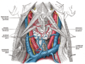
The neck is the part of the body on many vertebrates that connects the head with the torso. The neck supports the weight of the head and protects the nerves that carry sensory and motor information from the brain down to the rest of the body. In addition, the neck is highly flexible and allows the head to turn and flex in all directions. The structures of the human neck are anatomically grouped into four compartments: vertebral, visceral and two vascular compartments. Within these compartments, the neck houses the cervical vertebrae and cervical part of the spinal cord, upper parts of the respiratory and digestive tracts, endocrine glands, nerves, arteries and veins. Muscles of the neck are described separately from the compartments. They bound the neck triangles.

<span class="mw-page-title-main">Sternocleidomastoid muscle</span> Cervical muscle

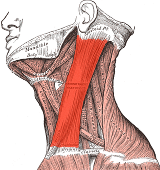
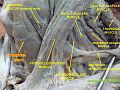
The sternocleidomastoid muscle is one of the largest and most superficial cervical muscles. The primary actions of the muscle are rotation of the head to the opposite side and flexion of the neck. The sternocleidomastoid is innervated by the accessory nerve.

<span class="mw-page-title-main">Infrahyoid muscles</span> Group of muscles

The infrahyoid muscles, or strap muscles, are a group of four pairs of muscles in the anterior (frontal) part of the neck. The four infrahyoid muscles are the sternohyoid, sternothyroid, thyrohyoid and omohyoid muscles.

<span class="mw-page-title-main">Omohyoid muscle</span> Human neck muscle

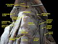
The omohyoid muscle is a muscle in the neck. It is one of the infrahyoid muscles. It consists of two bellies separated by an intermediate tendon. Its inferior belly is attached to the scapula; its superior belly is attached to the hyoid bone. Its intermediate tendon is anchored to the clavicle and first rib by a fascial sling. The omohyoid is innervated by the ansa cervicalis of the cervical plexus. It acts to depress the hyoid bone.

<span class="mw-page-title-main">Cervical plexus</span> Network of nerves in the neck

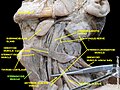
The cervical plexus is a nerve plexus of the anterior rami of the first four cervical spinal nerves C1-C4. The cervical plexus provides motor innervation to some muscles of the neck, and the diaphragm; it provides sensory innervation to parts of the head, neck, and chest.

<span class="mw-page-title-main">Digastric muscle</span> Small muscle located under the jaw in mammals

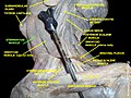
The digastric muscle is a bilaterally paired suprahyoid muscle located under the jaw. Its posterior belly is attached to the mastoid notch of temporal bone, and its anterior belly is attached to the digastric fossa of mandible; the two bellies are united by an intermediate tendon which is held in a loop that attaches to the hyoid bone. The anterior belly is innervated via the mandibular nerve, and the posterior belly is innervated via the facial nerve. It may act to depress the mandible or elevate the hyoid bone.

<span class="mw-page-title-main">Stylohyoid muscle</span> Muscle

The stylohyoid muscle is one of the suprahyoid muscles. Its originates from the styloid process of the temporal bone; it inserts onto hyoid bone. It is innervated by a branch of the facial nerve. It acts draw the hyoid bone upwards and backwards.

<span class="mw-page-title-main">Common carotid artery</span> One of the two arteries that supply the head and neck with blood

In anatomy, the left and right common carotid arteries (carotids) are arteries that supply the head and neck with oxygenated blood; they divide in the neck to form the external and internal carotid arteries.

<span class="mw-page-title-main">Sternothyroid muscle</span>

The sternothyroid muscle is an infrahyoid muscle of the neck. It acts to depress the hyoid bone.

<span class="mw-page-title-main">Thyrohyoid muscle</span> Neck muscle that depresses the hyoid bone and elevates the larynx

The thyrohyoid muscle is a small skeletal muscle of the neck. Above, it attaches onto the greater cornu of the hyoid bone; below, it attaches onto the oblique line of the thyroid cartilage. It is innervated by fibres derived from the cervical spinal nerve 1 that run with the hypoglossal nerve to reach this muscle. The thyrohyoid muscle depresses the hyoid bone and elevates the larynx during swallowing. By controlling the position and shape of the larynx, it aids in making sound.

<span class="mw-page-title-main">Shoulder girdle</span> Set of bones which connects the arm to the axial skeleton on each side

The shoulder girdle or pectoral girdle is the set of bones in the appendicular skeleton which connects to the arm on each side. In humans it consists of the clavicle and scapula; in those species with three bones in the shoulder, it consists of the clavicle, scapula, and coracoid. Some mammalian species have only the scapula.

<span class="mw-page-title-main">Superior thyroid artery</span>

The superior thyroid artery arises from the external carotid artery just below the level of the greater cornu of the hyoid bone and ends in the thyroid gland.

<span class="mw-page-title-main">Anterior triangle of the neck</span>

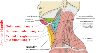
The anterior triangle is a region of the neck.

<span class="mw-page-title-main">Deep cervical fascia</span>

The deep cervical fascia lies under cover of the platysma, and invests the muscles of the neck; it also forms sheaths for the carotid vessels, and for the structures situated in front of the vertebral column. Its attachment to the hyoid bone prevents the formation of a dewlap.

<span class="mw-page-title-main">Sternoclavicular joint</span> Joint between the manubrium of the sternum and the clavicle bone

The sternoclavicular joint or sternoclavicular articulation is a synovial saddle joint between the manubrium of the sternum, and the clavicle, and the first costal cartilage. The joint possesses a joint capsule, and an articular disc, and is reinforced by multiple ligaments.

<span class="mw-page-title-main">Carotid triangle</span>

The carotid triangle is a portion of the anterior triangle of the neck.

<span class="mw-page-title-main">Muscular triangle</span>

The inferior carotid triangle, is bounded, in front, by the median line of the neck from the hyoid bone to the sternum; behind, by the anterior margin of the sternocleidomastoid; above, by the superior belly of the omohyoid.

<span class="mw-page-title-main">Cervical lymph nodes</span> Lymph nodes found in the neck

Cervical lymph nodes are lymph nodes found in the neck. Of the 800 lymph nodes in the human body, 300 are in the neck. Cervical lymph nodes are subject to a number of different pathological conditions including tumours, infection and inflammation.

<span class="mw-page-title-main">Investing layer of deep cervical fascia</span>

The investing layer of deep cervical fascia is the most superficial part of the deep cervical fascia, and encloses the whole neck.

<span class="mw-page-title-main">Outline of human anatomy</span> Overview of and topical guide to human anatomy

The following outline is provided as an overview of and topical guide to human anatomy:

References

PD-icon.svgThis article incorporates text in the public domain from page 393 of the 20th edition of Gray's Anatomy (1918)

  1. 1 2 3 4 5 Kim, Jong Seung; Hong, Ki Hwan; Hong, Yong Tae; Han, Baek Hwa (2015-03-01). "Sternohyoid muscle syndrome". American Journal of Otolaryngology. 36 (2): 190–194. doi:10.1016/j.amjoto.2014.10.028. ISSN   0196-0709. PMID   25484367.
  2. 1 2 3 4 5 6 7 8 Standring, Susan (2020). Gray's Anatomy: The Anatomical Basis of Clinical Practice (42th ed.). New York. p. 581. ISBN   978-0-7020-7707-4. OCLC   1201341621.{{cite book}}: CS1 maint: location missing publisher (link)
  3. Nguyen, John D.; Duong, Hieu (2024), "Anatomy, Head and Neck, Sternohyoid Muscle", StatPearls, Treasure Island (FL): StatPearls Publishing, PMID   31613473 , retrieved 2024-02-21
  4. Chokroverty, Sudhansu (2009-01-01), Chokroverty, Sudhansu (ed.), "Chapter 7 - Physiologic Changes in Sleep", Sleep Disorders Medicine (Third Edition), Philadelphia: W.B. Saunders, pp. 80–104, doi:10.1016/b978-0-7506-7584-0.00007-0, ISBN   978-0-7506-7584-0 , retrieved 2020-11-25
  5. 1 2 Kim, Jong Seung; Hong, Ki Hwan; Hong, Yong Tae; Han, Baek Hwa (2015-03-01). "Sternohyoid muscle syndrome". American Journal of Otolaryngology. 36 (2): 190–194. doi:10.1016/j.amjoto.2014.10.028. ISSN   0196-0709. PMID   25484367.
  6. Hirano, M.; Koike, Y.; Leden, H. von (1967-01-01). "The Sternohyoid Muscle During Phonation: Electromyographic Studies". Acta Oto-Laryngologica. 64 (1–6): 500–507. doi:10.3109/00016486709139135. ISSN   0001-6489. PMID   6083377.