Inferior colliculus

Last updated
Inferior colliculus
Inferior colliculus - sagittal cut.jpg
Inferior colliculus (red dot) in human brain, sagittal section.
Gray711.png
Transverse section of mid-brain at level of inferior colliculi
Details
Part of Tectum
System Auditory system
Identifiers
Latin colliculus inferior
MeSH D007245
NeuroNames 476
NeuroLex ID birnlex_806
TA98 A14.1.06.014
TA2 5916
FMA 62404
Anatomical terms of neuroanatomy

The inferior colliculus (IC) (Latin for lower hill) is the principal midbrain nucleus of the auditory pathway and receives input from several peripheral brainstem nuclei in the auditory pathway, as well as inputs from the auditory cortex. [1] The inferior colliculus has three subdivisions: the central nucleus, a dorsal cortex by which it is surrounded, and an external cortex which is located laterally. [2] Its bimodal neurons are implicated in auditory-somatosensory interaction, receiving projections from somatosensory nuclei. This multisensory integration may underlie a filtering of self-effected sounds from vocalization, chewing, or respiration activities. [3]

Contents

The inferior colliculi are part of the tectum of the midbrain, and together with the superior colliculi form the corpora quadrigemina. An inferior colliculus lies caudal/inferior to the ipsilateral superior colliculus, rostral/superior to the superior cerebellar peduncle [4] and the trochlear nerve, and at the base of the projection of the medial geniculate nucleus and the lateral geniculate nucleus.[ citation needed ]

Subdivisions

The inferior colliculus has three subdivisions – the central nucleus, the dorsal cortex by which it is surrounded, and an external cortex which is located laterally. [5]

Relationship to auditory system

The inferior colliculi of the midbrain are located just below the visual processing centers known as the superior colliculi. The inferior colliculus is the first place where vertically orienting data from the fusiform cells in the dorsal cochlear nucleus can finally synapse with horizontally orienting data. Sound location data thus becomes fully integrated by the inferior colliculus.

IC are large auditory nuclei on the right and left sides of the midbrain. Of the three subdivisions the central nucleus of IC (CNIC) is the principal way station for ascending auditory information in the IC.

Input and output connections of IC

The input connections to the inferior colliculus are composed of many brainstem nuclei. All nuclei except the contralateral ventral nucleus of the lateral lemniscus send projections to the central nucleus (CNIC) bilaterally. It has been shown that great majority of auditory fibers ascending in the lateral lemniscus terminate in the CNIC. In addition, the IC receives inputs from the auditory cortex, the medial division of the medial geniculate body, the posterior limitans, suprapeduncular nucleus and subparafascicular intralaminar nuclei of the thalamus, the substantia nigra pars compacta lateralis, the dorsolateral periaqueductal gray, the nucleus of the brachium of the inferior colliculus (or inferior brachium) and deep layers of the superior colliculus. The inferior brachium carries auditory afferent fibers from the inferior colliculus of the mesencephalon to the medial geniculate nucleus. [6]

The inferior colliculus receives input from both the ipsilateral and contralateral cochlear nucleus and respectively the corresponding ears. There is some lateralization, the dorsal projections (containing vertical data) only project to the contralateral inferior colliculus. This inferior colliculus contralateral to the ear it is receiving the most information from, then projects to its ipsilateral medial geniculate nucleus.

The inferior colliculus also receives descending inputs from the auditory cortex and auditory thalamus (or medial geniculate nucleus). [7]

The medial geniculate body (MGB) is the output connection from inferior colliculus and the last subcortical way station. The MGB is composed of ventral, dorsal, and medial divisions, which are relatively similar in humans and other mammals. The ventral division receives auditory signals from the central nucleus of the IC. [8]

Function of IC

The majority of the ascending fibers from the lateral lemniscus project to IC, which means major ascending auditory pathways converge here. IC appears as an integrative station and switchboard as well. It is involved in the integration and routing of multi-modal sensory perception, mainly the startle response and vestibulo-ocular reflex. It is also responsive to specific amplitude modulation frequencies and this might be responsible for detection of pitch. In addition, spatial localization by binaural hearing is a related function of IC as well.

The inferior colliculus has a relatively high metabolism in the brain. The Conrad Simon Memorial Research Initiative measured the blood flow of the IC and put a number at 1.80 cc/g/min in the cat brain. For reference, the runner up in the included measurements was the somatosensory cortex at 1.53. This indicates that the inferior colliculus is metabolically more active than many other parts of the brain. The hippocampus, normally considered to use up a disproportionate amount of energy, was not measured or compared. [9]

Skottun et al. measured the interaural time difference sensitivity of single neurons in the inferior colliculus, and used these to predict behavioural performance. The predicted just noticeable difference was comparable to that achieved by humans in behavioral tests. [10] This suggested that by the level of the inferior colliculus, integration of information over multiple neurons is unnecessary (see population code).

Axiomatically determined functional models of spectro-temporal receptive fields in inferior colliculus have been determined by Lindeberg and Friberg [11] in terms of derivatives of Gaussian functions over the log-spectral domain and either Gaussian kernels over time in the case of non-causal time or first-order integrators (truncated exponential kernels) coupled in cascade in the case of truly time-causal operations, optionally in combination with local glissando transformations to account for variations in frequencies over time. The shapes of the receptive field functions in these models can be determined by necessity from structural properties of the environment combined with requirements about the internal structure of the auditory system to enable theoretically well-founded processing of sound signals at different temporal and log-spectral scales. Thereby, the receptive fields in inferior colliculus can be seen as well adapted to handling natural sound transformations.

Additional images

See also

Related Research Articles

Articles related to anatomy include:

<span class="mw-page-title-main">Brainstem</span> Posterior part of the brain, adjoining and structurally continuous

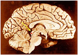
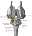
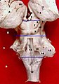
The brainstem is the stalk-like part of the brain that interconnects the cerebrum and diencephalon with the spinal cord. In the human brain, the brainstem is composed of the midbrain, the pons, and the medulla oblongata. The midbrain is continuous with the thalamus of the diencephalon through the tentorial notch.

<span class="mw-page-title-main">Lateral geniculate nucleus</span> Component of the visual system in the brains thalamus

In neuroanatomy, the lateral geniculate nucleus is a structure in the thalamus and a key component of the mammalian visual pathway. It is a small, ovoid, ventral projection of the thalamus where the thalamus connects with the optic nerve. There are two LGNs, one on the left and another on the right side of the thalamus. In humans, both LGNs have six layers of neurons alternating with optic fibers.

<span class="mw-page-title-main">Midbrain</span> Forward-most portion of the brainstem

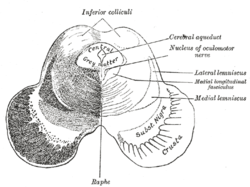
The midbrain or mesencephalon is the rostral-most portion of the brainstem connecting the diencephalon and cerebrum with the pons. It consists of the cerebral peduncles, tegmentum, and tectum.

<span class="mw-page-title-main">Spinothalamic tract</span> Sensory pathway from the skin to the thalamus

The spinothalamic tract is a part of the anterolateral system or the ventrolateral system, a sensory pathway to the thalamus. From the ventral posterolateral nucleus in the thalamus, sensory information is relayed upward to the somatosensory cortex of the postcentral gyrus.

<span class="mw-page-title-main">Auditory system</span> Sensory system used for hearing

The auditory system is the sensory system for the sense of hearing. It includes both the sensory organs and the auditory parts of the sensory system.

<span class="mw-page-title-main">Medial lemniscus</span> Ascending bundle of axons which cross in the brainstem

In neuroanatomy, the medial lemniscus, also known as Reil's band or Reil's ribbon, is a large ascending bundle of heavily myelinated axons that decussate (cross) in the brainstem, specifically in the medulla oblongata. The medial lemniscus is formed by the crossings of the internal arcuate fibers. The internal arcuate fibers are composed of axons of nucleus gracilis and nucleus cuneatus. The axons of the nucleus gracilis and nucleus cuneatus in the medial lemniscus have cell bodies that lie contralaterally.

<span class="mw-page-title-main">Lateral lemniscus</span>

The lateral lemniscus is a tract of axons in the brainstem that carries information about sound from the cochlear nucleus to various brainstem nuclei and ultimately the contralateral inferior colliculus of the midbrain. Three distinct, primarily inhibitory, cellular groups are located interspersed within these fibers, and are thus named the nuclei of the lateral lemniscus.

<span class="mw-page-title-main">Superior colliculus</span> Structure in the midbrain

In neuroanatomy, the superior colliculus is a structure lying on the roof of the mammalian midbrain. In non-mammalian vertebrates, the homologous structure is known as the optic tectum or optic lobe. The adjective form tectal is commonly used for both structures.

<span class="mw-page-title-main">Pretectal area</span> Structure in the midbrain which mediates responses to ambient light

In neuroanatomy, the pretectal area, or pretectum, is a midbrain structure composed of seven nuclei and comprises part of the subcortical visual system. Through reciprocal bilateral projections from the retina, it is involved primarily in mediating behavioral responses to acute changes in ambient light such as the pupillary light reflex, the optokinetic reflex, and temporary changes to the circadian rhythm. In addition to the pretectum's role in the visual system, the anterior pretectal nucleus has been found to mediate somatosensory and nociceptive information.

<span class="mw-page-title-main">Spinocerebellar tract</span> Nerve tract in humans

The spinocerebellar tract is a nerve tract originating in the spinal cord and terminating in the same side (ipsilateral) of the cerebellum.

<span class="mw-page-title-main">Medial geniculate nucleus</span>

The medial geniculate nucleus (MGN) or medial geniculate body (MGB) is part of the auditory thalamus and represents the thalamic relay between the inferior colliculus (IC) and the auditory cortex (AC). It is made up of a number of sub-nuclei that are distinguished by their neuronal morphology and density, by their afferent and efferent connections, and by the coding properties of their neurons. It is thought that the MGN influences the direction and maintenance of attention.

<span class="mw-page-title-main">Corpora quadrigemina</span> Part of the brain

In the brain, the corpora quadrigemina are the four colliculi—two inferior, two superior—located on the tectum of the dorsal aspect of the midbrain. They are respectively named the inferior and superior colliculus.

<span class="mw-page-title-main">Cochlear nucleus</span> Two cranial nerve nuclei of the human brainstem

The cochlear nuclear (CN) complex comprises two cranial nerve nuclei in the human brainstem, the ventral cochlear nucleus (VCN) and the dorsal cochlear nucleus (DCN). The ventral cochlear nucleus is unlayered whereas the dorsal cochlear nucleus is layered. Auditory nerve fibers, fibers that travel through the auditory nerve carry information from the inner ear, the cochlea, on the same side of the head, to the nerve root in the ventral cochlear nucleus. At the nerve root the fibers branch to innervate the ventral cochlear nucleus and the deep layer of the dorsal cochlear nucleus. All acoustic information thus enters the brain through the cochlear nuclei, where the processing of acoustic information begins. The outputs from the cochlear nuclei are received in higher regions of the auditory brainstem.

<span class="mw-page-title-main">Superior olivary complex</span> Collection of brainstem nuclei related to hearing

The superior olivary complex (SOC) or superior olive is a collection of brainstem nuclei that is located in pons, functions in multiple aspects of hearing and is an important component of the ascending and descending auditory pathways of the auditory system. The SOC is intimately related to the trapezoid body: most of the cell groups of the SOC are dorsal to this axon bundle while a number of cell groups are embedded in the trapezoid body. Overall, the SOC displays a significant interspecies variation, being largest in bats and rodents and smaller in primates.

<span class="mw-page-title-main">Interaural time difference</span> Difference in time that it takes a sound to travel between two ears

The interaural time difference when concerning humans or animals, is the difference in arrival time of a sound between two ears. It is important in the localization of sounds, as it provides a cue to the direction or angle of the sound source from the head. If a signal arrives at the head from one side, the signal has further to travel to reach the far ear than the near ear. This pathlength difference results in a time difference between the sound's arrivals at the ears, which is detected and aids the process of identifying the direction of sound source.

<span class="mw-page-title-main">Cerebellar peduncle</span> Structure connecting the cerebellum to the brainstem

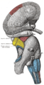
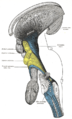
Cerebellar peduncles connect the cerebellum to the brain stem. There are six cerebellar peduncles in total, three on each side:

<span class="mw-page-title-main">Superior cerebellar peduncle</span>

In the human brain, the superior cerebellar peduncle is a paired structure of white matter that connects the cerebellum to the midbrain. It consists mainly of efferent fibers, the cerebellothalamic tract that runs from a cerebellar hemisphere to the contralateral thalamus, and the cerebellorubral tract that runs from a cerebellar hemisphere to the red nucleus. It also contains afferent tracts, most prominent of which is the ventral spinocerebellar tract. Other afferent tracts are the trigeminothalamic fibers, tectocerebellar fibers, and noradrenergic fibers from the locus coeruleus. The superior peduncle emerges from the upper and medial parts of the white matter of each hemisphere and is placed under cover of the upper part of the cerebellum.

<span class="mw-page-title-main">Ventral cochlear nucleus</span>

In the ventral cochlear nucleus (VCN), auditory nerve fibers enter the brain via the nerve root in the VCN. The ventral cochlear nucleus is divided into the anterior ventral (anteroventral) cochlear nucleus (AVCN) and the posterior ventral (posteroventral) cochlear nucleus (PVCN). In the VCN, auditory nerve fibers bifurcate, the ascending branch innervates the AVCN and the descending branch innervates the PVCN and then continue to the dorsal cochlear nucleus. The orderly innervation by auditory nerve fibers gives the AVCN a tonotopic organization along the dorsoventral axis. Fibers that carry information from the apex of the cochlea that are tuned to low frequencies contact neurons in the ventral part of the AVCN; those that carry information from the base of the cochlea that are tuned to high frequencies contact neurons in the dorsal part of the AVCN. Several populations of neurons populate the AVCN. Bushy cells receive input from auditory nerve fibers through particularly large endings called end bulbs of Held. They contact stellate cells through more conventional boutons.

References

  1. Shore, S. E.: Auditory/Somatosensory Interactions. In: Squire (Ed.): Encyclopedia of Neuroscience, Academic Press, 2009, p. 693 f.
  2. Shore, S. E.: Auditory/Somatosensory Interactions. In: Squire (Ed.): Encyclopedia of Neuroscience, Academic Press, 2009, p. 694.
  3. Shore, S. E.: Auditory/Somatosensory Interactions. In: Squire (Ed.): Encyclopedia of Neuroscience, Academic Press, 2009, pp. 691–695.
  4. Sinnatamby, Chummy S. (2011). Last's Anatomy (12th ed.). p. 476. ISBN   978-0-7295-3752-0.
  5. Shore, S. E.: Auditory/Somatosensory Interactions. In: Squire (Ed.): Encyclopedia of Neuroscience, Academic Press, 2009, p. 694.
  6. Neuroanatomy 5th edition, Nolte. Mosby 2002.
  7. Schreiner, Christoph (2005). The inferior colliculus. Springer Science+ Business Media, Incorporated.
  8. Gelfand, Stanley A.: Hearing, an Introduction to Psychological and Physiological Acoustics, 4th Ed., Marcel Dekker, 2004, pp. 71-75.
  9. Conrad Simon Memorial Research Initiative homepage. http://www.conradsimon.org/InferiorColliculus.shtml. MIME type: application/octet-stream.
  10. Skottun, Bernt C. et al.: The ability of inferior colliculus neurons to signal differences in interaural delay. PNAS November 20, 2001 vol. 98, no. 24, pp. 14050-14054.
  11. T. Lindeberg and A. Friberg "Idealized computational models of auditory receptive fields", PLOS ONE, 10(3): e0119032, pages 1-58, 2015