Ventricular system

Last updated
Ventricular system
1317 CFS Circulation.jpg
The ventricular system accounts for the production and circulation of cerebrospinal fluid.
Human Ventricular system colored and animated.gif
Rotating 3D rendering of the four ventricles and connections. From top to bottom:
Blue - lateral ventricles
Cyan - interventricular foramina (Monro)
Yellow - third ventricle
Red - cerebral aqueduct (Sylvius)
Purple - fourth ventricle
Green - continuous with the central canal

(Parts between median aperture and subarachnoid space are not shown)
Details
Identifiers
Latin ventriculi cerebri
MeSH D002552
NeuroNames 2497
FMA 242787
Anatomical terms of neuroanatomy

In neuroanatomy, the ventricular system is a set of four interconnected cavities known as cerebral ventricles in the brain. [1] [2] Within each ventricle is a region of choroid plexus which produces the circulating cerebrospinal fluid (CSF). The ventricular system is continuous with the central canal of the spinal cord from the fourth ventricle, [3] allowing for the flow of CSF to circulate. [3] [4]

Contents

All of the ventricular system and the central canal of the spinal cord are lined with ependyma, a specialised form of epithelium connected by tight junctions that make up the blood–cerebrospinal fluid barrier. [2]

Structure

Size and location of the ventricular system in the human head. Human ventricular system - animation.gif
Size and location of the ventricular system in the human head.

The system comprises four ventricles: [5]

There are several foramina, openings acting as channels, that connect the ventricles. The interventricular foramina (also called the foramina of Monro) connect the lateral ventricles to the third ventricle through which the cerebrospinal fluid can flow.

NameFromTo
interventricular foramina (Monro)lateral ventriclesthird ventricle
Cerebral aqueduct (Sylvius)third ventriclefourth ventricle
Median aperture (Magendie)fourth ventricle subarachnoid space via the cisterna magna
Right and left lateral apertures (Luschka)fourth ventriclesubarachnoid space via the cistern of great cerebral vein

Ventricles

3D rendering of ventricles (lateral and anterior views). Blausen 0896 Ventricles Brain.png
3D rendering of ventricles (lateral and anterior views).
Ventricular system anatomy. Brain-ventricle-anatomy-diagram.jpg
Ventricular system anatomy.

The four cavities of the human brain are called ventricles. [6] The two largest are the lateral ventricles in the cerebrum, the third ventricle is in the diencephalon of the forebrain between the right and left thalamus, and the fourth ventricle is located at the back of the pons and upper half of the medulla oblongata of the hindbrain. The ventricles are concerned with the production and circulation of cerebrospinal fluid. [7]

Development

The structures of the ventricular system are embryologically derived from the neural canal, the centre of the neural tube.[ citation needed ]

As the part of the primitive neural tube that will develop into the brainstem, the neural canal expands dorsally and laterally, creating the fourth ventricle, whereas the neural canal that does not expand and remains the same at the level of the midbrain superior to the fourth ventricle forms the cerebral aqueduct. The fourth ventricle narrows at the obex (in the caudal medulla), to become the central canal of the spinal cord.[ citation needed ]

In more detail, around the third week of development, the embryo is a three-layered disc. The embryo is covered on the dorsal surface by a layer of cells called ectoderm. In the middle of the dorsal surface of the embryo is a linear structure called the notochord. As the ectoderm proliferates, the notochord is dragged into the middle of the developing embryo. [8]

As the brain develops, by the fourth week of embryological development three swellings known as brain vesicles have formed within the embryo around the canal, near where the head will develop. The three primary brain vesicles represent different components of the central nervous system: the prosencephalon, mesencephalon and rhombencephalon. These in turn divide into five secondary vesicles. As these sections develop around the neural canal, the inner neural canal becomes known as primitive ventricles. These form the ventricular system of the brain: [8] The neural stem cells of the developing brain, principally radial glial cells, line the developing ventricular system in a transient zone called the ventricular zone. [9]

Separating the anterior horns of the lateral ventricles is the septum pellucidum: a thin, triangular, vertical membrane which runs as a sheet from the corpus callosum down to the fornix. During the third month of fetal development, a space forms between two septal laminae, known as the cave of septum pellucidum (CSP), which is a marker for fetal neural maldevelopment. During the fifth month of development, the laminae start to close and this closure completes from about three to six months after birth. Fusion of the septal laminae is attributed to rapid development of the alvei of the hippocampus, amygdala, septal nuclei, fornix, corpus callosum and other midline structures. Lack of such limbic development interrupts this posterior-to-anterior fusion, resulting in the continuation of the CSP into adulthood. [10]

Function

Flow of cerebrospinal fluid

MRI showing flow of CSF NPH MRI 272 GILD.gif
MRI showing flow of CSF
The cerebrospinal fluid passes out through arachnoid villi into the venous sinuses of the skull. 1316 Meningeal LayersN.jpg
The cerebrospinal fluid passes out through arachnoid villi into the venous sinuses of the skull.
A schematic illustration of the venous sinuses surrounding the brain. 1315 Brain Sinuses.jpg
A schematic illustration of the venous sinuses surrounding the brain.

The ventricles are filled with cerebrospinal fluid (CSF) which bathes and cushions the brain and spinal cord within their bony confines. CSF is produced by modified ependymal cells of the choroid plexus found in all components of the ventricular system except for the cerebral aqueduct and the posterior and anterior horns of the lateral ventricles. CSF flows from the lateral ventricles via the interventricular foramina into the third ventricle, and then the fourth ventricle via the cerebral aqueduct in the midbrain. From the fourth ventricle it can pass into the central canal of the spinal cord or into the subarachnoid cisterns via three small foramina: the central median aperture and the two lateral apertures. According to the traditional understanding of cerebrospinal fluid (CSF) physiology, the majority of CSF is produced by the choroid plexus, circulates through the ventricles, the cisterns, and the subarachnoid space to be absorbed into the blood by the arachnoid villi.[ citation needed ]

The fluid then flows around the superior sagittal sinus to be reabsorbed via the arachnoid granulations (or arachnoid villi) into the venous sinuses, after which it passes through the jugular vein and major venous system. CSF within the spinal cord can flow all the way down to the lumbar cistern at the end of the cord around the cauda equina where lumbar punctures are performed.

The cerebral aqueduct between the third and fourth ventricles is very small, as are the foramina, which means that they can be easily blocked.

Protection of the brain

The brain and spinal cord are covered by the meninges, the three protective membranes of the tough dura mater, the arachnoid mater and the pia mater. The cerebrospinal fluid (CSF) within the skull and spine provides further protection and also buoyancy, and is found in the subarachnoid space between the pia mater and the arachnoid mater.[ citation needed ]

The CSF that is produced in the ventricular system is also necessary for chemical stability, and the provision of nutrients needed by the brain. The CSF helps to protect the brain from jolts and knocks to the head and also provides buoyancy and support to the brain against gravity. (Since the brain and CSF are similar in density, the brain floats in neutral buoyancy, suspended in the CSF.) This allows the brain to grow in size and weight without resting on the floor of the cranium, which would destroy nervous tissue. [11] [12]

Clinical significance

The narrowness of the cerebral aqueduct and foramina means that they can become blocked, for example, by blood following a hemorrhagic stroke. As cerebrospinal fluid is continually produced by the choroid plexus within the ventricles, a blockage of outflow leads to increasingly high pressure in the lateral ventricles. As a consequence, this commonly leads in turn to hydrocephalus. Medically one would call this post-haemorrhagic acquired hydrocephalus, but is often referred to colloquially by the layperson as "water on the brain". This is an extremely serious condition regardless of the cause of blockage. An endoscopic third ventriculostomy is a surgical procedure for the treatment of hydrocephalus in which an opening is created in the floor of the third ventricle using an endoscope placed within the ventricular system through a burr hole. This allows the cerebrospinal fluid to flow directly to the basal cisterns, thereby bypassing any obstruction. A surgical procedure to make an entry hole to access any of the ventricles is called a ventriculostomy. This is done to drain accumulated cerebrospinal fluid either through a temporary catheter or a permanent shunt.[ citation needed ]

Other diseases of the ventricular system include inflammation of the membranes (meningitis) or of the ventricles (ventriculitis) caused by infection or the introduction of blood following trauma or haemorrhage (cerebral haemorrhage or subarachnoid haemorrhage).

During embryogenesis in the choroid plexus of the ventricles, choroid plexus cysts can form.

The scientific study of CT scans of the ventricles in the late 1970s gave new insight into the study of mental disorders. Researchers found that individuals with schizophrenia had (in terms of group averages) larger than usual ventricles. This became the first "evidence" that schizophrenia was biological in origin and led to a renewed interest in its study via the use of imaging techniques. Magnetic resonance imaging (MRI) has superseded the use of CT in research in the role of detecting ventricular abnormalities in psychiatric illness.

Whether the enlarged ventricles is a cause or a result of schizophrenia has not yet been established. Enlarged ventricles are also found in organic dementia and have been explained largely in terms of environmental factors. [13] They have also been found to be extremely diverse between individuals, such that the percentage difference in group averages in schizophrenia studies (+16%) has been described as "not a very profound difference in the context of normal variation" (ranging from 25% to 350% of the mean average). [14]

The cave of septum pellucidum has been loosely associated with schizophrenia, [15] post-traumatic stress disorder, [16] traumatic brain injury, [17] as well as with antisocial personality disorder. [10] CSP is one of the distinguishing features of individuals displaying symptoms of dementia pugilistica. [18]

Additional media

See also

Related Research Articles

<span class="mw-page-title-main">Cerebrospinal fluid</span> Clear, colorless bodily fluid found in the brain and spinal cord

Cerebrospinal fluid (CSF) is a clear, colorless body fluid found within the tissue that surrounds the brain and spinal cord of all vertebrates.

<span class="mw-page-title-main">Hydrocephalus</span> Abnormal increase in cerebrospinal fluid in the ventricles of the brain

Hydrocephalus is a condition in which an accumulation of cerebrospinal fluid (CSF) occurs within the brain. This typically causes increased pressure inside the skull. Older people may have headaches, double vision, poor balance, urinary incontinence, personality changes, or mental impairment. In babies, it may be seen as a rapid increase in head size. Other symptoms may include vomiting, sleepiness, seizures, and downward pointing of the eyes.

<span class="mw-page-title-main">Pia mater</span> Delicate innermost layer of the meninges, the membranes surrounding the brain and spinal cord

Pia mater, often referred to as simply the pia, is the delicate innermost layer of the meninges, the membranes surrounding the brain and spinal cord. Pia mater is medieval Latin meaning "tender mother". The other two meningeal membranes are the dura mater and the arachnoid mater. Both the pia and arachnoid mater are derivatives of the neural crest while the dura is derived from embryonic mesoderm. The pia mater is a thin fibrous tissue that is permeable to water and small solutes. The pia mater allows blood vessels to pass through and nourish the brain. The perivascular space between blood vessels and pia mater is proposed to be part of a pseudolymphatic system for the brain. When the pia mater becomes irritated and inflamed the result is meningitis.

<span class="mw-page-title-main">Third ventricle</span> Ventricle of the brain located between the two thalami

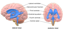
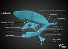
The third ventricle is one of the four connected ventricles of the ventricular system within the mammalian brain. It is a slit-like cavity formed in the diencephalon between the two thalami, in the midline between the right and left lateral ventricles, and is filled with cerebrospinal fluid (CSF).

<span class="mw-page-title-main">Choroid plexus</span> Structure in the ventricles of the brain

The choroid plexus, or plica choroidea, is a plexus of cells that arises from the tela choroidea in each of the ventricles of the brain. Regions of the choroid plexus produce and secrete most of the cerebrospinal fluid (CSF) of the central nervous system. The choroid plexus consists of modified ependymal cells surrounding a core of capillaries and loose connective tissue. Multiple cilia on the ependymal cells move to circulate the cerebrospinal fluid.

<span class="mw-page-title-main">Cerebral aqueduct</span> Conduit for CSF to the brain

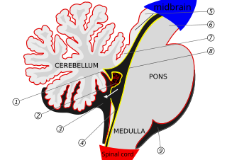
The cerebral aqueduct is a narrow 15 mm conduit for cerebrospinal fluid (CSF) that connects the third ventricle to the fourth ventricle of the ventricular system of the brain. It is located in the midbrain dorsal to the pons and ventral to the cerebellum. The cerebral aqueduct is surrounded by an enclosing area of gray matter called the periaqueductal gray, or central gray. It was first named after Franciscus Sylvius.

<span class="mw-page-title-main">Fourth ventricle</span> Ventricle in front of the cerebellum

The fourth ventricle is one of the four connected fluid-filled cavities within the human brain. These cavities, known collectively as the ventricular system, consist of the left and right lateral ventricles, the third ventricle, and the fourth ventricle. The fourth ventricle extends from the cerebral aqueduct to the obex, and is filled with cerebrospinal fluid (CSF).

<span class="mw-page-title-main">Ependyma</span> Lining of the ventricular system of the brain

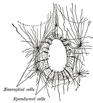
The ependyma is the thin neuroepithelial lining of the ventricular system of the brain and the central canal of the spinal cord. The ependyma is one of the four types of neuroglia in the central nervous system (CNS). It is involved in the production of cerebrospinal fluid (CSF), and is shown to serve as a reservoir for neuroregeneration.

<span class="mw-page-title-main">Median aperture</span>

The median aperture is an opening of the fourth ventricle at the caudal portion of the roof of the fourth ventricle. It allows flow of cerebrospinal fluid (CSF) from the fourth ventricle into the cisterna magna. The other two openings of the fourth ventricle are the lateral apertures - one on either side. Nonetheless, the median aperture accounts for most of the outflow of CSF out of the fourth ventricle. The median aperture varies in size.

<span class="mw-page-title-main">Lateral ventricles</span> Two largest ventricles in each cerebral hemisphere

The lateral ventricles are the two largest ventricles of the brain and contain cerebrospinal fluid. Each cerebral hemisphere contains a lateral ventricle, known as the left or right lateral ventricle, respectively.

<span class="mw-page-title-main">Interventricular foramina (neuroanatomy)</span> It is part of diencephalon that makes connection between lateral and third ventricular

In the brain, the interventricular foramina are channels that connect the paired lateral ventricles with the third ventricle at the midline of the brain. As channels, they allow cerebrospinal fluid (CSF) produced in the lateral ventricles to reach the third ventricle and then the rest of the brain's ventricular system. The walls of the interventricular foramina also contain choroid plexus, a specialized CSF-producing structure, that is continuous with that of the lateral and third ventricles above and below it.

<span class="mw-page-title-main">Subarachnoid cisterns</span> Spaces around the brain filled with cerebrospinal fluid

The subarachnoid cisterns are spaces formed by openings in the subarachnoid space, an anatomic space in the meninges of the brain. The space is situated between the two meninges, the arachnoid mater and the pia mater. These cisterns are filled with cerebrospinal fluid (CSF).

<span class="mw-page-title-main">Arachnoid mater</span> Web-like middle layer of the three meninges

The arachnoid mater is one of the three meninges, the protective membranes that cover the brain and spinal cord. It is so named because of its resemblance to a spider web. The arachnoid mater is a derivative of the neural crest mesoectoderm in the embryo.

<span class="mw-page-title-main">Dandy–Walker malformation</span> Congenital malformation of the cerebellar vermis

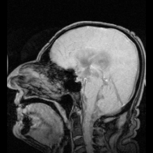
Dandy–Walker malformation (DWM), also known as Dandy–Walker syndrome (DWS), is a rare congenital brain malformation in which the part joining the two hemispheres of the cerebellum does not fully form, and the fourth ventricle and space behind the cerebellum are enlarged with cerebrospinal fluid. Most of those affected develop hydrocephalus within the first year of life, which can present as increasing head size, vomiting, excessive sleepiness, irritability, downward deviation of the eyes and seizures. Other, less common symptoms are generally associated with comorbid genetic conditions and can include congenital heart defects, eye abnormalities, intellectual disability, congenital tumours, other brain defects such as agenesis of the corpus callosum, skeletal abnormalities, an occipital encephalocele or underdeveloped genitalia or kidneys. It is sometimes discovered in adolescents or adults due to mental health problems.

<span class="mw-page-title-main">Tela choroidea</span>

The tela choroidea is a region of meningeal pia mater that adheres to the underlying ependyma, and gives rise to the choroid plexus in each of the brain’s four ventricles. Tela is Latin for woven and is used to describe a web-like membrane or layer. The tela choroidea is a very thin part of the loose connective tissue of pia mater overlying and closely adhering to the ependyma. It has a rich blood supply. The ependyma and vascular pia mater – the tela choroidea, form regions of minute projections known as a choroid plexus that projects into each ventricle. The choroid plexus produces most of the cerebrospinal fluid of the central nervous system that circulates through the ventricles of the brain, the central canal of the spinal cord, and the subarachnoid space. The tela choroidea in the ventricles forms from different parts of the roof plate in the development of the embryo.

<span class="mw-page-title-main">Superior medullary velum</span> Thin layer between the superior cerebellar peduncles

The superior medullary velum is a thin, transparent lamina of white matter which - together with the inferior medullary velum - forms the roof of the fourth ventricle. It extends between the two superior cerebellar peduncles. The lingula of cerebellum covers - and adheres to - its dorsal surface.

Cisternography is a medical imaging technique to examine the flow of cerebrospinal fluid (CSF) in the brain, and spinal cord. The gold standard for diagnosis of a cranial cerebrospinal fluid leak is CT cisternography. For the diagnosis of a spinal CSF leak radionuclide cisternography also known as radioisotope cisternography is used. The third type of cisternography is MR cisternography.

Bobble-head doll syndrome is a rare neurological movement disorder in which patients, usually children around age 3, begin to bob their head and shoulders forward and back, or sometimes side-to-side, involuntarily, in a manner reminiscent of a bobblehead doll. The syndrome is related to cystic lesions and swelling of the third ventricle in the brain.

<span class="mw-page-title-main">Glymphatic system</span> System for waste clearance in the central nervous system of vertebrates

The glymphatic system is a system for waste clearance in the central nervous system (CNS) of vertebrates. According to this model, cerebrospinal fluid (CSF) flows into the paravascular space around cerebral arteries, combining with interstitial fluid (ISF) and parenchymal solutes, and exiting down venous paravascular spaces. The pathway consists of a para-arterial influx route for CSF to enter the brain parenchyma, coupled to a clearance mechanism for the removal of interstitial fluid (ISF) and extracellular solutes from the interstitial compartments of the brain and spinal cord. Exchange of solutes between CSF and ISF is driven primarily by arterial pulsation and regulated during sleep by the expansion and contraction of brain extracellular space. Clearance of soluble proteins, waste products, and excess extracellular fluid is accomplished through convective bulk flow of ISF, facilitated by astrocytic aquaporin 4 (AQP4) water channels.

References

  1. Grow, W.A. (2018). "Development of the Nervous System". Fundamental Neuroscience for Basic and Clinical Applications. Elsevier. pp. 72–90.e1. doi:10.1016/b978-0-323-39632-5.00005-0. ISBN   978-0-323-39632-5. The ventricular system is an elaboration of the lumen of cephalic portions of the neural tube, and its development parallels that of the brain.
  2. 1 2 Shoykhet, Mish; Clark, Robert S.B. (2011). "Structure, Function, and Development of the Nervous System". Pediatric Critical Care. Elsevier. pp. 783–804. doi:10.1016/b978-0-323-07307-3.10057-6. ISBN   978-0-323-07307-3. The ventricles contain the choroid plexus, which produces CSF, and serve as conduits for CSF flow in the CNS. Ventricular walls are lined with ependymal cells, which are connected by tight junctions and constitute a CSF-brain barrier.
  3. 1 2 Shoykhet, Mish; Clark, Robert S.B. (2011). "Structure, Function, and Development of the Nervous System". Pediatric Critical Care. Elsevier. pp. 783–804. doi:10.1016/b978-0-323-07307-3.10057-6. ISBN   978-0-323-07307-3. The ventricular system arises from the hollow space within the developing neural tube and gives rise to cisterns within the CNS, from the brain to the spinal cord.
  4. Vernau, William; Vernau, Karen A.; Sue Bailey, Cleta (2008). "Cerebrospinal Fluid". Clinical Biochemistry of Domestic Animals. Elsevier. pp. 769–819. doi:10.1016/b978-0-12-370491-7.00026-x. ISBN   978-0-12-370491-7. S2CID   71013935. Cerebrospinal fluid flows in bulk from sites of production to sites of absorption. Fluid formed in the lateral ventricles flows through the paired interventricular foramina (foramen of Monro) into the third ventricle, then through the mesencephalic aqueduct (aqueduct of Sylvius) into the fourth ventricle. The majority of CSF exits from the fourth ventricle into the subarachnoid space; a small amount may enter the central canal of the spinal cord.
  5. Creative Commons by small.svg  This article incorporates text available under the CC BY 4.0 license.Betts, J Gordon; Desaix, Peter; Johnson, Eddie; Johnson, Jody E; Korol, Oksana; Kruse, Dean; Poe, Brandon; Wise, James; Womble, Mark D; Young, Kelly A (July 16, 2023). Anatomy & Physiology. Houston: OpenStax CNX. 13.3 Circulation and the Central Nervous System. ISBN   978-1-947172-04-3.
  6. National Institutes of Health (December 13, 2011). "Ventricles of the brain". nih.gov.
  7. International school of medicine and applied sciences kisumu library
  8. 1 2 Schoenwolf, Gary C. (2009). ""Development of the Brain and Cranial Nerves"". Larsen's human embryology (4th ed.). Philadelphia: Churchill Livingstone/Elsevier. ISBN   9780443068119.
  9. Rakic, P (October 2009). "Evolution of the neocortex: a perspective from developmental biology". Nature Reviews. Neuroscience. 10 (10): 724–35. doi:10.1038/nrn2719. PMC   2913577 . PMID   19763105.
  10. 1 2 Raine, Adrian; Lee, Lydia; Yang, Yaling; Colletti, Patrick (2010). "Neurodevelopmental marker for limbic maldevelopment in antisocial personality disorder and psychopathy". BJPsych". The British Journal of Psychiatry. 197 (3): 186–192. doi:10.1192/bjp.bp.110.078485. PMC   2930915 . PMID   20807962.
  11. Klein, S.B., & Thorne, B.M. Biological Psychology. Worth Publishers: New York. 2007.
  12. Saladin, Kenneth S. Anatomy & Physiology. The Unit of Form and Function. 5th Edition. McGraw-Hill: New York. 2007
  13. Peper, Jiska S.; Brouwer, RM; Boomsma, DI; Kahn, RS; Hulshoff Pol, HE (2007). "Genetic influences on human brain structure: A review of brain imaging studies in twins". Human Brain Mapping. 28 (6): 464–73. doi:10.1002/hbm.20398. PMC   6871295 . PMID   17415783.
  14. Allen JS, Damasio H, Grabowski TJ (August 2002). "Normal neuroanatomical variation in the human brain: an MRI-volumetric study". American Journal of Physical Anthropology. 118 (4): 341–58. doi:10.1002/ajpa.10092. PMID   12124914.
  15. Galarza M, Merlo A, Ingratta A, Albanese E, Albanese A (2004). "Cavum septum pellucidum and its increased prevalence in schizophrenia: a neuroembryological classification". The Journal of Neuropsychiatry and Clinical Neurosciences. 16 (1): 41–6. doi:10.1176/appi.neuropsych.16.1.41. PMID   14990758.
  16. May F, Chen Q, Gilbertson M, Shenton M, Pitman R (2004). "Cavum septum pellucidum in monozygotic twins discordant for combat exposure: relationship to posttraumatic stress disorder" (PDF). Biol. Psychiatry. 55 (6): 656–8. doi:10.1016/j.biopsych.2003.09.018. PMC   2794416 . PMID   15013837.
  17. Zhang L, Ravdin L, Relkin N, Zimmerman R, Jordan B, Lathan W, Uluğ A (2003). "Increased diffusion in the brain of professional boxers: a preclinical sign of traumatic brain injury?". American Journal of Neuroradiology. 24 (1): 52–7. PMC   8148951 . PMID   12533327.
  18. McKee, AC; Cantu, RC; Nowinski, CJ; Hedley-Whyte, ET; Gavett, BE; Budson, AE; Santini, VE; Lee, HS; Kubilus, CA; Stern, RA (Jul 2009). "Chronic traumatic encephalopathy in athletes: progressive tauopathy after repetitive head injury". Neuropathol Exp Neurol. 68 (7): 709–35. doi:10.1097/NEN.0b013e3181a9d503. PMC   2945234 . PMID   19535999.