The hippocampus is a major component of the brain of humans and other vertebrates. Humans and other mammals have two hippocampi, one in each side of the brain. The hippocampus is part of the limbic system, and plays important roles in the consolidation of information from short-term memory to long-term memory, and in spatial memory that enables navigation. The hippocampus is located in the allocortex, with neural projections into the neocortex in humans, as well as primates. The hippocampus, as the medial pallium, is a structure found in all vertebrates. In humans, it contains two main interlocking parts: the hippocampus proper, and the dentate gyrus.

The abducens nerve or abducent nerve, also known as the sixth cranial nerve, cranial nerve VI, or simply CN VI, is a cranial nerve in humans and various other animals that controls the movement of the lateral rectus muscle, one of the extraocular muscles responsible for outward gaze. It is a somatic efferent nerve.

In neuroanatomy, the mandibular nerve (V3) is the largest of the three divisions of the trigeminal nerve, the fifth cranial nerve (CN V). Unlike the other divisions of the trigeminal nerve (ophthalmic nerve, maxillary nerve) which contain only afferent fibers, the mandibular nerve contains both afferent and efferent fibers. These nerve fibers innervate structures of the lower jaw and face, such as the tongue, lower lip, and chin. The mandibular nerve also innervates the muscles of mastication.

Pia mater, often referred to as simply the pia, is the delicate innermost layer of the meninges, the membranes surrounding the brain and spinal cord. Pia mater is medieval Latin meaning "tender mother". The other two meningeal membranes are the dura mater and the arachnoid mater. Both the pia and arachnoid mater are derivatives of the neural crest while the dura is derived from embryonic mesoderm. The pia mater is a thin fibrous tissue that is permeable to water and small solutes. The pia mater allows blood vessels to pass through and nourish the brain. The perivascular space between blood vessels and pia mater is proposed to be part of a pseudolymphatic system for the brain. When the pia mater becomes irritated and inflamed the result is meningitis.

The temporal lobe is one of the four major lobes of the cerebral cortex in the brain of mammals. The temporal lobe is located beneath the lateral fissure on both cerebral hemispheres of the mammalian brain.

The fornix is a C-shaped bundle of nerve fibers in the brain that acts as the major output tract of the hippocampus. The fornix also carries some afferent fibers to the hippocampus from structures in the diencephalon and basal forebrain. The fornix is part of the limbic system. While its exact function and importance in the physiology of the brain are still not entirely clear, it has been demonstrated in humans that surgical transection—the cutting of the fornix along its body—can cause memory loss. There is some debate over what type of memory is affected by this damage, but it has been found to most closely correlate with recall memory rather than recognition memory. This means that damage to the fornix can cause difficulty in recalling long-term information such as details of past events, but it has little effect on the ability to recognize objects or familiar situations.

The sella turcica is a saddle-shaped depression in the body of the sphenoid bone of the human skull and of the skulls of other hominids including chimpanzees, gorillas and orangutans. It serves as a cephalometric landmark. The pituitary gland or hypophysis is located within the most inferior aspect of the sella turcica, the hypophyseal fossa.

The lateral ventricles are the two largest ventricles of the brain and contain cerebrospinal fluid (CSF). Each cerebral hemisphere contains a lateral ventricle, known as the left or right lateral ventricle, respectively.

The cavernous sinus within the human head is one of the dural venous sinuses creating a cavity called the lateral sellar compartment bordered by the temporal bone of the skull and the sphenoid bone, lateral to the sella turcica.

The calcarine sulcus is an anatomical landmark located at the caudal end of the medial surface of the brain of humans and other primates. Its name comes from the Latin "calcar" meaning "spur". It is very deep, and known as a complete sulcus.

The lesser petrosal nerve is the general visceral efferent (GVE) nerve conveying pre-ganglionic parasympathetic secretomotor fibers for the parotid gland from the tympanic plexus to the otic ganglion. It passes out of the tympanic cavity through the petrous part of the temporal bone into the middle cranial fossa of the cranial cavity, then exits the cranial cavity through its own canaliculus to reach the infratemporal fossa.

The occipital condyles are undersurface protuberances of the occipital bone in vertebrates, which function in articulation with the superior facets of the atlas vertebra.

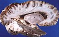
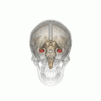
Hippocampus anatomy describes the physical aspects and properties of the hippocampus, a neural structure in the medial temporal lobe of the brain. It has a distinctive, curved shape that has been likened to the sea-horse monster of Greek mythology and the ram's horns of Amun in Egyptian mythology. This general layout holds across the full range of mammalian species, from hedgehog to human, although the details vary. For example, in the rat, the two hippocampi look similar to a pair of bananas, joined at the stems. In primate brains, including humans, the portion of the hippocampus near the base of the temporal lobe is much broader than the part at the top. Due to the three-dimensional curvature of this structure, two-dimensional sections such as shown are commonly seen. Neuroimaging pictures can show a number of different shapes, depending on the angle and location of the cut.
The Great Hippocampus Question was a 19th-century scientific controversy about the anatomy of ape and human uniqueness. The dispute between Thomas Henry Huxley and Richard Owen became central to the scientific debate on human evolution that followed Charles Darwin's publication of On the Origin of Species. The name comes from the title of a satire the Reverend Charles Kingsley wrote about the arguments, which in modified form appeared as "the great hippopotamus test" in Kingsley's 1863 book for children, The Water-Babies, A Fairy Tale for a Land Baby. Together with other humorous skits on the topic, this helped to spread and popularise Darwin's ideas on evolution.
In human neuroanatomy, the longitudinal striae are two bundles of fibres embedded in the indusium griseum running along the corpus callosum of the brain. They were originally described by Italian physician, epidemiologist and anatomist Giovanni Maria Lancisi. The striae are categorized as medial longitudinal stria and lateral longitudinal stria; the area between the striae is a useful neurosurgical mark of the middle of the corpus callosum.

Saleem Abdulrauf is an American physician specializing in neurosurgery in Washington, DC, who has helped develop high-flow brain bypass surgery, a less invasive procedure for treating intracranial aneurysm than methods used previously.
The Dextroscope is a medical equipment system that creates a virtual reality (VR) environment in which surgeons can plan neurosurgical and other surgical procedures.
The Dextrobeam is a highly interactive console that enables collaborative examination of three-dimensional (3-D) medical imaging data for planning, discussing, or teaching neurosurgical approaches and strategies. The console is designed to work in combination with a 3D stereoscopic display. The console enables two-handed interaction by means of two 6 Degree-of-Freedom motion tracking devices. A set of built-in software tools gives users the ability to manipulate and interact with patients’ imaging data in a natural and intuitive way.

Antonio Bernardo is an Italian-American neurosurgeon and academic physician. He is a professor of Neurological Surgery and the Director of the Neurosurgical Innovations and Training Center for Skull Base and Microneurosurgery in the Department of Neurological Surgery at Weill Cornell Medical College. He has gained significant notoriety for his expertise in skull base and cerebrovascular surgery, and has published extensively on minimally invasive neurosurgery. He is a pioneer in the use of 3D technology in neurosurgery and a strong advocate for competency-based training in surgery.