The medulla oblongata or simply medulla is a long stem-like structure which makes up the lower part of the brainstem. It is anterior and partially inferior to the cerebellum. It is a cone-shaped neuronal mass responsible for autonomic (involuntary) functions, ranging from vomiting to sneezing. The medulla contains the cardiac, respiratory, vomiting and vasomotor centers, and therefore deals with the autonomic functions of breathing, heart rate and blood pressure as well as the sleep–wake cycle.
Articles related to anatomy include:
The brainstem is the stalk-like part of the brain that interconnects the cerebrum and diencephalon with the spinal cord. In the human brain, the brainstem is composed of the midbrain, the pons, and the medulla oblongata. The midbrain is continuous with the thalamus of the diencephalon through the tentorial notch.

In anatomy, the orbit is the cavity or socket/hole of the skull in which the eye and its appendages are situated. "Orbit" can refer to the bony socket, or it can also be used to imply the contents. In the adult human, the volume of the orbit is 30 millilitres, of which the eye occupies 6.5 ml. The orbital contents comprise the eye, the orbital and retrobulbar fascia, extraocular muscles, cranial nerves II, III, IV, V, and VI, blood vessels, fat, the lacrimal gland with its sac and duct, the eyelids, medial and lateral palpebral ligaments, cheek ligaments, the suspensory ligament, septum, ciliary ganglion and short ciliary nerves.

The fourth ventricle is one of the four connected fluid-filled cavities within the human brain. These cavities, known collectively as the ventricular system, consist of the left and right lateral ventricles, the third ventricle, and the fourth ventricle. The fourth ventricle extends from the cerebral aqueduct to the obex, and is filled with cerebrospinal fluid (CSF).

The lateral ventricles are the two largest ventricles of the brain and contain cerebrospinal fluid (CSF). Each cerebral hemisphere contains a lateral ventricle, known as the left or right lateral ventricle, respectively.

The falx cerebri is a large, crescent-shaped fold of dura mater that descends vertically into the longitudinal fissure between the cerebral hemispheres of the human brain, separating the two hemispheres and supporting dural sinuses that provide venous and CSF drainage to the brain. It is attached to the crista galli anteriorly, and blends with the tentorium cerebelli posteriorly.

The middle cerebral artery (MCA) is one of the three major paired cerebral arteries that supply blood to the cerebrum. The MCA arises from the internal carotid artery and continues into the lateral sulcus where it then branches and projects to many parts of the lateral cerebral cortex. It also supplies blood to the anterior temporal lobes and the insular cortices.

The anterior choroidal artery is a bilaterally paired artery of the brain. It is typically a branch of the internal carotid artery which supplies the choroid plexus of lateral ventricle and third ventricle as well as numerous structures of the brain.

The posterior inferior cerebellar artery (PICA) is the largest branch of the vertebral artery. It is one of the three main arteries that supply blood to the cerebellum, a part of the brain. Blockage of the posterior inferior cerebellar artery can result in a type of stroke called lateral medullary syndrome.

The cerebellopontine angle (CPA) is located between the cerebellum and the pons. The cerebellopontine angle is the site of the cerebellopontine angle cistern.

The vestibular nuclei (VN) are the cranial nuclei for the vestibular nerve located in the brainstem.

In neuroanatomy, a sulcus is a depression or groove in the cerebral cortex. It surrounds a gyrus, creating the characteristic folded appearance of the brain in humans and other mammals. The larger sulci are usually called fissures.

The rhomboid fossa is a rhombus-shaped depression that is the anterior part of the fourth ventricle. Its anterior wall, formed by the back of the pons and the medulla oblongata, constitutes the floor of the fourth ventricle.

The tela choroidea is a region of meningeal pia mater that adheres to the underlying ependyma, and gives rise to the choroid plexus in each of the brain’s four ventricles. Tela is Latin for woven and is used to describe a web-like membrane or layer. The tela choroidea is a very thin part of the loose connective tissue of pia mater overlying and closely adhering to the ependyma. It has a rich blood supply. The ependyma and vascular pia mater – the tela choroidea, form regions of minute projections known as a choroid plexus that projects into each ventricle. The choroid plexus produces most of the cerebrospinal fluid of the central nervous system that circulates through the ventricles of the brain, the central canal of the spinal cord, and the subarachnoid space. The tela choroidea in the ventricles forms from different parts of the roof plate in the development of the embryo.

The basal vein is a vein in the brain. It is formed at the anterior perforated substance by the union of

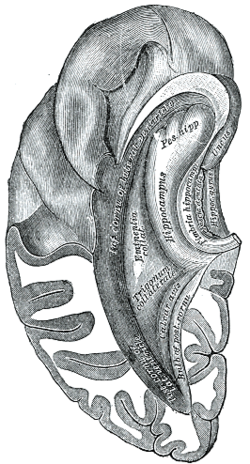
The calcar avis, previously known as the hippocampus minor, is an involution of the wall of the lateral ventricle's posterior cornu produced by the calcarine fissure.

The collateral fissure is on the tentorial surface of the hemisphere and extends from near the occipital pole to within a short distance of the temporal pole.

The following outline is provided as an overview of and topical guide to human anatomy:
Within anatomy for the circulatory system, the plexal point or ventral choroidal point is the proposed naming for the point at which the anterior choroidal artery enters into the lateral ventricle of the brain at the tela choroidea. The choroid fissure, in this sense, is the narrow cleft along the medial wall of the lateral ventricle, where the choroid plexus is attached at the margins. Not to be confused with the choroidal fissure of the eye.