Julius Caesar Aranzi was a leading figure in the history of the science of human anatomy.

A heart valve normally allows blood to flow in only one direction through the heart. The four valves are commonly represented in a mammalian heart that determines the pathway of blood flow through the heart. A heart valve opens or closes incumbent on differential blood pressure on each side.

The aortic valve is a valve in the human heart between the left ventricle and the aorta. It is one of the two semilunar valves of the heart, the other being the pulmonary valve. The heart has four valves and the other two are the mitral and the tricuspid valves. The aortic valve normally has three cusps or leaflets, although in 1–2% of the population it is found to congenitally have two leaflets. The aortic valve is the last structure in the heart the blood travels through before flowing through the systemic circulation.

The cerebral aqueduct,, is within the midbrain. It contains cerebrospinal fluid (CSF), and connects the third ventricle to the fourth ventricle, located dorsal to the pons and ventral to the cerebellum.

The diencephalon is a division of the forebrain, and is situated between the telencephalon and the midbrain. It consists of structures that are on either side of the third ventricle, including the thalamus, the hypothalamus, the epithalamus and the subthalamus.
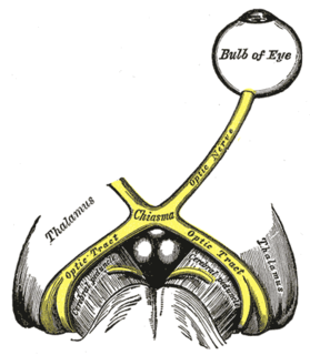
The optic tract is a part of the visual system in the brain. It is a continuation of the optic nerve that relays information from the optic chiasm to the ipsilateral lateral geniculate nucleus (LGN), pretectal nuclei, and superior colliculus.

The septum pellucidum is a thin, triangular, vertical double membrane separating the anterior horns of the left and right lateral ventricles of the brain. It runs as a sheet from the corpus callosum down to the fornix.

In the brain, the interventricular foramina are channels that connect the paired lateral ventricles with the third ventricle at the midline of the brain. As channels, they allow cerebrospinal fluid (CSF) produced in the lateral ventricles to reach the third ventricle and then the rest of the brain's ventricular system. The walls of the interventricular foramina also contain choroid plexus, a specialized CSF-producing structure, that is continuous with that of the lateral and third ventricles above and below it.

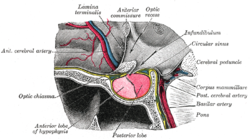
The pituitary stalk is the connection between the hypothalamus and the posterior pituitary. The floor of the third ventricle is prolonged downward as a funnel-shaped recess—the infundibular recess—into the infundibulum, where the apex of the pituitary is attached. It passes through the dura mater of the diaphragma sellae as it carries axons from the magnocellular neurosecretory cells of the hypothalamus down to the posterior pituitary where they release their neurohypophysial hormones, oxytocin and vasopressin, into the blood.

The anterior choroidal artery originates from the internal carotid artery, though it also rarely arises from the middle cerebral artery.
The tuber cinereum is a hollow eminence of the middle–ventral hypothalamus, specifically the arcuate nucleus, situated between the mammillary bodies and the optic chiasm. In addition to the ventral hypothalamus, the tuber cinereum includes the median eminence and pituitary gland. Together with the hollow itself, it is sometimes referred to as the pituitary stalk.

The lateral aperture is a paired structure in human anatomy. It is an opening in each lateral extremity of the lateral recess of the fourth ventricle of the human brain, which also has a single median aperture. The two lateral apertures provide a conduit for cerebrospinal fluid to flow from the brain's ventricular system into the subarachnoid space; specifically into the pontocerebellar cistern at the cerebellopontine angle. The structure is also called the lateral aperture of the fourth ventricle or the foramen of Luschka after anatomist Hubert von Luschka. Gross total resection of tumours that extend through foramen of Lushka is sometimes not possible due to bradycardia.

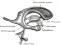
The rhomboid fossa is a rhombus-shaped depression that is the anterior part of the fourth ventricle. Its anterior wall, formed by the back of the pons and the medulla oblongata, constitutes the floor of the fourth ventricle.

In the brain, the taenia of the fourth ventricle are two narrow bands of white matter, one on either side, which complete the lower part of the roof of the fourth ventricle.
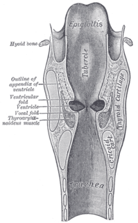
The laryngeal ventricle, is a fusiform fossa, situated between the vestibular and vocal folds on either side, and extending nearly their entire length. There is also a sinus of Morgagni in the pharynx.
In the upper part of the medulla oblongata, the hypoglossal nucleus approaches the rhomboid fossa, where it lies close to the middle line, under an eminence named the hypoglossal trigone. It is a slight elevation in the floor of the inferior recess of the fourth ventricle, beneath which is the nucleus of origin of the twelfth cranial nerve.

The lateral recess is a projection of the fourth ventricle which extends into, or rather below, the inferior cerebellar peduncle of the brainstem.
The chiasmatic cistern is formed as the interpeduncular cistern extends forward across the optic chiasm and onto the upper surface of the corpus callosum – the arachnoid stretches across from one cerebral hemisphere to the other immediately beneath the free border of the falx cerebri, and thus leaves a space in which the anterior cerebral arteries are contained. The "leaf" or extension of the chiasmatic cistern above the chiasma, which is separated from the optic recess of the third ventricle by the thin lamina terminalis, has been called the suprachiasmatic cistern. As spaces filled with freely circulating cerebrospinal fluid, cisterns receive little direct study, but are mentioned in pathological conditions. Cysts and tumors of the lamina terminalis extend into the suprachiasmatic cistern, as can pituitary tumors, or the cistern can be partially or completely effaced by injury and hematoma or by blockage of the cerebral aqueduct.
Noradrenergic cell group A4 is a group of cells exhibiting noradrenergic fluorescence that, in the rat, are located in the Tegmen ventriculi quarti ventral to the cerebellar nuclei, and in the macaque, are found at the edge of the lateral recess of the fourth ventricle caudally, extending to beneath the floor of the ventricle where they merge with the noradrenergic group A6, the locus ceruleus.