Radial glial cell

Last updated
Radial glial cell
G-CSF receptor is expressed in the embryonic nervous system radial glia.jpg
G-CSF receptor expression clearly delineates cells of radial glia in the embryonic murine brain. From Kirsch et al., 2008. [1]
Details
Identifiers
Latin gliocytus radialis
TH H3.11.08.3.01098
Anatomical terms of microanatomy

Radial glial cells, or radial glial progenitor cells (RGPs), are bipolar-shaped progenitor cells that are responsible for producing all of the neurons in the cerebral cortex. RGPs also produce certain lineages of glia, including astrocytes and oligodendrocytes. [2] [3] [4] Their cell bodies (somata) reside in the embryonic ventricular zone, which lies next to the developing ventricular system.

Contents

During development, newborn neurons use radial glia as scaffolds, traveling along the radial glial fibers in order to reach their final destinations. [3] [5] [6] Despite the various possible fates of the radial glial population, it has been demonstrated through clonal analysis that most radial glia have restricted, unipotent or multipotent, fates. Radial glia can be found during the neurogenic phase in all vertebrates (studied to date). [7]

The term "radial glia" refers to the morphological characteristics of these cells that were first observed: namely, their radial processes and their similarity to astrocytes, another member of the glial cell family. [8]

Structure

Müller glia

Müller glia are radial glial cells that are present in the developing, as well as the adult, retina. As in the cortex, Müller glia have long processes that span the entire width of the retina, from the basal cell layer to the apical layer. However, unlike cortical radial glia, Müller glia do not appear in the retina until after the first rounds of neurogenesis have occurred. Studies suggest that Müller glia can dedifferentiate into readily dividing neural progenitors in response to injury. [9]

Slcla3 in Bergmann Glia Slcla3 in Bergmann Glia.jpg
Slcla3 in Bergmann Glia

The characteristics that truly set Müller glia apart from radial glia in other areas of the brain, is their possession of optical properties. The majority of the retina is actually largely light scattering, suggesting that Müller glia serve as the main fiber responsible for the relay of light to the photoreceptors in the rear of the retina. Properties that help Müller glia achieve this function include a limited number mitochondria (which are very light scattering), as well as a specialized arrangement of internal protein filaments. [9]

Müller glia are the predominant type of macroglia in the retina, so they take on many of the supportive functions that astrocytes and oligodendrocytes usually handle in the rest of the central nervous system. [9]

Bergmann glia

Micrograph showing Bergmann gliosis. H&E stain. Bergmann gliosis - high mag.jpg
Micrograph showing Bergmann gliosis. H&E stain.

Bergmann glia (also known as radial epithelial cells, Golgi epithelial cells, or radial astrocytes) are unipolar astrocytes derived from radial glia that are intimately associated with Purkinje cells in the cerebellum. [10] Since bergmann glia appear to persist in the cerebellum, and perform many of the roles characteristic of astrocytes, they have also been called "specialized astrocytes." [9] Bergmann glia have multiple radial processes that extend across the molecular layer of the cerebellar cortex and terminate at the pial surface as a bulbous endfoot. [11] Bergmann glial cells assist with the migration of granule cells, guiding the small neurons from the external granular layer down to the internal granular layer along their extensive radial processes. [12] [13] Besides their role in early development of the cerebellum, Bergmann glia are also required for synaptic pruning. [14] Following Purkinje cell death induced by CNS injury, Bergmann glia undergo extensive proliferative changes so as to replace lost or damaged tissue in a process known as gliosis. [15] [16]

Development

Radial glial cells originate from the transformation of neuroepithelial cells that form the neural plate during neurogenesis in early embryonic development. [8] [9] [17] This process is mediated through the down-regulation of epithelium-related protein expression (such as tight junctions) and an up-regulation of glial-specific features such as glycogen granules, the astrocyte glutamate aspartate transporter (GLAST), the intermediate filament vimentin, and, in some instances, including humans, glial fibrillary acidic protein (GFAP). [7]

After this transition, radial glia retain many of the original characteristics of neuroepithelial cells including: their apical-basal polarity, their position along the lateral ventricles of the developing cortex, and the phasic migration of their nuclei depending on their location with the cell cycle (termed “interkinetic nuclear migration”). [9] [18] [19]

Function

Progenitors

Interneuron-radial glial interactions in the developing cerebral cortex Interneuron-radial glial interactions in the developing cerebral cortex.png
Interneuron-radial glial interactions in the developing cerebral cortex

Radial glia are now recognized as key progenitor cells in the developing nervous system. During the late stages of neurogenesis, radial glial cells divide asymmetrically in the ventricular zone, generating a new radial glial cell, as well as a postmitotic neuron or an intermediate progenitor (IPC) daughter cell. Intermediate progenitor cells then divide symmetrically in the subventricular zone to generate neurons. [18] Local environmental cues such as Notch and fibroblast growth factor (FGF) signaling, developmental period, and differing abilities of radial glia to respond to environmental cues have all been shown to influence the type of radial glia and radial glia-derived daughter cells that will be produced. FGF and Notch signaling regulate the proliferation of radial glia and the rate of neurogenesis, which affects the surface area expansion of the cerebral cortex and its ability to form surface convolutions known as gyri (see gyrification). [9] [20] [21] Radial glial cells show high levels of calcium transient activity, which is transmitted between RGCs in the ventricular zone and along the radial fibers bidirectionally to/from the cortical plate. [22] [23] The calcium activity is thought to promote RGC proliferation and could be involved in radial communication before synapses are present in the brain. Additionally, recent evidence suggests that cues from the external sensory environment can also influence the proliferation and neural differentiation of radial glia. [9] [24]

At the conclusion of cortical development, most radial glia lose their attachment to the ventricles, and migrate towards the surface of the cortex, where, in mammals, most will become astrocytes during the process of gliogenesis. [18]

While it has been suggested that radial glia most likely give rise to oligodendrocytes, through the generation of oligodendrocyte progenitor cells (OPCs), and OPCs can be generated from radial glial cells in vitro , more evidence is yet needed to conclude whether this process also occurs in the developing brain. [18] [25]

Recently, radial glia that exclusively generate upper-layer cortical neurons have also been discovered. [8] Since upper cortical layers have expanded greatly in recent evolution, and are associated with higher-level information processing and thinking, radial glia have been implicated as important mediators of brain evolution. [26]

Migration Pattern

The best characterized and first widely accepted function of radial glia is their role as scaffolds for neuronal migration in the cerebral and cerebellar cortexes. This role can be easily visualized using the electron microscope or high-resolution time-lapse microscopy, through which neurons can be seen tightly wrapped around radial glia as they travel upwards through the cortex. [8] Additional evidence suggests that many neurons may move between neighboring radial glial fibers during migration. [9]

While excitatory neuronal migration is largely radial, inhibitory, GABAergic neurons have been shown to undergo tangential migration. Tangentially migrating neurons also appear to initiate contact with radial glial fibers in the developing cortex of ferrets, implicating radial glial cells in both of these forms of migration. [9]

As radial glia seem to differentiate late in spinal cord development, near the onset of gliogenesis, it is unclear whether they are involved in spinal cord neurogenesis or migration. [8]

Compartmentalization

Radial glia have also been implicated in forming boundaries between different axonal tracts and white matter areas of the brain. [8] [27]

Clinical significance

As radial glia serve as the primary neural and glial progenitors in the brain, as well as being crucial for proper neuronal migration, defects in radial glial function can have profound effects in the development of the nervous system.

Mutations in either Lis1 or Nde1, essential proteins for radial glial differentiation and stabilization, cause the associated neurodevelopmental diseases Lissencephaly and microlissencephaly (which literally translate to “smooth brain”). Patients with these diseases are characterized by a lack of cortical folds (sulci and gyri) and reduced brain volume. Extreme cases of Lissencephaly cause death a few months after birth, while patients with milder forms may experience mental retardation, difficulty balancing, motor and speech deficits, and epilepsy. [8]

Death of neural progenitor cells has recently been linked the mosquito-borne virus, Zika. [28] Epidemiological evidence indicates infection of the embryo within the first two trimesters of pregnancy has potential to cause fetal birth defects and microcephaly, [29] possibly due to the death of progenitor cells. Further, mutations in microcephaly associated genes which encode proteins such as WDR62 can lead to radial glial depletion during brain development which ultimately leads to a smaller brain size and mental disabilities. [30]

History

Camillo Golgi, using his silver staining technique (later deemed the Golgi method), first described radially oriented cells spanning from the central canal to the outer surface of the embryonic chick spinal cord, in 1885. [31]

Using the Golgi method, Giuseppe Magini then studied the mammalian fetal cerebral cortex in 1888, confirming the similar presence of elongated radial cells in the cortex (also described by Kölliker just before him), and observing “various varicosities or swellings” on the radial fibers. Intrigued, Magini also observed that the size and number of these varicosities increased later in development, and were absent in the adult nervous system. Based on these findings, Magini then hypothesized that these varicosities could be developing neurons. Using a combination Golgi and hematoxylin staining method, Magini was able to identify these varicosities as cells, some of which were very closely associated with the radial fibers. [31]

Additional early works that were important in elucidating the identity and function of radial glia, were completed by Ramón y Cajal, who first suggested that the radial cells were a type of glia through their similarities to astrocytes; [8] and Wilhelm His, who also proposed the idea that growing axons may use radial cells for orientation and guidance during development. [31]

Despite the initial period of interest in radial glia, little additional information was learned about these cells until the electron microscope and immunohistochemistry became available some 60 years later. [31]

See also

List of distinct cell types in the adult human body

Related Research Articles

<span class="mw-page-title-main">Cerebral cortex</span> Outer layer of the cerebrum of the mammalian brain

The cerebral cortex, also known as the cerebral mantle, is the outer layer of neural tissue of the cerebrum of the brain in humans and other mammals. The cerebral cortex mostly consists of the six-layered neocortex, with just 10% consisting of the allocortex. It is separated into two cortices, by the longitudinal fissure that divides the cerebrum into the left and right cerebral hemispheres. The two hemispheres are joined beneath the cortex by the corpus callosum. The cerebral cortex is the largest site of neural integration in the central nervous system. It plays a key role in attention, perception, awareness, thought, memory, language, and consciousness. The cerebral cortex is part of the brain responsible for cognition.

The development of the nervous system, or neural development (neurodevelopment), refers to the processes that generate, shape, and reshape the nervous system of animals, from the earliest stages of embryonic development to adulthood. The field of neural development draws on both neuroscience and developmental biology to describe and provide insight into the cellular and molecular mechanisms by which complex nervous systems develop, from nematodes and fruit flies to mammals.

<span class="mw-page-title-main">Glia</span> Support cells in the nervous system

Glia, also called glial cells(gliocytes) or neuroglia, are non-neuronal cells in the central nervous system (brain and spinal cord) and the peripheral nervous system that do not produce electrical impulses. The neuroglia make up more than one half the volume of neural tissue in our body. They maintain homeostasis, form myelin in the peripheral nervous system, and provide support and protection for neurons. In the central nervous system, glial cells include oligodendrocytes, astrocytes, ependymal cells and microglia, and in the peripheral nervous system they include Schwann cells and satellite cells.

In vertebrates, a neuroblast or primitive nerve cell is a postmitotic cell that does not divide further, and which will develop into a neuron after a migration phase. In invertebrates such as Drosophila, neuroblasts are neural progenitor cells which divide asymmetrically to produce a neuroblast, and a daughter cell of varying potency depending on the type of neuroblast. Vertebrate neuroblasts differentiate from radial glial cells and are committed to becoming neurons. Neural stem cells, which only divide symmetrically to produce more neural stem cells, transition gradually into radial glial cells. Radial glial cells, also called radial glial progenitor cells, divide asymmetrically to produce a neuroblast and another radial glial cell that will re-enter the cell cycle.

<span class="mw-page-title-main">Astrocyte</span> Type of brain cell

Astrocytes, also known collectively as astroglia, are characteristic star-shaped glial cells in the brain and spinal cord. They perform many functions, including biochemical control of endothelial cells that form the blood–brain barrier, provision of nutrients to the nervous tissue, maintenance of extracellular ion balance, regulation of cerebral blood flow, and a role in the repair and scarring process of the brain and spinal cord following infection and traumatic injuries. The proportion of astrocytes in the brain is not well defined; depending on the counting technique used, studies have found that the astrocyte proportion varies by region and ranges from 20% to around 40% of all glia. Another study reports that astrocytes are the most numerous cell type in the brain. Astrocytes are the major source of cholesterol in the central nervous system. Apolipoprotein E transports cholesterol from astrocytes to neurons and other glial cells, regulating cell signaling in the brain. Astrocytes in humans are more than twenty times larger than in rodent brains, and make contact with more than ten times the number of synapses.

Oligodendrocyte progenitor cells (OPCs), also known as oligodendrocyte precursor cells, NG2-glia, O2A cells, or polydendrocytes, are a subtype of glia in the central nervous system named for their essential role as precursors to oligodendrocytes. They are typically identified in the human by co-expression of PDGFRA and CSPG4.

Neural stem cells (NSCs) are self-renewing, multipotent cells that firstly generate the radial glial progenitor cells that generate the neurons and glia of the nervous system of all animals during embryonic development. Some neural progenitor stem cells persist in highly restricted regions in the adult vertebrate brain and continue to produce neurons throughout life. Differences in the size of the central nervous system are among the most important distinctions between the species and thus mutations in the genes that regulate the size of the neural stem cell compartment are among the most important drivers of vertebrate evolution.

<span class="mw-page-title-main">Subventricular zone</span> Region outside each lateral ventricle of the brain

The subventricular zone (SVZ) is a region situated on the outside wall of each lateral ventricle of the vertebrate brain. It is present in both the embryonic and adult brain. In embryonic life, the SVZ refers to a secondary proliferative zone containing neural progenitor cells, which divide to produce neurons in the process of neurogenesis. The primary neural stem cells of the brain and spinal cord, termed radial glial cells, instead reside in the ventricular zone (VZ).

<span class="mw-page-title-main">Müller glia</span> Glial cell type in the retina

Müller glia, or Müller cells, are a type of retinal glial cells, first recognized and described by Heinrich Müller. They are found in the vertebrate retina, where they serve as support cells for the neurons, as all glial cells do. They are the most common type of glial cell found in the retina. While their cell bodies are located in the inner nuclear layer of the retina, they span across the entire retina.

<span class="mw-page-title-main">Subgranular zone</span>

The subgranular zone (SGZ) is a brain region in the hippocampus where adult neurogenesis occurs. The other major site of adult neurogenesis is the subventricular zone (SVZ) in the brain.

Gyrification is the process of forming the characteristic folds of the cerebral cortex.

<span class="mw-page-title-main">Eomesodermin</span> Protein-coding gene in the species Homo sapiens

Eomesodermin also known as T-box brain protein 2 (Tbr2) is a protein that in humans is encoded by the EOMES gene.

Endogenous regeneration in the brain is the ability of cells to engage in the repair and regeneration process. While the brain has a limited capacity for regeneration, endogenous neural stem cells, as well as numerous pro-regenerative molecules, can participate in replacing and repairing damaged or diseased neurons and glial cells. Another benefit that can be achieved by using endogenous regeneration could be avoiding an immune response from the host.

The development of the cerebral cortex, known as corticogenesis is the process during which the cerebral cortex of the brain is formed as part of the development of the nervous system of mammals including its development in humans. The cortex is the outer layer of the brain and is composed of up to six layers. Neurons formed in the ventricular zone migrate to their final locations in one of the six layers of the cortex. The process occurs from embryonic day 10 to 17 in mice and between gestational weeks seven to 18 in humans.

Proneural genes encode transcription factors of the basic helix-loop-helix (bHLH) class which are responsible for the development of neuroectodermal progenitor cells. Proneural genes have multiple functions in neural development. They integrate positional information and contribute to the specification of progenitor-cell identity. From the same ectodermal cell types, neural or epidermal cells can develop based on interactions between proneural and neurogenic genes. Neurogenic genes are so called because loss of function mutants show an increase number of developed neural precursors. On the other hand, proneural genes mutants fail to develop neural precursor cells.

<span class="mw-page-title-main">Neuronal lineage marker</span> Endogenous tag expressed in different cells along neurogenesis and differentiated cells

A neuronal lineage marker is an endogenous tag that is expressed in different cells along neurogenesis and differentiated cells such as neurons. It allows detection and identification of cells by using different techniques. A neuronal lineage marker can be either DNA, mRNA or RNA expressed in a cell of interest. It can also be a protein tag, as a partial protein, a protein or an epitope that discriminates between different cell types or different states of a common cell. An ideal marker is specific to a given cell type in normal conditions and/or during injury. Cell markers are very valuable tools for examining the function of cells in normal conditions as well as during disease. The discovery of various proteins specific to certain cells led to the production of cell-type-specific antibodies that have been used to identify cells.

<span class="mw-page-title-main">Ventricular zone</span> Transient embryonic layer of tissue containing neural stem cells

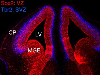
In vertebrates, the ventricular zone (VZ) is a transient embryonic layer of tissue containing neural stem cells, principally radial glial cells, of the central nervous system (CNS). The VZ is so named because it lines the ventricular system, which contains cerebrospinal fluid (CSF). The embryonic ventricular system contains growth factors and other nutrients needed for the proper function of neural stem cells. Neurogenesis, or the generation of neurons, occurs in the VZ during embryonic and fetal development as a function of the Notch pathway, and the newborn neurons must migrate substantial distances to their final destination in the developing brain or spinal cord where they will establish neural circuits. A secondary proliferative zone, the subventricular zone (SVZ), lies adjacent to the VZ. In the embryonic cerebral cortex, the SVZ contains intermediate neuronal progenitors that continue to divide into post-mitotic neurons. Through the process of neurogenesis, the parent neural stem cell pool is depleted and the VZ disappears. The balance between the rates of stem cell proliferation and neurogenesis changes during development, and species from mouse to human show large differences in the number of cell cycles, cell cycle length, and other parameters, which is thought to give rise to the large diversity in brain size and structure.

Neurogenesis is the process by which nervous system cells, the neurons, are produced by neural stem cells (NSCs). In short, it is brain growth in relation to its organization. This occurs in all species of animals except the porifera (sponges) and placozoans. Types of NSCs include neuroepithelial cells (NECs), radial glial cells (RGCs), basal progenitors (BPs), intermediate neuronal precursors (INPs), subventricular zone astrocytes, and subgranular zone radial astrocytes, among others.

Intermediate progenitor cells (IPCs) are a type of progenitor cell in the developing cerebral cortex. They are multipolar cells produced by radial glial cells who have undergone asymmetric division. IPCs can produce neuron cells via neurogenesis and are responsible for ensuring the proper quantity of cortical neurons are produced. In mammals, neural stem cells are the primary progenitors during embryogenesis whereas intermediate progenitor cells are the secondary progenitors.

<span class="mw-page-title-main">Brain cell</span> Functional tissue of the brain

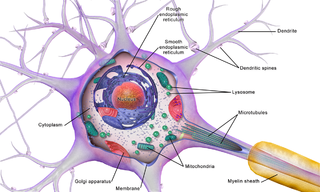
Brain cells make up the functional tissue of the brain. The rest of the brain tissue is structural or connective called the stroma which includes blood vessels. The two main types of cells in the brain are neurons, also known as nerve cells, and glial cells, also known as neuroglia.

References

  1. Kirsch F, Krüger C, Schneider A (March 2008). "The receptor for granulocyte-colony stimulating factor (G-CSF) is expressed in radial glia during development of the nervous system". BMC Developmental Biology. 8: 32. doi: 10.1186/1471-213X-8-32 . PMC   2329616 . PMID   18371196.
  2. Beattie, R; Hippenmeyer, S (December 2017). "Mechanisms of radial glia progenitor cell lineage progression". FEBS Letters. 591 (24): 3993–4008. doi:10.1002/1873-3468.12906. PMC   5765500 . PMID   29121403.
  3. 1 2 Rakic P (October 2009). "Evolution of the neocortex: a perspective from developmental biology". Nature Reviews. Neuroscience. 10 (10): 724–35. doi:10.1038/nrn2719. PMC   2913577 . PMID   19763105.
  4. Noctor SC, Flint AC, Weissman TA, Dammerman RS, Kriegstein AR (February 2001). "Neurons derived from radial glial cells establish radial units in neocortex". Nature. 409 (6821): 714–20. Bibcode:2001Natur.409..714N. doi:10.1038/35055553. PMID   11217860. S2CID   3041502.
  5. Rakic P (May 1972). "Mode of cell migration to the superficial layers of fetal monkey neocortex". The Journal of Comparative Neurology. 145 (1): 61–83. doi:10.1002/cne.901450105. PMID   4624784. S2CID   41001390.
  6. Shohayeb B, et al. (October 2021). "Conservation of neural progenitor identity and the emergence of neocortical neuronal diversity". Seminars in Cell and Developmental Biology. 118 (118): 4–13. doi:10.1016/j.semcdb.2021.05.024. PMID   34083116. S2CID   235336596.
  7. 1 2 Malatesta P, Appolloni I, Calzolari F (January 2008). "Radial glia and neural stem cells". Cell and Tissue Research. 331 (1): 165–78. doi:10.1007/s00441-007-0481-8. PMID   17846796. S2CID   1903664.
  8. 1 2 3 4 5 6 7 8 Barry DS, Pakan JM, McDermott KW (January 2014). "Radial glial cells: key organisers in CNS development". The International Journal of Biochemistry & Cell Biology. 46: 76–9. doi:10.1016/j.biocel.2013.11.013. hdl: 2262/68379 . PMID   24269781.
  9. 1 2 3 4 5 6 7 8 9 10 Sild M, Ruthazer ES (June 2011). "Radial glia: progenitor, pathway, and partner". The Neuroscientist. 17 (3): 288–302. doi:10.1177/1073858410385870. PMID   21558559. S2CID   1817598.
  10. Verkhratsky, Alexei; Butt, Arthur M. (2013). Glial Physiology and Pathophysiology. John Wiley and Sons, Inc. ISBN   9780470978535.
  11. Komine O, Nagaoka M, Watase K, Gutmann DH, Tanigaki K, Honjo T, Radtke F, Saito T, Chiba S, Tanaka K (November 2007). "The monolayer formation of Bergmann glial cells is regulated by Notch/RBP-J signaling". Developmental Biology. 311 (1): 238–50. doi:10.1016/j.ydbio.2007.08.042. PMID   17915208.
  12. Rubenstein, John; Rakic, Pasko (2013). Cellular Migration and Formation of Neuronal Connections: Comprehensive Developmental Neuroscience. Elsevier Science and Technology. ISBN   9780123972668.
  13. Sanes DH, Reh TA, Harris WA (2005). Development of the Nervous System. Elsevier Science and Technology. ISBN   9780126186215.
  14. "Bergmann Glial Cell". 14 Oct 2011. Retrieved 10 September 2014.
  15. Sofroniew MV (November 2014). "Astrogliosis". Cold Spring Harbor Perspectives in Biology. 7 (2): a020420. doi:10.1101/cshperspect.a020420. PMC   4315924 . PMID   25380660.
  16. Catherine., Haberland (2007). Clinical neuropathology : text and color atlas. New York: Demos. ISBN   9781934559529. OCLC   166267295.
  17. Shohayeb B, et al. (October 2021). "Conservation of neural progenitor identity and the emergence of neocortical neuronal diversity". Seminars in Cell and Developmental Biology. 118 (118): 4–13. doi:10.1016/j.semcdb.2021.05.024. PMID   34083116. S2CID   235336596.
  18. 1 2 3 4 Kriegstein A, Alvarez-Buylla A (2009). "The glial nature of embryonic and adult neural stem cells". Annual Review of Neuroscience. 32: 149–84. doi:10.1146/annurev.neuro.051508.135600. PMC   3086722 . PMID   19555289.
  19. Shohayeb B, et al. (October 2021). "Conservation of neural progenitor identity and the emergence of neocortical neuronal diversity". Seminars in Cell and Developmental Biology. 118 (118): 4–13. doi:10.1016/j.semcdb.2021.05.024. PMID   34083116. S2CID   235336596.
  20. Rash BG, Lim HD, Breunig JJ, Vaccarino FM (October 2011). "FGF signaling expands embryonic cortical surface area by regulating Notch-dependent neurogenesis". The Journal of Neuroscience. 31 (43): 15604–17. doi:10.1523/jneurosci.4439-11.2011. PMC   3235689 . PMID   22031906.
  21. Rash BG, Tomasi S, Lim HD, Suh CY, Vaccarino FM (June 2013). "Cortical gyrification induced by fibroblast growth factor 2 in the mouse brain". The Journal of Neuroscience. 33 (26): 10802–14. doi:10.1523/jneurosci.3621-12.2013. PMC   3693057 . PMID   23804101.
  22. Weissman TA, Riquelme PA, Ivic L, Flint AC, Kriegstein AR (September 2004). "Calcium waves propagate through radial glial cells and modulate proliferation in the developing neocortex". Neuron. 43 (5): 647–61. doi: 10.1016/j.neuron.2004.08.015 . PMID   15339647. S2CID   7094565.
  23. Rash BG, Ackman JB, Rakic P (February 2016). "Bidirectional radial Ca(2+) activity regulates neurogenesis and migration during early cortical column formation". Science Advances. 2 (2): e1501733. Bibcode:2016SciA....2E1733R. doi:10.1126/sciadv.1501733. PMC   4771444 . PMID   26933693.
  24. Sharma P, Cline HT (November 2010). "Visual activity regulates neural progenitor cells in developing xenopus CNS through musashi1". Neuron. 68 (3): 442–55. doi:10.1016/j.neuron.2010.09.028. PMC   3005332 . PMID   21040846.
  25. Mo Z, Zecevic N (April 2009). "Human fetal radial glia cells generate oligodendrocytes in vitro". Glia. 57 (5): 490–8. doi:10.1002/glia.20775. PMC   2644732 . PMID   18814269.
  26. "Scripps Research Neuroscientists Find Brain Stem Cells that May Be Responsible for Higher Functions, Bigger Brains". Scripps Research Institute. Retrieved 1 March 2014.
  27. Steindler DA (1993). "Glial boundaries in the developing nervous system". Annual Review of Neuroscience. 16: 445–70. doi:10.1146/annurev.ne.16.030193.002305. PMID   8460899.
  28. Tang H, Hammack C, Ogden SC, Wen Z, Qian X, Li Y, Yao B, Shin J, Zhang F, Lee EM, Christian KM, Didier RA, Jin P, Song H, Ming GL (May 2016). "Zika Virus Infects Human Cortical Neural Progenitors and Attenuates Their Growth". Cell Stem Cell. 18 (5): 587–90. doi:10.1016/j.stem.2016.02.016. PMC   5299540 . PMID   26952870.
  29. Mlakar J, Korva M, Tul N, Popović M, Poljšak-Prijatelj M, Mraz J, Kolenc M, Resman Rus K, Vesnaver Vipotnik T, Fabjan Vodušek V, Vizjak A, Pižem J, Petrovec M, Avšič Županc T (March 2016). "Zika Virus Associated with Microcephaly". The New England Journal of Medicine. 374 (10): 951–8. doi: 10.1056/NEJMoa1600651 . PMID   26862926.
  30. Shohayeb, B, et al. (January 2020). "The association of microcephaly protein WDR62 with CPAP/IFT88 is required for cilia formation and neocortical development". Human Molecular Genetics. 29 (2): 248–263. doi: 10.1093/hmg/ddz281 . PMID   31816041.
  31. 1 2 3 4 Bentivoglio M, Mazzarello P (July 1999). "The history of radial glia". Brain Research Bulletin. 49 (5): 305–15. doi:10.1016/s0361-9230(99)00065-9. PMID   10452351. S2CID   26003944.