Granule cell

Last updated
Drawing of Purkinje cells (A) and granule cells (B) from pigeon cerebellum by Santiago Ramon y Cajal, 1899. Instituto Santiago Ramon y Cajal, Madrid, Spain. PurkinjeCell.jpg
Drawing of Purkinje cells (A) and granule cells (B) from pigeon cerebellum by Santiago Ramón y Cajal, 1899. Instituto Santiago Ramón y Cajal, Madrid, Spain.

The name granule cell has been used for a number of different types of neurons whose only common feature is that they all have very small cell bodies. Granule cells are found within the granular layer of the cerebellum, the dentate gyrus of the hippocampus, the superficial layer of the dorsal cochlear nucleus, the olfactory bulb, and the cerebral cortex.

Contents

Cerebellar granule cells account for the majority of neurons in the human brain. [1] These granule cells receive excitatory input from mossy fibers originating from pontine nuclei. Cerebellar granule cells project up through the Purkinje layer into the molecular layer where they branch out into parallel fibers that spread through Purkinje cell dendritic arbors. These parallel fibers form thousands of excitatory granule-cell–Purkinje-cell synapses onto the intermediate and distal dendrites of Purkinje cells using glutamate as a neurotransmitter.

Layer 4 granule cells of the cerebral cortex receive inputs from the thalamus and send projections to supragranular layers 2–3, but also to infragranular layers of the cerebral cortex.

Structure

Granule cells in different brain regions are both functionally and anatomically diverse: the only thing they have in common is smallness. For instance, olfactory bulb granule cells are GABAergic and axonless, while granule cells in the dentate gyrus have glutamatergic projection axons. These two populations of granule cells are also the only major neuronal populations that undergo adult neurogenesis, while cerebellar and cortical granule cells do not. Granule cells (save for those of the olfactory bulb) have a structure typical of a neuron consisting of dendrites, a soma (cell body) and an axon.

Dendrites: Each granule cell has 3 – 4 stubby dendrites which end in a claw. Each of the dendrites are only about 15 μm in length.

Soma: Granule cells all have a small soma diameter of approximately 10 μm.

Axon: Each granule cell sends a single axon onto the Purkinje cell dendritic tree.[ citation needed ] The axon has an extremely narrow diameter: ½ micrometre.

Synapse: 100–300,000 granule cell axons synapse onto a single Purkinje cell.[ citation needed ]

The existence of gap junctions between granule cells allows multiple neurons to be coupled to one another allowing multiple cells to act in synchrony and to allow signalling functions necessary for granule cell development to occur. [2]

Cerebellar granule cell

The granule cells, produced by the rhombic lip, are found in the granule cell layer of the cerebellar cortex. They are small and numerous. They are characterized by a very small soma and several short dendrites which terminate with claw-shaped endings. In the transmission electron microscope, these cells are characterized by a darkly stained nucleus surrounded by a thin rim of cytoplasm. The axon ascends into the molecular layer where it splits to form parallel fibers. [1]

Dentate gyrus granule cell

The principal cell type of the dentate gyrus is the granule cell. The dentate gyrus granule cell has an elliptical cell body with a width of approximately 10 μm and a height of 18μm. [3]

The granule cell has a characteristic cone-shaped tree of spiny apical dendrites. The dendrite branches project throughout the entire molecular layer and the furthest tips of the dendritic tree end just at the hippocampal fissure or at the ventricular surface. [4] The granule cells are tightly packed in the granular cell layer of the dentate gyrus.

Dorsal cochlear nucleus granule cell

The granule cells in the dorsal cochlear nucleus are small neurons with two or three short dendrites that give rise to a few branches with expansions at the terminals. The dendrites are short with claw-like endings that form glomeruli to receive mossy fibers, similar to cerebellar granule cells. [5] Its axon projects to the molecular layer of the dorsal cochlear nucleus where it forms parallel fibers, also similar to cerebellar granule cells. [6] The dorsal cochlear granule cells are small excitatory interneurons which are developmentally related and thus resemble the cerebellar granule cell.

Olfactory bulb granule cell

The main intrinsic granule cell in the vertebrate olfactory bulb lacks an axon (as does the accessory neuron). Each cell gives rise to short central dendrites and a single long apical dendrite that expands into the granule cell layer and enters the mitral cell body layer. The dendrite branches terminate within the outer plexiform layer among the dendrites in the olfactory tract. [7] In the mammalian olfactory bulb, granule cells can process both synaptic input and output due to the presence of large spines. [8]

Function

Neural pathways and circuits of the cerebellum

Neural pathways and circuits in the cerebellum. (+) represent excitatory synapse, while (-) represent inhibitory synapses. Architecture of the Cerebellar Cortex.svg
Neural pathways and circuits in the cerebellum. (+) represent excitatory synapse, while (-) represent inhibitory synapses.

Cerebellar granule cells receive excitatory input from 3 or 4 mossy fibers originating from pontine nuclei. Mossy fibers make an excitatory connection onto granule cells which cause the granule cell to fire an action potential.

The axon of a cerebellar granule cell splits to form a parallel fiber which innervates Purkinje cells. The vast majority of granule cell axonal synapses are found on the parallel fibers. [9]

The parallel fibers are sent up through the Purkinje layer into the molecular layer where they branch out and spread through Purkinje cell dendritic arbors. These parallel fibers form thousands of excitatory Granule-cell-Purkinje-cell synapses onto the dendrites of Purkinje cells.

This connection is excitatory as glutamate is released.

The parallel fibers and ascending axon synapses from the same granule cell fire in synchrony which results in excitatory signals. In the cerebellar cortex there are a variety of inhibitory neurons (interneurons). The only excitatory neurons present in the cerebellar cortex are granule cells. [10]

Plasticity of the synapse between a parallel fiber and a Purkinje cell is believed to be important for motor learning. [11] The function of cerebellar circuits is entirely dependent on processes carried out by the granular layer. Therefore, the function of granule cells determines the cerebellar function as a whole. [12]

Mossy fiber input on cerebellar granule cells

Granule cell dendrites also synapse with distinctive unmyelinated axons which Santiago Ramón y Cajal called mossy fibers [4] Mossy fibers and golgi cells both make synaptic connections with granule cells. Together these cells form the glomeruli. [10]

Granule cells are subject to feed-forward inhibition: granule cells excite Purkinje cells but also excite GABAergic interneurons that inhibit Purkinje cells.

Granule cells are also subject to feedback inhibition: Golgi cells receive excitatory stimuli from granule cells and in turn send back inhibitory signals to the granule cell. [13]

Mossy fiber input codes are conserved during synaptic transmission between granule cells, suggesting that innervation is specific to the input that is received. [14] Granule cells do not just relay signals from mossy fibers, rather they perform various, intricate transformations which are required in the spatiotemporal domain. [10]

Each granule cell is receiving an input from two different mossy fiber inputs. The input is thus coming from two different places as opposed to the granule cell receiving multiple inputs from the same source.

The differences in mossy fibers that are sending signals to the granule cells directly effects the type of information that granule cells translate to Purkinje cells. The reliability of this translation will depend on the reliability of synaptic activity in granule cells and on the nature of the stimulus being received. [15] The signal a granule cell receives from a Mossy fiber depends on the function of the mossy fiber itself. Therefore, granule cells are able to integrate information from the different mossy fibers and generate new patterns of activity. [15]

Climbing fiber input on cerebellar granule cells

Different patterns of mossy fiber input will produce unique patterns of activity in granule cells that can be modified by a teaching signal conveyed by the climbing fiber input. David Marr and James Albus suggested that the cerebellum operates as an adaptive filter, altering motor behaviour based on the nature of the sensory input.

Since multiple (~200,000) granule cells synapse onto a single Purkinje cell, the effects of each parallel fiber can be altered in response to a “teacher signal” from the climbing fiber input.

Specific functions of different granule cells

Cerebellum granule cells

David Marr suggested that the granule cells encode combinations of mossy fiber inputs. In order for the granule cell to respond, it needs to receive active inputs from multiple mossy fibers. The combination of multiple inputs results in the cerebellum being able to make more precise distinctions between input patterns than a single mossy fiber would allow. [16] The cerebellar granule cells also play a role in orchestrating the tonic conductances which control sleep in conjunction with the ambient levels of GABA which are found in the brain.

Dentate granule cells

Loss of dentate gyrus neurons from the hippocampus results in spatial memory deficits. Therefore, dentate granule cells are thought to function in the formation of spatial memories [17] and of episodic memories. [18] Immature and mature dentate granule cells have distinct roles in memory function. Adult-born granule cells are thought to be involved in pattern separation whereas old granule cells contribute to rapid pattern completion. [19]

Dorsal cochlear granule cells

Pyramidal cells from the primary auditory cortex project directly on to the cochlear nucleus. This is important in the acoustic startle reflex, in which the pyramidal cells modulate the secondary orientation reflex and the granule cell input is responsible for appropriate orientation. [20] This is because the signals received by the granule cells contain information about the head position. Granule cells in the dorsal cochlear nucleus play a role in the perception and response to sounds in our environment.

Olfactory bulb granule cells

Inhibition generated by granule cells, the most common GABAergic cell type in the olfactory bulb, plays a critical role in shaping the output of the olfactory bulb. [21] There are two types of excitatory inputs received by GABAergic granule cells; those activated by an AMPA receptor and those activated by a NMDA receptor. This allows the granule cells to regulate the processing of the sensory input in the olfactory bulb. [21] The olfactory bulb transmits smell information from the nose to the brain, and is thus necessary for a proper sense of smell. Granule cells in the olfactory bulb have also been found to be important in forming memories linked with scents. [22]

Critical factors for function

Calcium

Calcium dynamics are essential for several functions of granule cells such as changing membrane potential, synaptic plasticity, apoptosis, and regulation of gene transcription. [10] The nature of the calcium signals that control the presynaptic and postsynaptic function of the olfactory bulb granule cells spines is mostly unknown. [8]

Nitric oxide

Granule neurons have high levels of the neuronal isoform of nitric oxide synthase. This enzyme is dependent on the presence of calcium and is responsible for the production of nitric oxide (NO). This neurotransmitter is a negative regulator of granule cell precursor proliferation which promotes the differentiation of different granule cells. NO regulates interactions between granule cells and glia [10] and is essential for protecting the granule cells from damage. NO is also responsible for neuroplasticity and motor learning. [23]

Role in disease

Altered morphology of dentate granule cells

TrkB is responsible for the maintenance of normal synaptic connectivity of the dentate granule cells. TrkB also regulates the specific morphology (biology) of the granule cells and is thus said to be important in regulating neuronal development, neuronal plasticity, learning, and the development of epilepsy. [24] The TrkB regulation of granule cells is important in preventing memory deficits and limbic epilepsy. This is due to the fact that dentate granule cells play a critical role in the function of the entorhinal-hippocampal circuitry in health and disease. Dentate granule cells are situated to regulate the flow of information into the hippocampus, a structure required for normal learning and memory. [24]

Decreased granule cell neurogenesis

Both epilepsy and depression show a disrupted production of adult-born hippocampal granule cells. [25] Epilepsy is associated with increased production - but aberrant integration - of new cells early in the disease and decreased production late in the disease. [25] Aberrant integration of adult-generated cells during the development of epilepsy may impair the ability of the dentate gyrus to prevent excess excitatory activity from reaching hippocampal pyramidal cells, thereby promoting seizures. [25] Long-lasting epileptic seizure stimulate dentate granule cell neurogenesis. These newly born dentate granule cells may result in aberrant connections that result in the hippocampal network plasticity associated with epileptogenesis. [26]

Shorter granule cell dendrites

Patients with Alzheimer's have shorter granule cell dendrites. Furthermore, the dendrites were less branched and had fewer spines than those in patients not with Alzheimer's. [27] However, granule cell dendrites are not an essential component of senile plaques and these plaques have no direct effect on granule cells in the dentate gyrus. The specific neurofibrillary changes of dentate granule cells occur in patients with Alzheimer's, Lewy body variant and progressive supranuclear palsy. [28]

See also

List of distinct cell types in the adult human body

Related Research Articles

<span class="mw-page-title-main">Cerebellum</span> Structure at the rear of the vertebrate brain, beneath the cerebrum

The cerebellum is a major feature of the hindbrain of all vertebrates. Although usually smaller than the cerebrum, in some animals such as the mormyrid fishes it may be as large as it or even larger. In humans, the cerebellum plays an important role in motor control. It may also be involved in some cognitive functions such as attention and language as well as emotional control such as regulating fear and pleasure responses, but its movement-related functions are the most solidly established. The human cerebellum does not initiate movement, but contributes to coordination, precision, and accurate timing: it receives input from sensory systems of the spinal cord and from other parts of the brain, and integrates these inputs to fine-tune motor activity. Cerebellar damage produces disorders in fine movement, equilibrium, posture, and motor learning in humans.

<span class="mw-page-title-main">Dentate gyrus</span> Region of the hippocampus in the brain

The dentate gyrus (DG) is part of the hippocampal formation in the temporal lobe of the brain, which also includes the hippocampus and the subiculum. The dentate gyrus is part of the hippocampal trisynaptic circuit and is thought to contribute to the formation of new episodic memories, the spontaneous exploration of novel environments and other functions.

<span class="mw-page-title-main">Olfactory bulb</span> Neural structure

The olfactory bulb is a neural structure of the vertebrate forebrain involved in olfaction, the sense of smell. It sends olfactory information to be further processed in the amygdala, the orbitofrontal cortex (OFC) and the hippocampus where it plays a role in emotion, memory and learning. The bulb is divided into two distinct structures: the main olfactory bulb and the accessory olfactory bulb. The main olfactory bulb connects to the amygdala via the piriform cortex of the primary olfactory cortex and directly projects from the main olfactory bulb to specific amygdala areas. The accessory olfactory bulb resides on the dorsal-posterior region of the main olfactory bulb and forms a parallel pathway. Destruction of the olfactory bulb results in ipsilateral anosmia, while irritative lesions of the uncus can result in olfactory and gustatory hallucinations.

<span class="mw-page-title-main">Neural pathway</span> Connection formed between neurons that allows neurotransmission

In neuroanatomy, a neural pathway is the connection formed by axons that project from neurons to make synapses onto neurons in another location, to enable neurotransmission. Neurons are connected by a single axon, or by a bundle of axons known as a nerve tract, or fasciculus. Shorter neural pathways are found within grey matter in the brain, whereas longer projections, made up of myelinated axons, constitute white matter.

<span class="mw-page-title-main">Pyramidal cell</span> Projection neurons in the cerebral cortex and hippocampus

Pyramidal cells, or pyramidal neurons, are a type of multipolar neuron found in areas of the brain including the cerebral cortex, the hippocampus, and the amygdala. Pyramidal cells are the primary excitation units of the mammalian prefrontal cortex and the corticospinal tract. One of the main structural features of the pyramidal neuron is the conic shaped soma, or cell body, after which the neuron is named. Other key structural features of the pyramidal cell are a single axon, a large apical dendrite, multiple basal dendrites, and the presence of dendritic spines.

<span class="mw-page-title-main">Purkinje cell</span> Specialized neuron in the cerebellum

Purkinje cells or Purkinje neurons, named for Czech physiologist Jan Evangelista Purkyně who identified them in 1837, are a unique type of prominent large neurons located in the cerebellar cortex of the brain. With their flask-shaped cell bodies, many branching dendrites, and a single long axon, these cells are essential for controlling motor activity. Purkinje cells mainly release GABA neurotransmitter, which inhibits some neurons to reduce nerve impulse transmission. Purkinje cells efficiently control and coordinate the body's motor motions through these inhibitory actions.

An apical dendrite is a dendrite that emerges from the apex of a pyramidal cell. Apical dendrites are one of two primary categories of dendrites, and they distinguish the pyramidal cells from spiny stellate cells in the cortices. Pyramidal cells are found in the prefrontal cortex, the hippocampus, the entorhinal cortex, the olfactory cortex, and other areas. Dendrite arbors formed by apical dendrites are the means by which synaptic inputs into a cell are integrated. The apical dendrites in these regions contribute significantly to memory, learning, and sensory associations by modulating the excitatory and inhibitory signals received by the pyramidal cells.

<span class="mw-page-title-main">Golgi cell</span>

In neuroscience, Golgi cells are the most abundant inhibitory interneurons found within the granular layer of the cerebellum. Golgi cells can be found in the granular layer at various layers. The Golgi cell is essential for controlling the activity of the granular layer. They were first identified as inhibitory in 1964. It was also the first example of an inhibitory feedback network in which the inhibitory interneuron was identified anatomically. Golgi cells produce a wide lateral inhibition that reaches beyond the afferent synaptic field and inhibit granule cells via feedforward and feedback inhibitory loops. These cells synapse onto the dendrite of granule cells and unipolar brush cells. They receive excitatory input from mossy fibres, also synapsing on granule cells, and parallel fibers, which are long granule cell axons. Thereby this circuitry allows for feed-forward and feed-back inhibition of granule cells.

<span class="mw-page-title-main">Mitral cell</span> Neurons that are part of the olfactory system

Mitral cells are neurons that are part of the olfactory system. They are located in the olfactory bulb in the mammalian central nervous system. They receive information from the axons of olfactory receptor neurons, forming synapses in neuropils called glomeruli. Axons of the mitral cells transfer information to a number of areas in the brain, including the piriform cortex, entorhinal cortex, and amygdala. Mitral cells receive excitatory input from olfactory sensory neurons and external tufted cells on their primary dendrites, whereas inhibitory input arises either from granule cells onto their lateral dendrites and soma or from periglomerular cells onto their dendritic tuft. Mitral cells together with tufted cells form an obligatory relay for all olfactory information entering from the olfactory nerve. Mitral cell output is not a passive reflection of their input from the olfactory nerve. In mice, each mitral cell sends a single primary dendrite into a glomerulus receiving input from a population of olfactory sensory neurons expressing identical olfactory receptor proteins, yet the odor responsiveness of the 20-40 mitral cells connected to a single glomerulus is not identical to the tuning curve of the input cells, and also differs between sister mitral cells. Odorant response properties of individual neurons in an olfactory glomerular module. The exact type of processing that mitral cells perform with their inputs is still a matter of controversy. One prominent hypothesis is that mitral cells encode the strength of an olfactory input into their firing phases relative to the sniff cycle. A second hypothesis is that the olfactory bulb network acts as a dynamical system that decorrelates to differentiate between representations of highly similar odorants over time. Support for the second hypothesis comes primarily from research in zebrafish.

The stratum lucidum of the hippocampus is a layer of the hippocampus between the stratum pyramidale and the stratum radiatum. It is the tract of the mossy fiber projections, both inhibitory and excitatory from the granule cells of the dentate gyrus. One mossy fiber may make up to 37 connections to a single pyramidal cell, and innervate around 12 pyramidal cells on top of that. Any given pyramidal cell in the stratum lucidum may get input from as many as 50 granule cells.

<span class="mw-page-title-main">Mossy fiber (hippocampus)</span> Pathway in the hippocampus

In the hippocampus, the mossy fiber pathway consists of unmyelinated axons projecting from granule cells in the dentate gyrus that terminate on modulatory hilar mossy cells and in Cornu Ammonis area 3 (CA3), a region involved in encoding short-term memory. These axons were first described as mossy fibers by Santiago Ramón y Cajal as they displayed varicosities along their lengths that gave them a mossy appearance. The axons that make up the pathway emerge from the basal portions of the granule cells and pass through the hilus of the dentate gyrus before entering the stratum lucidum of CA3. Granule cell synapses tend to be glutamatergic, though immunohistological data has indicated that some synapses contain neuropeptidergic elements including opiate peptides such as dynorphin and enkephalin. There is also evidence for co-localization of both GABAergic and glutamatergic neurotransmitters within mossy fiber terminals. GABAergic and glutamatergic co-localization in mossy fiber boutons has been observed primarily in the developing hippocampus, but in adulthood, evidence suggests that mossy fiber synapses may alternate which neurotransmitter is released through activity-dependent regulation.

<span class="mw-page-title-main">Mossy fiber (cerebellum)</span> Major input to cerebellum

Mossy fibers are one of the major inputs to cerebellum. There are many sources of this pathway, the largest of which is the cerebral cortex, which sends input to the cerebellum via the pontocerebellar pathway. Other contributors include the vestibular nerve and nuclei, the spinal cord, the reticular formation, and feedback from deep cerebellar nuclei. Axons enter the cerebellum via the middle and inferior cerebellar peduncles, where some branch to make contact with deep cerebellar nuclei. They ascend into the white matter of the cerebellum, where each axon branches to innervate granule cells in several cerebellar folia.

<span class="mw-page-title-main">Hippocampus anatomy</span> Component of brain anatomy

Hippocampus anatomy describes the physical aspects and properties of the hippocampus, a neural structure in the medial temporal lobe of the brain. It has a distinctive, curved shape that has been likened to the sea-horse monster of Greek mythology and the ram's horns of Amun in Egyptian mythology. This general layout holds across the full range of mammalian species, from hedgehog to human, although the details vary. For example, in the rat, the two hippocampi look similar to a pair of bananas, joined at the stems. In primate brains, including humans, the portion of the hippocampus near the base of the temporal lobe is much broader than the part at the top. Due to the three-dimensional curvature of this structure, two-dimensional sections such as shown are commonly seen. Neuroimaging pictures can show a number of different shapes, depending on the angle and location of the cut.

<span class="mw-page-title-main">Anatomy of the cerebellum</span> Structures in the cerebellum, a part of the brain

The anatomy of the cerebellum can be viewed at three levels. At the level of gross anatomy, the cerebellum consists of a tightly folded and crumpled layer of cortex, with white matter underneath, several deep nuclei embedded in the white matter, and a fluid-filled ventricle in the middle. At the intermediate level, the cerebellum and its auxiliary structures can be broken down into several hundred or thousand independently functioning modules or compartments known as microzones. At the microscopic level, each module consists of the same small set of neuronal elements, laid out with a highly stereotyped geometry.

<span class="mw-page-title-main">Cerebellar granule cell</span> Thick granular layer of the cerebellar cortex

Cerebellar granule cells form the thick granular layer of the cerebellar cortex and are among the smallest neurons in the brain. Cerebellar granule cells are also the most numerous neurons in the brain: in humans, estimates of their total number average around 50 billion, which means that they constitute about 3/4 of the brain's neurons.

<span class="mw-page-title-main">Unipolar brush cell</span>

Unipolar brush cells (UBCs) are a class of excitatory glutamatergic interneuron found in the granular layer of the cerebellar cortex and also in the granule cell domain of the cochlear nucleus.

<span class="mw-page-title-main">Glomerulus (cerebellum)</span>

The cerebellar glomerulus is a small, intertwined mass of nerve fiber terminals in the granular layer of the cerebellar cortex. It consists of post-synaptic granule cell dendrites and pre-synaptic terminals of mossy fibers.

Dendrodendritic synapses are connections between the dendrites of two different neurons. This is in contrast to the more common axodendritic synapse (chemical synapse) where the axon sends signals and the dendrite receives them. Dendrodendritic synapses are activated in a similar fashion to axodendritic synapses in respects to using a chemical synapse. An incoming action potential permits the release of neurotransmitters to propagate the signal to the post synaptic cell. There is evidence that these synapses are bi-directional, in that either dendrite can signal at that synapse. Ordinarily, one of the dendrites will display inhibitory effects while the other will display excitatory effects. The actual signaling mechanism utilizes Na+ and Ca2+ pumps in a similar manner to those found in axodendritic synapses.

<span class="mw-page-title-main">Hippocampus proper</span> Part of the brain of mammals

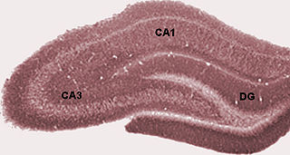
The hippocampus proper refers to the actual structure of the hippocampus which is made up of three regions or subfields. The subfields CA1, CA2, and CA3 use the initials of cornu Ammonis, an earlier name of the hippocampus.

An axo-axonic synapse is a type of synapse, formed by one neuron projecting its axon terminals onto another neuron's axon.

References

  1. 1 2 Llinás, Rodolfo; Walton, Kerry; Lang, Eric (2004). "Chapter 7: Cerebellum". In Shepherd, Gordon (ed.). The synaptic organization of the brain. Oxford, New York: Oxford University Press. pp. 271–309. ISBN   9780195159561.
  2. C. Reyher; J Liibke; W Larsen; G Hendrix; M Shipley; H Baumgarten (1991). "Olfactory Bulb Granule Cell Aggregates: Morphological Evidence for lnterperikaryal[sic] Electrotonic Coupling via Gap Junctions" (PDF). The Journal of Neuroscience . 11 (6): 1465–495. doi: 10.1523/JNEUROSCI.11-06-01485.1991 . PMC   6575397 . PMID   1904478. Archived (PDF) from the original on 14 August 2021. Retrieved 3 February 2016.
  3. Claiborne BJ, Amaral DG, Cowan WM (1990). "A quantitative three-dimensional analysis of granule cell dendrites in the rat dentate gyrus". The Journal of Comparative Neurology . 302 (2): 206–219. doi:10.1002/cne.903020203. PMID   2289972. S2CID   1841245.
  4. 1 2 David G. Amaral; Helen E. Scharfman; Pierre Lavenex (2007). "The dentate gyrus: Fundamental neuroanatomical organization (Dentate gyrus for dummies)". The Dentate Gyrus: A Comprehensive Guide to Structure, Function, and Clinical Implications. Progress in Brain Research. Vol. 163. pp. 3–22. doi:10.1016/S0079-6123(07)63001-5. ISBN   978-0-444-53015-8. PMC   2492885 . PMID   17765709.
  5. Mugnaini E, Osen KK, Dahl AL, Friedrich VL Jr, Korte G (1980). "Fine structure of granule cells and related interneurons (termed Golgi cells) in the cochlear nuclear complex of cat, rat and mouse". Journal of Neurocytology. 9 (4): 537–70. doi:10.1007/BF01204841. PMID   7441303. S2CID   19588537.
  6. Young, Eric; Oertel, Donata (2004). "Chapter 4: Cochlear Nucleus". In Shepherd, Gordon (ed.). The synaptic organization of the brain. Oxford, New York: Oxford University Press. pp. 125–163. ISBN   9780195159561.
  7. Neville, Kevin; Haberly, Lewis (2004). "Chapter 10: Olfactory Cortex". In Shepherd, Gordon (ed.). The synaptic organization of the brain. Oxford, New York: Oxford University Press. pp. 415–453. ISBN   9780195159561.
  8. 1 2 V Egger; K Svoboda; Z Mainen (2005). "Dendrodendritic Synaptic Signals in Olfactory Bulb Granule Cells: Local Spine Boost and Global Low-Threshold Spike". The Journal of Neuroscience . 25 (14): 3521–3530. doi:10.1523/JNEUROSCI.4746-04.2005. PMC   6725376 . PMID   15814782.
  9. Huang CM, Wang L, Huang RH (2006). "Cerebellar granule cell: ascending axon and parallel fiber". European Journal of Neuroscience . 23 (7): 1731–1737. doi:10.1111/j.1460-9568.2006.04690.x. PMID   16623829. S2CID   19270731.
  10. 1 2 3 4 5 M Manto; C De Zeeuw (2012). "Diversity and Complexity of Roles of Granule Cells in the Cerebellar Cortex". The Cerebellum. 11 (1): 1–4. doi:10.1007/s12311-012-0365-7. PMID   22396329. S2CID   7270048.
  11. M. Bear; M. Paradiso (2006). Neuroscience: Exploring the Brain. Lippincott Williams & Wilkins. p.  855. ISBN   9780781760034.
  12. P. Seja; M. Schonewille; G. Spitzmaul; A. Badura; I. Klein; Y. Rudhard; W. Wisden; C.A. Hübner; C.I. De Zeeuw; T.J. Jentsch (Mar 7, 2012). "Raising cytosolic Cl(-) in cerebellar granule cells affects their excitability and vestibulo-ocular learning". The EMBO Journal . 31 (5): 1217–30. doi:10.1038/emboj.2011.488. PMC   3297995 . PMID   22252133.
  13. Eccles JC, Ito M, Szentagothai J (1967). The cerebellum as a neural machine. Springer-Verlag. p. 56. doi:10.1007/978-3-662-13147-3. ISBN   978-3-662-13149-7. S2CID   20690516.
  14. Bengtssona, F; Jörntell, H (2009). "Sensory transmission in cerebellar granule cells relies on similarly coded mossy fiber inputs". PNAS . 106 (7): 2389–2394. Bibcode:2009PNAS..106.2389B. doi: 10.1073/pnas.0808428106 . PMC   2650166 . PMID   19164536.
  15. 1 2 A Arenz; E Bracey; T Margrie (2009). "Sensory representations in cerebellar granule cells". Current Opinion in Neurobiology . 19 (4): 445–451. doi:10.1016/j.conb.2009.07.003. PMID   19651506. S2CID   30942776.
  16. Marr D (1969). "A theory of cerebellar cortex". The Journal of Physiology. 202 (2): 437–70. doi:10.1113/jphysiol.1969.sp008820. PMC   1351491 . PMID   5784296.
  17. M Colicos; P Dash (1996). "Apoptotic morphology of dentate gyrus granule cells following experimental cortical impact injury in rats: possible role in spatial memory deficits". Brain Research . 739 (1–2): 120–131. doi:10.1016/S0006-8993(96)00824-4. PMID   8955932. S2CID   26445302.
  18. Kovács KA (September 2020). "Episodic Memories: How do the Hippocampus and the Entorhinal Ring Attractors Cooperate to Create Them?". Frontiers in Systems Neuroscience. 14: 68. doi: 10.3389/fnsys.2020.559186 . PMC   7511719 . PMID   33013334.
  19. T Nakashiba; J Cushman; K Pelkey; S Renaudineau; D Buhl; T McHugh; V Rodriguez Barrera; R Chittajallu; K Iwamoto; C McBain; M Fanselow; S Tonegawa (2012). "Young Dentate Granule Cells Mediate Pattern Separation, whereas Old Granule Cells Facilitate Pattern Completion". Cell . 149 (1): 188–201. doi:10.1016/j.cell.2012.01.046. PMC   3319279 . PMID   22365813.
  20. Weedman DL, Ryugo DK (1996). "Projections from auditory cortex to the cochlear nucleus in rats: synapses on granule cell dendrites". The Journal of Comparative Neurology . 371 (2): 311–324. doi:10.1002/(SICI)1096-9861(19960722)371:2<311::AID-CNE10>3.0.CO;2-V. PMID   8835735. S2CID   18382868.
  21. 1 2 R Balu; R Pressler; B Strowbridge (2007). "Multiple Modes of Synaptic Excitation of Olfactory Bulb Granule Cells". The Journal of Neuroscience . 27 (21): 5621–5632. doi:10.1523/JNEUROSCI.4630-06.2007. PMC   6672747 . PMID   17522307.
  22. Jansen, Jaclyn. "First glimpse of brain circuit that helps experience to shape perception". ScienceDaily. Archived from the original on 6 March 2014. Retrieved 2 March 2014.
  23. R Feil; J Hartmann; C Luo; W Wolfsgruber; K Schilling; S Feil; et al. (2003). "Impairment of LTD and cerebellar learning by Purkinje cell specific ablation of cGMP-dependent protein kinase I." The Journal of Cell Biology . 163 (2): 295–302. doi:10.1083/jcb.200306148. PMC   2173527 . PMID   14568994.
  24. 1 2 S Danzer; R Kotloski; C Walter; Maya Hughes; J McNamara (2008). "Altered morphology of hippocampal dentate granule cell presynaptic and postsynaptic terminals following conditional deletion of TrkB". Hippocampus . 18 (7): 668–678. doi:10.1002/hipo.20426. PMC   3377475 . PMID   18398849.
  25. 1 2 3 S Danzer (2012). "Depression, stress, epilepsy and adult neurogenesis". Experimental Neurology . 233 (1): 22–32. doi:10.1016/j.expneurol.2011.05.023. PMC   3199026 . PMID   21684275.
  26. J. Parent; T Yu; R Leibowitz; D Geschwind; R Sloviter; D Lowenstein (1997). "Dentate Granule Cell Neurogenesis Is Increased by Seizures and Contributes to Aberrant Network Reorganization in the Adult Rat Hippocampus". The Journal of Neuroscience . 17 (10): 3727–3738. doi:10.1523/JNEUROSCI.17-10-03727.1997. PMC   6573703 . PMID   9133393.
  27. Einstein G, Buranosky R, Crain BJ (1994). "Dendritic pathology of granule cells in Alzheimer's disease is unrelated to neuritic plaques". The Journal of Neuroscience . 14 (8): 5077–5088. doi:10.1523/JNEUROSCI.14-08-05077.1994. PMC   6577187 . PMID   8046469.
  28. Wakabayashi K, Hansen LA, Vincent I, Mallory M, Masliah E (1997). "Neurofibrillary tangles in the dentate granule cells of patients with Alzheimer's disease, Lewy body disease and progressive supranuclear palsy". Acta Neuropathologica . 93 (1): 7–12. doi:10.1007/s004010050576. PMID   9006651. S2CID   10136450.