Synapse

Last updated
Diagram of a chemical synaptic connection. Synapse figure.png
Diagram of a chemical synaptic connection.

In the nervous system, a synapse [1] is a structure that permits a neuron (or nerve cell) to pass an electrical or chemical signal to another neuron or to the target effector cell.

Contents

Synapses are essential to the transmission of nervous impulses from one neuron to another, [2] playing a key role in enabling rapid and direct communication by creating circuits. In addition, a synapse serves as a junction where both the transmission and processing of information occur, making it a vital means of communication between neurons. [3] Neurons are specialized to pass signals to individual target cells, and synapses are the means by which they do so. At a synapse, the plasma membrane of the signal-passing neuron (the presynaptic neuron) comes into close apposition with the membrane of the target (postsynaptic) cell. Both the presynaptic and postsynaptic sites contain extensive arrays of molecular machinery that link the two membranes together and carry out the signaling process. In many synapses, the presynaptic part is located on an axon and the postsynaptic part is located on a dendrite or soma. Astrocytes also exchange information with the synaptic neurons, responding to synaptic activity and, in turn, regulating neurotransmission. [2] Synapses (at least chemical synapses) are stabilized in position by synaptic adhesion molecules (SAMs) projecting from both the pre- and post-synaptic neuron and sticking together where they overlap; SAMs may also assist in the generation and functioning of synapses. [4] Moreover, SAMs coordinate the formation of synapses, with various types working together to achieve the remarkable specificity of synapses. [3] [5] In essence, SAMs function in both excitatory and inhibitory synapses, likely serving as devices for signal transmission. [3]

History

Santiago Ramón y Cajal proposed that neurons are not continuous throughout the body, yet still communicate with each other, an idea known as the neuron doctrine. [6] The word "synapse" was introduced in 1897 by the English neurophysiologist Charles Sherrington in Michael Foster's Textbook of Physiology. [1] Sherrington struggled to find a good term that emphasized a union between two separate elements, and the actual term "synapse" was suggested by the English classical scholar Arthur Woollgar Verrall, a friend of Foster. [7] [8] The word was derived from the Greek synapsis (σύναψις), meaning "conjunction", which in turn derives from synaptein (συνάπτειν), from syn (σύν) "together" and haptein (ἅπτειν) "to fasten". [7] [9]

However, while the synaptic gap remained a theoretical construct, and was sometimes reported as a discontinuity between contiguous axonal terminations and dendrites or cell bodies, histological methods using the best light microscopes of the day could not visually resolve their separation which is now known to be about 20 nm. It needed the electron microscope in the 1950s to show the finer structure of the synapse with its separate, parallel pre- and postsynaptic membranes and processes, and the cleft between the two. [10] [11] [12]

Types

An example of chemical synapse by the release of neurotransmitters like acetylcholine or glutamic acid. Neuro Muscular Junction.png
An example of chemical synapse by the release of neurotransmitters like acetylcholine or glutamic acid.

Chemical and electrical synapses are two ways of synaptic transmission.

The formation of neural circuits in nervous systems, appears to heavily depend on the crucial interactions between chemical and electrical synapses. Thus, these interactions govern the generation of synaptic transmission. [14] Synaptic communication is distinct from an ephaptic coupling, in which communication between neurons occurs via indirect electric fields. An autapse is a chemical or electrical synapse that forms when the axon of one neuron synapses onto dendrites of the same neuron.

Excitatory and inhibitory

  1. Excitatory synapse: Enhances the probability of depolarization in postsynaptic neurons and the initiation of an action potential.
  2. Inhibitory Synapse: Diminishes the probability of depolarization in postsynaptic neurons and the initiation of an action potential.

An influx of Na+ driven by excitatory neurotransmitters opens cation channels, depolarizing the postsynaptic membrane toward the action potential threshold. In contrast, inhibitory neurotransmitters cause the postsynaptic membrane to become less depolarized by opening either Cl- or K+ channels, reducing firing. Depending on their release location, the receptors they bind to, and the ionic circumstances they encounter, various transmitters can be either excitatory or inhibitory. For instance, acetylcholine can either excite or inhibit depending on the type of receptors it binds to. [18] For example, glutamate serves as an excitatory neurotransmitter, in contrast to GABA, which acts as an inhibitory neurotransmitter. Additionally, dopaminergic is a neurotransmitter that exerts dual effects, displaying both excitatory and inhibitory impacts through binding to distinct receptors. [19]

The membrane potential prevents Cl- from entering the cell, even when its concentration is much higher outside than inside. The resting potential for Cl- in many neurons is quite negative, nearly equal to the resting potential. Opening Cl- channels tends to buffer the membrane potential, but this effect is countered when the membrane starts to depolarize, allowing more negatively charged Cl- ions to enter the cell. Consequently, it becomes more difficult to depolarize the membrane and excite the cell when Cl- channels are open. Similar effects result from the opening of K+ channels. The significance of inhibitory neurotransmitters is evident from the effects of toxins that impede their activity. For instance, strychnine binds to glycine receptors, blocking the action of glycine and leading to muscle spasms, convulsions, and death. [18]

Interfaces

Synapses can be classified by the type of cellular structures serving as the pre- and post-synaptic components. The vast majority of synapses in the mammalian nervous system are classical axo-dendritic synapses (axon synapsing upon a dendrite), however, a variety of other arrangements exist. These include but are not limited to[ clarification needed ] axo-axonic, dendro-dendritic, axo-secretory, axo-ciliary, [20] somato-dendritic, dendro-somatic, and somato-somatic synapses.[ citation needed ]

In fact, the axon can synapse onto a dendrite, onto a cell body, or onto another axon or axon terminal, as well as into the bloodstream or diffusely into the adjacent nervous tissue.

Different types of synapses Blausen 0843 SynapseTypes.png
Different types of synapses

Conversion of chemical into electrical signals

Neurotransmitters are tiny signal molecules stored in membrane-enclosed synaptic vesicles and released via exocytosis. Indeed, a change in electrical potential in the presynaptic cell triggers the release of these molecules. By attaching to transmitter-gated ion channels, the neurotransmitter causes an electrical alteration in the postsynaptic cell and rapidly diffuses across the synaptic cleft. Once released, the neurotransmitter is swiftly eliminated, either by being absorbed by the nerve terminal that produced it, taken up by nearby glial cells, or broken down by specific enzymes in the synaptic cleft. Numerous Na+-dependent neurotransmitter carrier proteins recycle the neurotransmitters and enable the cells to maintain rapid rates of release.

At chemical synapses, transmitter-gated ion channels play a vital role in rapidly converting extracellular chemical impulses into electrical signals. These channels are located in the postsynaptic cell's plasma membrane at the synapse region, and they temporarily open in response to neurotransmitter molecule binding, causing a momentary alteration in the membrane's permeability. Additionally, transmitter-gated channels are comparatively less sensitive to the membrane potential than voltage-gated channels, which is why they are unable to generate self-amplifying excitement on their own. However, they result in graded variations in membrane potential due to local permeability, influenced by the amount and duration of neurotransmitter released at the synapse. [18]

Recently, mechanical tension, a phenomenon never thought relevant to synapse function has been found to be required for those on hippocampal neurons to fire. [21]

Release of neurotransmitters

Neurotransmitters bind to ionotropic receptors on postsynaptic neurons, either causing their opening or closing. [19] The variations in the quantities of neurotransmitters released from the presynaptic neuron may play a role in regulating the effectiveness of synaptic transmission. In fact, the concentration of cytoplasmic calcium is involved in regulating the release of neurotransmitters from presynaptic neurons. [22]

The chemical transmission involves several sequential processes:

  1. Synthesizing neurotransmitters within the presynaptic neuron.
  2. Loading the neurotransmitters into secretory vesicles.
  3. Controlling the release of neurotransmitters into the synaptic cleft.
  4. Binding of neurotransmitters to postsynaptic receptors.
  5. Ceasing the activity of the released neurotransmitters. [23]

Synaptic polarization

The function of neurons depends upon cell polarity. The distinctive structure of nerve cells allows action potentials to travel directionally (from dendrites to cell body down the axon), and for these signals to then be received and carried on by post-synaptic neurons or received by effector cells. Nerve cells have long been used as models for cellular polarization, and of particular interest are the mechanisms underlying the polarized localization of synaptic molecules. PIP2 signaling regulated by IMPase plays an integral role in synaptic polarity.

Phosphoinositides (PIP, PIP2, and PIP3) are molecules that have been shown to affect neuronal polarity. [24] A gene (ttx-7) was identified in Caenorhabditis elegans that encodes myo-inositol monophosphatase (IMPase), an enzyme that produces inositol by dephosphorylating inositol phosphate. Organisms with mutant ttx-7 genes demonstrated behavioral and localization defects, which were rescued by expression of IMPase. This led to the conclusion that IMPase is required for the correct localization of synaptic protein components. [25] [26] The egl-8 gene encodes a homolog of phospholipase Cβ (PLCβ), an enzyme that cleaves PIP2. When ttx-7 mutants also had a mutant egl-8 gene, the defects caused by the faulty ttx-7 gene were largely reversed. These results suggest that PIP2 signaling establishes polarized localization of synaptic components in living neurons. [25]

Presynaptic modulation

Modulation of neurotransmitter release by G-protein-coupled receptors (GPCRs) is a prominent presynaptic mechanism for regulation of synaptic transmission. The activation of GPCRs located at the presynaptic terminal, can decrease the probability of neurotransmitter release. This presynaptic depression involves activation of Gi/o-type G-proteins that mediate different inhibitory mechanisms, including inhibition of voltage-gated calcium channels, activation of potassium channels, and direct inhibition of the vesicle fusion process. Endocannabinoids, synthesized in and released from postsynaptic neuronal elements, and their cognate receptors, including the (GPCR) CB1 receptor, located at the presynaptic terminal, are involved in this modulation by a retrograde signaling process, in which these compounds are synthesized in and released from postsynaptic neuronal elements, and travel back to the presynaptic terminal to act on the CB1 receptor for short-term or long-term synaptic depression, that cause a short or long lasting decrease in neurotransmitter release. [27]

Effects of drugs on ligand-gated ion channels

Drugs have long been considered crucial targets for transmitter-gated ion channels. The majority of medications utilized to treat schizophrenia, anxiety, depression, and sleeplessness work at chemical synapses, and many of these pharmaceuticals function by binding to transmitter-gated channels. For instance, some drugs like barbiturates and tranquilizers bind to GABA receptors and enhance the inhibitory effect of GABA neurotransmitter. Thus, reduced concentration of GABA enables the opening of Cl- channels.

Furthermore, psychoactive drugs could potentially target many other synaptic signalling machinery components. In fact, numerous neurotransmitters are released by Na+-driven carriers and are subsequently removed from the synaptic cleft. By inhibiting such carriers, synaptic transmission is strengthened as the action of the transmitter is prolonged. For example, Prozac is one of antidepressant medications that works by preventing the absorption of serotonin neurotransmitter. Also, other antidepressants operate by inhibiting the reabsorption of both serotonin and norepinephrine. [18]

Biogenesis

In nerve terminals, synaptic vesicles are produced quickly to compensate for their rapid depletion during neurotransmitter release. Their biogenesis involves segregating synaptic vesicle membrane proteins from other cellular proteins and packaging those distinct proteins into vesicles of appropriate size. Besides, it entails the endocytosis of synaptic vesicle membrane proteins from the plasma membrane. [28]

Synaptoblastic and synaptoclastic refer to synapse-producing and synapse-removing activities within the biochemical signalling chain. This terminology is associated with the Bredesen Protocol for treating Alzheimer's disease, which conceptualizes Alzheimer's as an imbalance between these processes. As of October 2023, studies concerning this protocol remain small and few results have been obtained within a standardized control framework.

Role in memory

Potentiation and depression

It is widely accepted that the synapse plays a key role in the formation of memory. [29] The stability of long-term memory can persist for many years; nevertheless, synapses, the neurological basis of memory, are very dynamic. [30] The formation of synaptic connections significantly depends on activity-dependent synaptic plasticity observed in various synaptic pathways. Indeed, the connection between memory formation and alterations in synaptic efficacy enables the reinforcement of neuronal interactions between neurons. As neurotransmitters activate receptors across the synaptic cleft, the connection between the two neurons is strengthened when both neurons are active at the same time, as a result of the receptor's signaling mechanisms. The strength of two connected neural pathways is thought to result in the storage of information, resulting in memory. This process of synaptic strengthening is known as long-term potentiation (LTP). [29]

By altering the release of neurotransmitters, the plasticity of synapses can be controlled in the presynaptic cell. The postsynaptic cell can be regulated by altering the function and number of its receptors. Changes in postsynaptic signaling are most commonly associated with a N-methyl-d-aspartic acid receptor (NMDAR)-dependent LTP and long-term depression (LTD) due to the influx of calcium into the post-synaptic cell, which are the most analyzed forms of plasticity at excitatory synapses. [31]

Mechanism of protein kinase

Moreover, Ca2+/calmodulin (CaM)-dependent protein kinase II (CaMKII) is best recognized for its roles in the brain, particularly in the neocortex and hippocampal regions because it serves as a ubiquitous mediator of cellular Ca2+ signals. CaMKII is abundant in the nervous system, mainly concentrated in the synapses in the nerve cells. Indeed, CaMKII has been definitively identified as a key regulator of cognitive processes, such as learning, and neural plasticity. The first concrete experimental evidence for the long-assumed function of CaMKII in memory storage was demonstrated

While Ca2+/CaM binding stimulates CaMKII activity, Ca2+-independent autonomous CaMKII activity can also be produced by a number of other processes. CaMKII becomes active by autophosphorylating itself upon Ca2+/calmodulin binding. CaMKII is still active and phosphorylates itself even after Ca2+ is cleaved; as a result, the brain stores long-term memories using this mechanism. Nevertheless, when the CaMKII enzyme is dephosphorylated by a phosphatase enzyme, it becomes inactive, and memories are lost. Hence, CaMKII plays a vital role in both the induction and maintenance of LTP. [32]

Experimental models

For technical reasons, synaptic structure and function have been historically studied at unusually large model synapses, for example:

Synapses and Diseases

Synapses function as ensembles within particular brain networks to control the amount of neuronal activity, which is essential for memory, learning, and behavior. Consequently, synaptic disruptions might have negative effects. In fact, alterations in cell-intrinsic molecular systems or modifications to environmental biochemical processes can lead to synaptic dysfunction. The synapse is the primary unit of information transfer in the nervous system, and correct synaptic contact creation during development is essential for normal brain function. In addition, several mutations have been connected to neurodevelopmental disorders, and that compromised function at different synapse locations is a hallmark of neurodegenerative diseases.

Synaptic defects are causally associated with early appearing neurological diseases, including autism spectrum disorders (ASD), schizophrenia (SCZ), and bipolar disorder (BP). On the other hand, in late-onset degenerative pathologies, such as Alzheimer's (AD), Parkinson's (PD), and Huntington's (HD) diseases, synaptopathy is thought to be the inevitable end-result of an ongoing pathophysiological cascade. These diseases are identified by a gradual loss in cognitive and behavioral function and a steady loss of brain tissue. Moreover, these deteriorations have been mostly linked to the gradual build-up of protein aggregates in neurons, the composition of which may vary based on the pathology; all have the same deleterious effects on neuronal integrity. Furthermore, the high number of mutations linked to synaptic structure and function, as well as dendritic spine alterations in post-mortem tissue, has led to the association between synaptic defects and neurodevelopmental disorders, such as ASD and SCZ, characterized by abnormal behavioral or cognitive phenotypes.

Nevertheless, due to limited access to human tissue at late stages and a lack of thorough assessment of the essential components of human diseases in the available experimental animal models, it has been difficult to fully grasp the origin and role of synaptic dysfunction in neurological disorders. [34]

Additional images

See also

Related Research Articles

<span class="mw-page-title-main">Neuron</span> Electrically excitable cell found in the nervous system of animals

Within a nervous system, a neuron, neurone, or nerve cell is an electrically excitable cell that fires electric signals called action potentials across a neural network. Neurons communicate with other cells via synapses, which are specialized connections that commonly use minute amounts of chemical neurotransmitters to pass the electric signal from the presynaptic neuron to the target cell through the synaptic gap.

<span class="mw-page-title-main">Neurotransmitter</span> Chemical substance that enables neurotransmission

A neurotransmitter is a signaling molecule secreted by a neuron to affect another cell across a synapse. The cell receiving the signal, or target cell, may be another neuron, but could also be a gland or muscle cell.

<span class="mw-page-title-main">Nervous system</span> Part of an animal that coordinates actions and senses

In biology, the nervous system is the highly complex part of an animal that coordinates its actions and sensory information by transmitting signals to and from different parts of its body. The nervous system detects environmental changes that impact the body, then works in tandem with the endocrine system to respond to such events. Nervous tissue first arose in wormlike organisms about 550 to 600 million years ago. In vertebrates, it consists of two main parts, the central nervous system (CNS) and the peripheral nervous system (PNS). The CNS consists of the brain and spinal cord. The PNS consists mainly of nerves, which are enclosed bundles of the long fibers, or axons, that connect the CNS to every other part of the body. Nerves that transmit signals from the brain are called motor nerves (efferent), while those nerves that transmit information from the body to the CNS are called sensory nerves (afferent). The PNS is divided into two separate subsystems, the somatic and autonomic, nervous systems. The autonomic nervous system is further subdivided into the sympathetic, parasympathetic and enteric nervous systems. The sympathetic nervous system is activated in cases of emergencies to mobilize energy, while the parasympathetic nervous system is activated when organisms are in a relaxed state. The enteric nervous system functions to control the gastrointestinal system. Nerves that exit from the brain are called cranial nerves while those exiting from the spinal cord are called spinal nerves.

<span class="mw-page-title-main">Chemical synapse</span> Biological junctions through which neurons signals can be sent

Chemical synapses are biological junctions through which neurons' signals can be sent to each other and to non-neuronal cells such as those in muscles or glands. Chemical synapses allow neurons to form circuits within the central nervous system. They are crucial to the biological computations that underlie perception and thought. They allow the nervous system to connect to and control other systems of the body.

An inhibitory postsynaptic potential (IPSP) is a kind of synaptic potential that makes a postsynaptic neuron less likely to generate an action potential. The opposite of an inhibitory postsynaptic potential is an excitatory postsynaptic potential (EPSP), which is a synaptic potential that makes a postsynaptic neuron more likely to generate an action potential. IPSPs can take place at all chemical synapses, which use the secretion of neurotransmitters to create cell-to-cell signalling. EPSPs and IPSPs compete with each other at numerous synapses of a neuron. This determines whether an action potential occurring at the presynaptic terminal produces an action potential at the postsynaptic membrane. Some common neurotransmitters involved in IPSPs are GABA and glycine.

<span class="mw-page-title-main">Excitatory postsynaptic potential</span> Process causing temporary increase in postsynaptic potential

In neuroscience, an excitatory postsynaptic potential (EPSP) is a postsynaptic potential that makes the postsynaptic neuron more likely to fire an action potential. This temporary depolarization of postsynaptic membrane potential, caused by the flow of positively charged ions into the postsynaptic cell, is a result of opening ligand-gated ion channels. These are the opposite of inhibitory postsynaptic potentials (IPSPs), which usually result from the flow of negative ions into the cell or positive ions out of the cell. EPSPs can also result from a decrease in outgoing positive charges, while IPSPs are sometimes caused by an increase in positive charge outflow. The flow of ions that causes an EPSP is an excitatory postsynaptic current (EPSC).

In neuroscience, a silent synapse is an excitatory glutamatergic synapse whose postsynaptic membrane contains NMDA-type glutamate receptors but no AMPA-type glutamate receptors. These synapses are named "silent" because normal AMPA receptor-mediated signaling is not present, rendering the synapse inactive under typical conditions. Silent synapses are typically considered to be immature glutamatergic synapses. As the brain matures, the relative number of silent synapses decreases. However, recent research on hippocampal silent synapses shows that while they may indeed be a developmental landmark in the formation of a synapse, that synapses can be "silenced" by activity, even once they have acquired AMPA receptors. Thus, silence may be a state that synapses can visit many times during their lifetimes.

<span class="mw-page-title-main">Excitatory synapse</span> Sort of synapse

An excitatory synapse is a synapse in which an action potential in a presynaptic neuron increases the probability of an action potential occurring in a postsynaptic cell. Neurons form networks through which nerve impulses travels, each neuron often making numerous connections with other cells of neurons. These electrical signals may be excitatory or inhibitory, and, if the total of excitatory influences exceeds that of the inhibitory influences, the neuron will generate a new action potential at its axon hillock, thus transmitting the information to yet another cell.

<span class="mw-page-title-main">Neuroeffector junction</span> Site where a motor neuron releases a neurotransmitter to affect a target cell

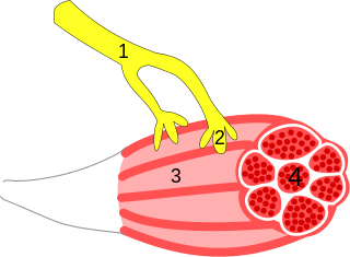
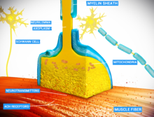
A neuroeffector junction is a site where a motor neuron releases a neurotransmitter to affect a target—non-neuronal—cell. This junction functions like a synapse. However, unlike most neurons, somatic efferent motor neurons innervate skeletal muscle, and are always excitatory. Visceral efferent neurons innervate smooth muscle, cardiac muscle, and glands, and have the ability to be either excitatory or inhibitory in function. Neuroeffector junctions are known as neuromuscular junctions when the target cell is a muscle fiber.

Synaptogenesis is the formation of synapses between neurons in the nervous system. Although it occurs throughout a healthy person's lifespan, an explosion of synapse formation occurs during early brain development, known as exuberant synaptogenesis. Synaptogenesis is particularly important during an individual's critical period, during which there is a certain degree of synaptic pruning due to competition for neural growth factors by neurons and synapses. Processes that are not used, or inhibited during their critical period will fail to develop normally later on in life.

Molecular neuroscience is a branch of neuroscience that observes concepts in molecular biology applied to the nervous systems of animals. The scope of this subject covers topics such as molecular neuroanatomy, mechanisms of molecular signaling in the nervous system, the effects of genetics and epigenetics on neuronal development, and the molecular basis for neuroplasticity and neurodegenerative diseases. As with molecular biology, molecular neuroscience is a relatively new field that is considerably dynamic.

Schaffer collaterals are axon collaterals given off by CA3 pyramidal cells in the hippocampus. These collaterals project to area CA1 of the hippocampus and are an integral part of memory formation and the emotional network of the Papez circuit, and of the hippocampal trisynaptic loop. It is one of the most studied synapses in the world and named after the Hungarian anatomist-neurologist Károly Schaffer.

<span class="mw-page-title-main">Neurotransmission</span> Impulse transmission between neurons

Neurotransmission is the process by which signaling molecules called neurotransmitters are released by the axon terminal of a neuron, and bind to and react with the receptors on the dendrites of another neuron a short distance away. A similar process occurs in retrograde neurotransmission, where the dendrites of the postsynaptic neuron release retrograde neurotransmitters that signal through receptors that are located on the axon terminal of the presynaptic neuron, mainly at GABAergic and glutamatergic synapses.

<span class="mw-page-title-main">Synaptic potential</span> Potential difference across the postsynaptic membrane

Synaptic potential refers to the potential difference across the postsynaptic membrane that results from the action of neurotransmitters at a neuronal synapse. In other words, it is the “incoming” signal that a neuron receives. There are two forms of synaptic potential: excitatory and inhibitory. The type of potential produced depends on both the postsynaptic receptor, more specifically the changes in conductance of ion channels in the post synaptic membrane, and the nature of the released neurotransmitter. Excitatory post-synaptic potentials (EPSPs) depolarize the membrane and move the potential closer to the threshold for an action potential to be generated. Inhibitory postsynaptic potentials (IPSPs) hyperpolarize the membrane and move the potential farther away from the threshold, decreasing the likelihood of an action potential occurring. The Excitatory Post Synaptic potential is most likely going to be carried out by the neurotransmitters glutamate and acetylcholine, while the Inhibitory post synaptic potential will most likely be carried out by the neurotransmitters gamma-aminobutyric acid (GABA) and glycine. In order to depolarize a neuron enough to cause an action potential, there must be enough EPSPs to both depolarize the postsynaptic membrane from its resting membrane potential to its threshold and counterbalance the concurrent IPSPs that hyperpolarize the membrane. As an example, consider a neuron with a resting membrane potential of -70 mV (millivolts) and a threshold of -50 mV. It will need to be raised 20 mV in order to pass the threshold and fire an action potential. The neuron will account for all the many incoming excitatory and inhibitory signals via summative neural integration, and if the result is an increase of 20 mV or more, an action potential will occur.

<span class="mw-page-title-main">Calyx of Held</span>

The Calyx of Held is a particularly large synapse in the mammalian auditory central nervous system, so named after Hans Held who first described it in his 1893 article Die centrale Gehörleitung because of its resemblance to the calyx of a flower. Globular bushy cells in the anteroventral cochlear nucleus (AVCN) send axons to the contralateral medial nucleus of the trapezoid body (MNTB), where they synapse via these calyces on MNTB principal cells. These principal cells then project to the ipsilateral lateral superior olive (LSO), where they inhibit postsynaptic neurons and provide a basis for interaural level detection (ILD), required for high frequency sound localization. This synapse has been described as the largest in the brain.

<span class="mw-page-title-main">Summation (neurophysiology)</span>

Summation, which includes both spatial summation and temporal summation, is the process that determines whether or not an action potential will be generated by the combined effects of excitatory and inhibitory signals, both from multiple simultaneous inputs, and from repeated inputs. Depending on the sum total of many individual inputs, summation may or may not reach the threshold voltage to trigger an action potential.

<span class="mw-page-title-main">Axon terminal</span> Nerve fiber part

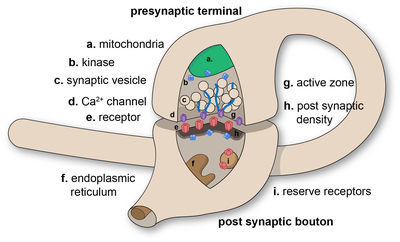
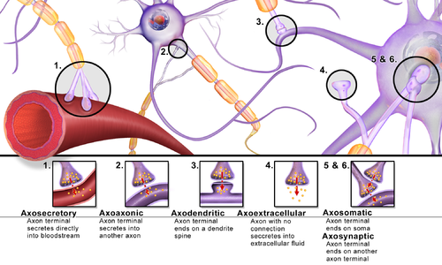
Axon terminals are distal terminations of the branches of an axon. An axon, also called a nerve fiber, is a long, slender projection of a nerve cell that conducts electrical impulses called action potentials away from the neuron's cell body in order to transmit those impulses to other neurons, muscle cells or glands. In the central nervous system, most presynaptic terminals are actually formed along the axons, not at their ends.

Cellular neuroscience is a branch of neuroscience concerned with the study of neurons at a cellular level. This includes morphology and physiological properties of single neurons. Several techniques such as intracellular recording, patch-clamp, and voltage-clamp technique, pharmacology, confocal imaging, molecular biology, two photon laser scanning microscopy and Ca2+ imaging have been used to study activity at the cellular level. Cellular neuroscience examines the various types of neurons, the functions of different neurons, the influence of neurons upon each other, and how neurons work together.

Neurotransmitters are released into a synapse in packaged vesicles called quanta. One quantum generates a miniature end plate potential (MEPP) which is the smallest amount of stimulation that one neuron can send to another neuron. Quantal release is the mechanism by which most traditional endogenous neurotransmitters are transmitted throughout the body. The aggregate sum of many MEPPs is an end plate potential (EPP). A normal end plate potential usually causes the postsynaptic neuron to reach its threshold of excitation and elicit an action potential. Electrical synapses do not use quantal neurotransmitter release and instead use gap junctions between neurons to send current flows between neurons. The goal of any synapse is to produce either an excitatory postsynaptic potential (EPSP) or an inhibitory postsynaptic potential (IPSP), which generate or repress the expression, respectively, of an action potential in the postsynaptic neuron. It is estimated that an action potential will trigger the release of approximately 20% of an axon terminal's neurotransmitter load.

Communication between neurons happens primarily through chemical neurotransmission at the synapse. Neurotransmitters are packaged into synaptic vesicles for release from the presynaptic cell into the synapse, from where they diffuse and can bind to postsynaptic receptors. While most presynaptic cells are historically thought to release one vesicle at a time per active site, more recent research has pointed towards the possibility of multiple vesicles being released from the same active site in response to an action potential.

References

  1. 1 2 Foster M, Sherrington CS (1897). Textbook of Physiology. Vol. 3 (7th ed.). London: Macmillan. p. 929. ISBN   978-1-4325-1085-5.
  2. 1 2 Perea G, Navarrete M, Araque A (August 2009). "Tripartite synapses: astrocytes process and control synaptic information". Trends in Neurosciences. 32 (8). Cell Press: 421–431. doi:10.1016/j.tins.2009.05.001. PMID   19615761. S2CID   16355401.
  3. 1 2 3 Südhof TC (July 2021). "The cell biology of synapse formation". The Journal of Cell Biology. 220 (7): e202103052. doi:10.1083/jcb.202103052. PMC   8186004 . PMID   34086051.
  4. Missler M, Südhof TC, Biederer T (April 2012). "Synaptic cell adhesion". Cold Spring Harbor Perspectives in Biology. 4 (4): a005694. doi:10.1101/cshperspect.a005694. PMC   3312681 . PMID   22278667.
  5. Hale WD, Südhof TC, Huganir RL (January 2023). "Engineered adhesion molecules drive synapse organization". Proceedings of the National Academy of Sciences of the United States of America. 120 (3): e2215905120. Bibcode:2023PNAS..12015905H. doi:10.1073/pnas.2215905120. PMC   9934208 . PMID   36638214.
  6. Elias LJ, Saucier DM (2006). Neuropsychology: Clinical and Experimental Foundations. Boston: Pearson/Allyn & Bacon. ISBN   978-0-20534361-4. LCCN   2005051341. OCLC   61131869.
  7. 1 2 Harper D. "synapse". Online Etymology Dictionary .
  8. Tansey EM (1997). "Not committing barbarisms: Sherrington and the synapse, 1897". Brain Research Bulletin. 44 (3). Elsevier: 211–212. doi:10.1016/S0361-9230(97)00312-2. PMID   9323432. S2CID   40333336. The word synapse first appeared in 1897, in the seventh edition of Michael Foster's Textbook of Physiology.
  9. σύναψις, συνάπτειν, σύν, ἅπτειν . Liddell, Henry George ; Scott, Robert ; A Greek–English Lexicon at the Perseus Project.
  10. De Robertis ED, Bennett HS (January 1955). "Some features of the submicroscopic morphology of synapses in frog and earthworm". The Journal of Biophysical and Biochemical Cytology. 1 (1): 47–58. doi:10.1083/jcb.1.1.47. PMC   2223594 . PMID   14381427.
  11. Palay SL, Palade GE (January 1955). "The fine structure of neurons". The Journal of Biophysical and Biochemical Cytology. 1 (1): 69–88. doi:10.1083/jcb.1.1.69. PMC   2223597 . PMID   14381429.
  12. Palay SL (July 1956). "Synapses in the central nervous system". The Journal of Biophysical and Biochemical Cytology. 2 (4 Suppl): 193–202. doi:10.1083/jcb.2.4.193. PMC   2229686 . PMID   13357542.
  13. 1 2 Silverthorn DU (2007). Human Physiology: An Integrated Approach (4th ed.). San Francisco: Pearson/Benjamin Cummings. p. 271. ISBN   978-0-8053-6851-2. LCCN   2005056517. OCLC   62742632.
  14. 1 2 Pereda AE (April 2014). "Electrical synapses and their functional interactions with chemical synapses". Nature Reviews. Neuroscience. 15 (4): 250–263. doi:10.1038/nrn3708. PMC   4091911 . PMID   24619342.
  15. Caire MJ, Reddy V, Varacallo M (2023). "Physiology, Synapse". StatPearls. Treasure Island (FL): StatPearls Publishing. PMID   30252303 . Retrieved 2024-01-01.
  16. Sotelo C, Palay SL (February 1970). "The fine structure of the later vestibular nucleus in the rat. II. Synaptic organization". Brain Research. 18 (1): 93–115. doi:10.1016/0006-8993(70)90459-2. PMID   4313893.
  17. Strausfeld NJ, Bassemir UK (December 1983). "Cobalt-coupled neurons of a giant fibre system in Diptera". Journal of Neurocytology. 12 (6): 971–91. doi:10.1007/BF01153345. PMID   6420522. S2CID   19764983.
  18. 1 2 3 4 Alberts B, Johnson A, Lewis J, Raff M, Roberts K, Walter P (2002). "Ion Channels and the Electrical Properties of Membranes". Molecular Biology of the Cell (4th ed.). Garland Science. Retrieved 2024-01-20.
  19. 1 2 Lasica A, Brewer C (2023). "Excitatory and Inhibitory Synaptic Signalling". TeachMePhysiology.
  20. Sheu SH, Upadhyayula S, Dupuy V, Pang S, Deng F, Wan J, et al. (September 2022). "A serotonergic axon-cilium synapse drives nuclear signaling to alter chromatin accessibility". Cell. 185 (18): 3390–3407.e18. doi: 10.1016/j.cell.2022.07.026 . PMC   9789380 . PMID   36055200. S2CID   251958800.
  21. Joy MS, Nall DL, Emon B, Lee KY, Barishman A, Ahmed M, Rahman S, Selvin PR, Saif MT (2023-12-26). "Synapses without tension fail to fire in an in vitro network of hippocampal neurons". Proceedings of the National Academy of Sciences. 120 (52). doi:10.1073/pnas.2311995120. ISSN   0027-8424. PMC   10756289 . PMID   38113266.
  22. Pitman RM (September 1984). "The versatile synapse". The Journal of Experimental Biology. 112 (1): 199–224. doi:10.1242/jeb.112.1.199. PMID   6150966.
  23. Holz RW, Fisher SK (1999). "Synaptic Transmission". In Siegel GJ, Agranoff BW, Albers RW, Fisher SK, Uhler MD (eds.). Basic Neurochemistry: Molecular, Cellular and Medical Aspects (6th ed.). Lippincott-Raven. Retrieved 2024-01-04.
  24. Arimura N, Kaibuchi K (December 2005). "Key regulators in neuronal polarity". Neuron. 48 (6). Cell Press: 881–884. doi: 10.1016/j.neuron.2005.11.007 . PMID   16364893.
  25. 1 2 Kimata T, Tanizawa Y, Can Y, Ikeda S, Kuhara A, Mori I (June 2012). "Synaptic polarity depends on phosphatidylinositol signaling regulated by myo-inositol monophosphatase in Caenorhabditis elegans". Genetics. 191 (2). Genetics Society of America: 509–521. doi:10.1534/genetics.111.137844. PMC   3374314 . PMID   22446320.
  26. Tanizawa Y, Kuhara A, Inada H, Kodama E, Mizuno T, Mori I (December 2006). "Inositol monophosphatase regulates localization of synaptic components and behavior in the mature nervous system of C. elegans". Genes & Development. 20 (23). Cold Spring Harbor Laboratory Press: 3296–3310. doi:10.1101/gad.1497806. PMC   1686606 . PMID   17158747.
  27. Lovinger DM (2008). "Presynaptic Modulation by Endocannabinoids". In Südhof TC, Starke K (eds.). Pharmacology of Neurotransmitter Release. Handbook of Experimental Pharmacology. Vol. 184. Springer Berlin Heidelberg. pp. 435–477. doi:10.1007/978-3-540-74805-2_14. ISBN   9783540748052. PMID   18064422.
  28. Desnos C, Clift-o'Grady L, Kelly RB (1995-09-01). "Biogenesis of synaptic vesicles in vitro". The Journal of Cell Biology. 130 (5): 1041–1049. doi:10.1083/jcb.130.5.1041. ISSN   0021-9525. PMC   2120557 . PMID   7544795.
  29. 1 2 Lynch MA (January 2004). "Long-term potentiation and memory". Physiological Reviews. 84 (1): 87–136. doi:10.1152/physrev.00014.2003. PMID   14715912.
  30. Yang Y, Lu J, Zuo Y (December 2018). "Changes of synaptic structures associated with learning, memory and diseases". Brain Science Advances. 4 (2): 99–117. doi: 10.26599/BSA.2018.2018.9050012 .
  31. Krugers HJ, Zhou M, Joëls M, Kindt M (October 11, 2011). "Regulation of excitatory synapses and fearful memories by stress hormones". Frontiers in Behavioral Neuroscience. 5. Frontiers Media SA: 62. doi: 10.3389/fnbeh.2011.00062 . PMC   3190121 . PMID   22013419.
  32. Bayer KU, Schulman H (August 2019). "CaM Kinase: Still Inspiring at 40". Neuron. 103 (3): 380–394. doi:10.1016/j.neuron.2019.05.033. PMC   6688632 . PMID   31394063.
  33. Stanley EF (1992). "The calyx-type synapse of the chick ciliary ganglion as a model of fast cholinergic transmission". Canadian Journal of Physiology and Pharmacology. 70 (Suppl): S73–S77. doi:10.1139/y92-246. PMID   1338300.
  34. Taoufik E, Kouroupi G, Zygogianni O, Matsas R (2018-09-05). "Synaptic dysfunction in neurodegenerative and neurodevelopmental diseases: an overview of induced pluripotent stem-cell-based disease models". Open Biology. 8 (9): 180138. doi:10.1098/rsob.180138. ISSN   2046-2441. PMC   6170506 . PMID   30185603.