Epineurium

Last updated
Epineurium
Gray636.png
Transverse section of human tibial nerve. (Epineurium labeled at upper right.)
Illu nerve structure.jpg
Nerve structure
Details
Identifiers
Latin Epineurium
TA98 A14.2.00.016
TA2 6155
FMA 12234
Anatomical terminology

The epineurium is the outermost layer of dense irregular connective tissue surrounding a peripheral nerve. [1] [2] It usually surrounds multiple nerve fascicles as well as blood vessels which supply the nerve. Smaller branches of these blood vessels penetrate into the perineurium. [3] In addition to blood vessels which supply the nerve, lymphocytes and fibroblasts are also present and contribute to the production of collagen fibers that form the backbone of the epineurium. In addition to providing structural support, lymphocytes and fibroblasts also play a vital role in maintenance and repair of the surrounding tissues. [4]

Contents

When the spinal nerve leaves the vertebral canal via an intervertebral foramen, two layers of the spinal meninges, the arachnoid and the dura invaginate the nerve to form a dural sleeve of connective tissue, which is the epineurium. The outer portion of this sleeve comprises the external epineurium which permits longitudinal nerve excursion and absorption of longitudinal stress. The layer of the epineurium that extends within the nerve to define the fascicles is termed the internal epineurium. Together, these two layers form the epineurium, which varies in thickness along the course of a nerve. [5] The epineurium is usually most abundant around joints, as its function is to protect the nerves from stretching and subsequent injury. Epineurium is primarily made from collagen. [3]

Clinical significance

A common surgical procedure to repair a torn nerve via the epineurium is termed an epineurial repair. [6]

Schwannomas often remain completely encapsulated by the epineurium as they grow. [7]

Epineurial barriers can be used to limit the formation of neuromas after neurotmesis. [8] The epineurium is an effective barrier against axon growth. [8]

See also

Related Research Articles

Pronator quadratus is a square-shaped muscle on the distal forearm that acts to pronate the hand.

The flexor digitorum profundus is a muscle in the forearm of humans that flexes the fingers. It is considered an extrinsic hand muscle because it acts on the hand while its muscle belly is located in the forearm.

Axillary nerve

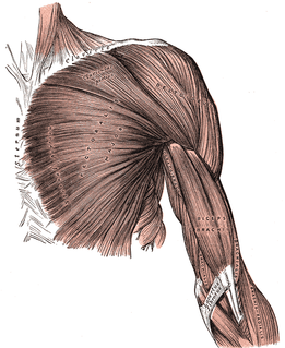
The axillary nerve or the circumflex nerve is a nerve of the human body, that originates from the brachial plexus at the level of the axilla (armpit) and carries nerve fibers from C5 and C6. The axillary nerve travels through the quadrangular space with the posterior circumflex humeral artery and vein to innervate the deltoid and teres minor.

Dorsal scapular nerve Branch of the brachial plexus that supplies rhomboid muscles and levator scapulae

The dorsal scapular nerve is a branch of the brachial plexus. It supplies rhomboid major muscle, rhomboid minor muscle, and levator scapulae muscle. It causes the scapula to be moved medially towards the vertebral column. Dorsal scapular nerve syndrome can cause a winged scapula, with pain and limited motion.

Long thoracic nerve Large nerve

The long thoracic nerve innervates the serratus anterior muscle.

Cervical plexus

The cervical plexus is a plexus of the anterior rami of the first four cervical spinal nerves which arise from C1 to C4 cervical segment in the neck. They are located laterally to the transverse processes between prevertebral muscles from the medial side and vertebral from lateral side. There is anastomosis with accessory nerve, hypoglossal nerve and sympathetic trunk.

Sternothyroid muscle

The sternothyroid muscle, or sternothyroideus, is an infrahyoid muscle in the neck. It acts to depress the hyoid bone. It is below the sternohyoid muscle. It is shorter and wider than the sternohyoid.

Medial pectoral nerve

The medial pectoral nerve arises from the medial cord of the brachial plexus, and through it from the eighth cervical and first thoracic roots (C8/T1).

Thoracodorsal nerve

The thoracodorsal nerve is a nerve present in humans and other animals, also known as the middle subscapular nerve or the long subscapular nerve. It supplies the latissimus dorsi muscle.

Posterior cutaneous nerve of thigh Sensory nerve that supplies the back of the thigh, leg, buttock, and perineum

The posterior cutaneous nerve of the thigh is a sensory nerve in the thigh. It supplies the skin of the posterior surface of the thigh, leg, buttock, and also the perineum.

Superior gluteal nerve

The superior gluteal nerve is a nerve that originates in the pelvis. It supplies the gluteus medius muscle, the gluteus minimus muscle, the tensor fasciae latae muscle, and the piriformis muscle.

Iliohypogastric nerve

The iliohypogastric nerve is a nerve that originates from the lumbar plexus that supplies sensation to skin over the lateral gluteal and hypogastric regions and motor to the internal oblique muscles and transverse abdominal muscles.

Anococcygeal nerve

The anococcygeal nerve is a nerve in the pelvis which provides sensory innervation to the skin over the coccyx.

Lower subscapular nerve

The lower subscapular nerve, also known as the inferior subscapular nerve, is the third branch of the posterior cord of the brachial plexus. It innervates the inferior portion of the subscapularis muscle and the teres major muscle.

Iliac fossa

The iliac fossa is a large, smooth, concave surface on the internal surface of the ilium.

Bicipital aponeurosis

The bicipital aponeurosis is a broad aponeurosis of the biceps brachii, which is located in the cubital fossa of the elbow. It separates superficial from deep structures in much of the fossa.

Lumbosacral trunk

The lumbosacral trunk is nervous tissue that connects the lumbar plexus with the sacral plexus.

Perineurium

The perineurium is a protective sheath that surrounds a nerve fascicle. This bundles together axons targeting the same anatomical location. The perineurium is composed from fibroblasts.

Endoneurium

The endoneurium is a layer of delicate connective tissue around the myelin sheath of each myelinated nerve fiber in the peripheral nervous system. Its component cells are called endoneurial cells. The endoneuria with their enclosed nerve fibers are bundled into groups called nerve fascicles, each fascicle within its own protective sheath called a perineurium. In sufficiently large nerves multiple fascicles, each with its blood supply and fatty tissue, may be bundled within yet another sheath, the epineurium.

Nerve fascicle Bundle of funiculi (small bundles of axons)

A nerve fascicle, or fasciculus is a bundle of funiculi. A funiculus is a bundle of axons.

References

  1. McCracken, Thomas (1999). New Atlas of Human Anatomy. China: Metro Books. pp. 96–97. ISBN   1-5866-3097-0.
  2. Howe, B. Matthew; Spinner, Robert J. (2015-01-01), Tubbs, R. Shane; Rizk, Elias; Shoja, Mohammadali M.; Loukas, Marios (eds.), "Chapter 54 - Perineural Spread of Cancers", Nerves and Nerve Injuries, San Diego: Academic Press, pp. 847–855, doi:10.1016/b978-0-12-802653-3.00103-2, ISBN   978-0-12-802653-3 , retrieved 2020-11-18
  3. 1 2 Reina, Miguel A.; Sala-Blanch, Xavier; Arriazu, Riánsares; Machés, Fabiola (2015-01-01), Tubbs, R. Shane; Rizk, Elias; Shoja, Mohammadali M.; Loukas, Marios (eds.), "Chapter 7 - Microscopic Morphology and Ultrastructure of Human Peripheral Nerves", Nerves and Nerve Injuries, San Diego: Academic Press, pp. 91–106, doi:10.1016/b978-0-12-410390-0.00007-x, ISBN   978-0-12-410390-0 , retrieved 2020-11-18
  4. Kulkarni, GS (2009). Textbook of Orthopaedics and Trauma. India: Jaypee Brothers Medical Publication. pp. 895–898. ISBN   978-9350908525.
  5. Payne, Samuel H. (2001). "Nerve Repair and Grafting in the Upper Extremity". Journal of the Southern Orthopaedic Association. Medscape. 10 (2): 173–189. PMID   12132829 . Retrieved 22 March 2015.
  6. Saladin, Kenneth (2014). Anatomy & Physiology: The Unity of Form and Function. USA: McGraw-Hill Science. pp. 484–485. ISBN   978-0073403717.
  7. LeGallo, Robin D.; Wick, Mark R. (2010-01-01), Gattuso, Paolo; Reddy, Vijaya B.; David, Odile; Spitz, Daniel J. (eds.), "17 - Soft Tissue", Differential Diagnosis in Surgical Pathology (Second Edition), Philadelphia: W.B. Saunders, pp. 889–948, doi:10.1016/b978-1-4160-4580-9.00017-4, ISBN   978-1-4160-4580-9 , retrieved 2020-11-18
  8. 1 2 Lee, Michelle; Guyuron, Bahman (2015-01-01), Tubbs, R. Shane; Rizk, Elias; Shoja, Mohammadali M.; Loukas, Marios (eds.), "Chapter 7 - Postoperative Neuromas", Nerves and Nerve Injuries, San Diego: Academic Press, pp. 99–112, doi:10.1016/b978-0-12-802653-3.00056-7, ISBN   978-0-12-802653-3 , retrieved 2020-11-18