Retrograde signaling

Last updated

Retrograde signaling in biology is the process where a signal travels backwards from a target source to its original source. For example, the nucleus of a cell is the original source for creating signaling proteins. During retrograde signaling, instead of signals leaving the nucleus, they are sent to the nucleus. [1] In cell biology, this type of signaling typically occurs between the mitochondria or chloroplast and the nucleus. Signaling molecules from the mitochondria or chloroplast act on the nucleus to affect nuclear gene expression. In this regard, the chloroplast or mitochondria act as a sensor for internal external stimuli which activate a signaling pathway. [2]

Contents

The postsynaptic dendrite (green) and presynaptic neuron (yellow) found in retrograde neurotransmission. Synapse Illustration2 tweaked.svg
The postsynaptic dendrite (green) and presynaptic neuron (yellow) found in retrograde neurotransmission.

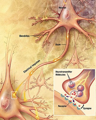
In neuroscience, retrograde signaling (or retrograde neurotransmission) refers more specifically to the process by which a retrograde messenger, such as anandamide or nitric oxide, is released by a postsynaptic dendrite or cell body, and travels "backwards" across a chemical synapse to bind to the axon terminal of a presynaptic neuron. [3]

In cell biology

Retrograde signals are transmitted from plastids to the nucleus in plants and eukaryotic algae, [4] [2] and from mitochondria to the nucleus in most eukaryotes. [5] Retrograde signals are generally considered to convey intracellular signals related to stress and environmental sensing. [6] Many of the molecules associated with retrograde signaling act on modifying the transcription or by directly binding and acting as a transcription factor. The outcomes of these signaling pathways vary by organism and by stimuli or stress. [4]

Evolution

Retrograde signaling is believed to have arisen after endocytosis of the mitochondria and chloroplast billions of years ago. [7] Originally believed to be photosynthetic bacteria, the mitochondria and chloroplast transferred some of their DNA to the membrane protected nucleus. [8] Thus, some of the proteins required for the mitochondria or chloroplast are within the nucleus. This transfer of DNA further required a network of communication to properly respond to external and internal signals and produce requisite proteins. [9]

In yeast

The first retrograde signaling pathways discovered in yeast is the RTG pathway. [10] [11] The RTG pathway plays an important role in maintaining the metabolic homeostasis of yeast. [11] Under limited resources the mitochondria must maintain a balance of glutamate for the citric acid cycle. [12] Retrograde signaling form the mitochondria initiates production precursor molecules of glutamate to properly balance supplies within the mitochondria. [13] Retrograde signaling can also act to arrest growth if problems are encountered. In Saccharomyces cerevisiae, if the mitochondria fails to develop properly, they will stop growing until the issue is addressed or cell death is induced. [13] These mechanism are vital to maintain homeostasis of the cell and ensure proper function of the mitochondria. [13]

In plants

One of the most studied retrograde signaling molecules in plants are reactive oxygen species (ROS). [14] These compounds, previously believed to be damaging to the cell, have since been discovered to act as a signaling molecule. [15] Reactive oxygen species are created as a by-product of aerobic respiration and act on genes involved in the stress response. [15] Depending on the stress, reactive oxygen species can act on neighboring cells to initiate a local signal. [16] By doing this, surrounding cells are "primed" to react to the stress because genes involved in stress response are initiated prior to encountering the stress. [16] The chloroplast can also act as a sensor for pathogen response and drought. Detection of these stresses in the cell will induce the formation of compounds that can then act on the nucleus to produce pathogen resistance genes or drought tolerance. [17]  

In neuroscience

Feedback loop found in retrograde neurological signaling. General Feedback Loop.svg
Feedback loop found in retrograde neurological signaling.

The primary purpose of retrograde neurotransmission is regulation of chemical neurotransmission. [3] For this reason, retrograde neurotransmission allows neural circuits to create feedback loops. In the sense that retrograde neurotransmission mainly serves to regulate typical, anterograde neurotransmission, rather than to actually distribute any information, it is similar to electrical neurotransmission.

In contrast to conventional (anterograde) neurotransmitters, retrograde neurotransmitters are synthesized in the postsynaptic neuron, and bind to receptors on the axon terminal of the presynaptic neuron. [18] Additionally, retrograde signaling initiates a signaling cascade that focuses on the presynaptic neuron. Once retrograde signaling is initiated, there is an increase in action potentials that begin in the presynaptic neuron, which directly impacts the postsynaptic neuron by increasing the number of its receptors. [19]

Endocannabinoids like anandamide are known to act as retrograde messengers, [20] [21] [22] as is nitric oxide. [23] [24]

Retrograde signaling may also play a role in long-term potentiation (LTP), a proposed mechanism of learning and memory, although this is controversial. [25] [26] [27]

Formal definition of a retrograde neurotransmitter

In 2009, Regehr et al. proposed criteria for defining retrograde neurotransmitters. According to their work, a signaling molecule can be considered a retrograde neurotransmitter if it satisfies all of the following criteria: [3]

Types of retrograde neurotransmitters

The most prevalent endogenous retrograde neurotransmitters are nitric oxide [23] [24] and various endocannabinoids, which are lipophilic ligands. [19] [28]

The retrograde neurotransmitter, nitric oxide (NO) is a soluble gas that can readily diffuse through various cell membranes. [29] Nitric oxide synthase is the enzyme responsible for the synthesis of NO in various presynaptic cells. [30] Specifically, NO is known to play a critical role in LTP, which plays an important role in memory storage within the hippocampus. [31] Additionally, literature suggests that NO can act as intracellular messengers in the brain and can also have an effect on the presynaptic glutamatergic and GABAergic synapses. [32]

Utilizing retrograde signaling, endocannabinoids, a type of retrograde neurotransmitter, are activated when they bind to G-protein coupled receptors on the presynaptic terminals of neurons. [33] The activation of endocannabinoids results in the release of particular neurotransmitters at the excitatory and inhibitory synapses of a neuron, ultimately impacting various forms of plasticity. [34] [19] [33]

Retrograde signaling in long-term potentiation

As it pertains to LTP, retrograde signaling is a hypothesis describing how events underlying LTP may begin in the postsynaptic neuron but be propagated to the presynaptic neuron, even though normal communication across a chemical synapse occurs in a presynaptic to postsynaptic direction. It is used most commonly by those who argue that presynaptic neurons contribute significantly to the expression of LTP. [35]

Background

Long-term potentiation is the persistent increase in the strength of a chemical synapse that lasts from hours to days. [36] It is thought to occur via two temporally separated events, with induction occurring first, followed by expression. [36] Most LTP investigators agree that induction is entirely postsynaptic, whereas there is disagreement as to whether expression is principally a presynaptic or postsynaptic event. [26] Some researchers believe that both presynaptic and postsynaptic mechanisms play a role in LTP expression. [26]

Were LTP entirely induced and expressed postsynaptically, there would be no need for the postsynaptic cell to communicate with the presynaptic cell following LTP induction. However, postsynaptic induction combined with presynaptic expression requires that, following induction, the postsynaptic cell must communicate with the presynaptic cell. Because normal synaptic transmission occurs in a presynaptic to postsynaptic direction, postsynaptic to presynaptic communication is considered a form of retrograde transmission. [25]

Mechanism

The retrograde signaling hypothesis proposes that during the early stages of LTP expression, the postsynaptic cell "sends a message" to the presynaptic cell to notify it that an LTP-inducing stimulus has been received postsynaptically. The general hypothesis of retrograde signaling does not propose a precise mechanism by which this message is sent and received. One mechanism may be that the postsynaptic cell synthesizes and releases a retrograde messenger upon receipt of LTP-inducing stimulation. [37] [38] Another is that it releases a preformed retrograde messenger upon such activation. Yet another mechanism is that synapse-spanning proteins may be altered by LTP-inducing stimuli in the postsynaptic cell, and that changes in conformation of these proteins propagates this information across the synapse and to the presynaptic cell. [39]

Identity of the messenger

Of these mechanisms, the retrograde messenger hypothesis has received the most attention. Among proponents of the model, there is disagreement over the identity of the retrograde messenger. A flurry of work in the early 1990s to demonstrate the existence of a retrograde messenger and to determine its identity generated a list of candidates including carbon monoxide, [40] platelet-activating factor, [41] [42] arachidonic acid, [43] and nitric oxide. Nitric oxide has received a great deal of attention in the past, but has recently been superseded by adhesion proteins that span the synaptic cleft to join the presynaptic and postsynaptic cells. [39] The endocannabinoids anandamide and/or 2-AG, acting through G-protein coupled cannabinoid receptors, may play an important role in retrograde signaling in LTP. [20] [21]

Related Research Articles

<span class="mw-page-title-main">Neurotransmitter</span> Chemical substance that enables neurotransmission

A neurotransmitter is a signaling molecule secreted by a neuron to affect another cell across a synapse. The cell receiving the signal, or target cell, may be another neuron, but could also be a gland or muscle cell.
<span class="mw-page-title-main">Chemical synapse</span> Biological junctions through which neurons signals can be sent

Chemical synapses are biological junctions through which neurons' signals can be sent to each other and to non-neuronal cells such as those in muscles or glands. Chemical synapses allow neurons to form circuits within the central nervous system. They are crucial to the biological computations that underlie perception and thought. They allow the nervous system to connect to and control other systems of the body.

<span class="mw-page-title-main">Long-term potentiation</span> Persistent strengthening of synapses based on recent patterns of activity

In neuroscience, long-term potentiation (LTP) is a persistent strengthening of synapses based on recent patterns of activity. These are patterns of synaptic activity that produce a long-lasting increase in signal transmission between two neurons. The opposite of LTP is long-term depression, which produces a long-lasting decrease in synaptic strength.

In neuroscience, synaptic plasticity is the ability of synapses to strengthen or weaken over time, in response to increases or decreases in their activity. Since memories are postulated to be represented by vastly interconnected neural circuits in the brain, synaptic plasticity is one of the important neurochemical foundations of learning and memory.

An inhibitory postsynaptic potential (IPSP) is a kind of synaptic potential that makes a postsynaptic neuron less likely to generate an action potential. The opposite of an inhibitory postsynaptic potential is an excitatory postsynaptic potential (EPSP), which is a synaptic potential that makes a postsynaptic neuron more likely to generate an action potential. IPSPs can take place at all chemical synapses, which use the secretion of neurotransmitters to create cell-to-cell signalling. EPSPs and IPSPs compete with each other at numerous synapses of a neuron. This determines whether an action potential occurring at the presynaptic terminal produces an action potential at the postsynaptic membrane. Some common neurotransmitters involved in IPSPs are GABA and glycine.

<span class="mw-page-title-main">Excitatory postsynaptic potential</span> Process causing temporary increase in postsynaptic potential

In neuroscience, an excitatory postsynaptic potential (EPSP) is a postsynaptic potential that makes the postsynaptic neuron more likely to fire an action potential. This temporary depolarization of postsynaptic membrane potential, caused by the flow of positively charged ions into the postsynaptic cell, is a result of opening ligand-gated ion channels. These are the opposite of inhibitory postsynaptic potentials (IPSPs), which usually result from the flow of negative ions into the cell or positive ions out of the cell. EPSPs can also result from a decrease in outgoing positive charges, while IPSPs are sometimes caused by an increase in positive charge outflow. The flow of ions that causes an EPSP is an excitatory postsynaptic current (EPSC).

In neurophysiology, long-term depression (LTD) is an activity-dependent reduction in the efficacy of neuronal synapses lasting hours or longer following a long patterned stimulus. LTD occurs in many areas of the CNS with varying mechanisms depending upon brain region and developmental progress.

In neuroscience, a silent synapse is an excitatory glutamatergic synapse whose postsynaptic membrane contains NMDA-type glutamate receptors but no AMPA-type glutamate receptors. These synapses are named "silent" because normal AMPA receptor-mediated signaling is not present, rendering the synapse inactive under typical conditions. Silent synapses are typically considered to be immature glutamatergic synapses. As the brain matures, the relative number of silent synapses decreases. However, recent research on hippocampal silent synapses shows that while they may indeed be a developmental landmark in the formation of a synapse, that synapses can be "silenced" by activity, even once they have acquired AMPA receptors. Thus, silence may be a state that synapses can visit many times during their lifetimes.

<span class="mw-page-title-main">Excitatory synapse</span> Sort of synapse

An excitatory synapse is a synapse in which an action potential in a presynaptic neuron increases the probability of an action potential occurring in a postsynaptic cell. Neurons form networks through which nerve impulses travels, each neuron often making numerous connections with other cells of neurons. These electrical signals may be excitatory or inhibitory, and, if the total of excitatory influences exceeds that of the inhibitory influences, the neuron will generate a new action potential at its axon hillock, thus transmitting the information to yet another cell.

Spike-timing-dependent plasticity (STDP) is a biological process that adjusts the strength of connections between neurons in the brain. The process adjusts the connection strengths based on the relative timing of a particular neuron's output and input action potentials. The STDP process partially explains the activity-dependent development of nervous systems, especially with regard to long-term potentiation and long-term depression.

Schaffer collaterals are axon collaterals given off by CA3 pyramidal cells in the hippocampus. These collaterals project to area CA1 of the hippocampus and are an integral part of memory formation and the emotional network of the Papez circuit, and of the hippocampal trisynaptic loop. It is one of the most studied synapses in the world and named after the Hungarian anatomist-neurologist Károly Schaffer.

<span class="mw-page-title-main">Neurotransmission</span> Impulse transmission between neurons

Neurotransmission is the process by which signaling molecules called neurotransmitters are released by the axon terminal of a neuron, and bind to and react with the receptors on the dendrites of another neuron a short distance away. A similar process occurs in retrograde neurotransmission, where the dendrites of the postsynaptic neuron release retrograde neurotransmitters that signal through receptors that are located on the axon terminal of the presynaptic neuron, mainly at GABAergic and glutamatergic synapses.

Depolarization-induced suppression of inhibition is the classical and original electrophysiological example of endocannabinoid function in the central nervous system. Prior to the demonstration that depolarization-induced suppression of inhibition was dependent on the cannabinoid CB1 receptor function, there was no way of producing an in vitro endocannabinoid mediated effect.

Neuromodulation is the physiological process by which a given neuron uses one or more chemicals to regulate diverse populations of neurons. Neuromodulators typically bind to metabotropic, G-protein coupled receptors (GPCRs) to initiate a second messenger signaling cascade that induces a broad, long-lasting signal. This modulation can last for hundreds of milliseconds to several minutes. Some of the effects of neuromodulators include: altering intrinsic firing activity, increasing or decreasing voltage-dependent currents, altering synaptic efficacy, increasing bursting activity and reconfigurating synaptic connectivity.

<span class="mw-page-title-main">Postsynaptic density</span>

The postsynaptic density (PSD) is a protein dense specialization attached to the postsynaptic membrane. PSDs were originally identified by electron microscopy as an electron-dense region at the membrane of a postsynaptic neuron. The PSD is in close apposition to the presynaptic active zone and ensures that receptors are in close proximity to presynaptic neurotransmitter release sites. PSDs vary in size and composition among brain regions, and have been studied in great detail at glutamatergic synapses. Hundreds of proteins have been identified in the postsynaptic density, including glutamate receptors, scaffold proteins, and many signaling molecules.

Metaplasticity is a term originally coined by W.C. Abraham and M.F. Bear to refer to the plasticity of synaptic plasticity. Until that time synaptic plasticity had referred to the plastic nature of individual synapses. However this new form referred to the plasticity of the plasticity itself, thus the term meta-plasticity. The idea is that the synapse's previous history of activity determines its current plasticity. This may play a role in some of the underlying mechanisms thought to be important in memory and learning such as long-term potentiation (LTP), long-term depression (LTD) and so forth. These mechanisms depend on current synaptic "state", as set by ongoing extrinsic influences such as the level of synaptic inhibition, the activity of modulatory afferents such as catecholamines, and the pool of hormones affecting the synapses under study. Recently, it has become clear that the prior history of synaptic activity is an additional variable that influences the synaptic state, and thereby the degree, of LTP or LTD produced by a given experimental protocol. In a sense, then, synaptic plasticity is governed by an activity-dependent plasticity of the synaptic state; such plasticity of synaptic plasticity has been termed metaplasticity. There is little known about metaplasticity, and there is much research currently underway on the subject, despite its difficulty of study, because of its theoretical importance in brain and cognitive science. Most research of this type is done via cultured hippocampus cells or hippocampal slices.

<span class="mw-page-title-main">Synapse</span> Structure connecting neurons in the nervous system

In the nervous system, a synapse is a structure that permits a neuron to pass an electrical or chemical signal to another neuron or to the target effector cell.

<span class="mw-page-title-main">Calyx of Held</span>

The Calyx of Held is a particularly large synapse in the mammalian auditory central nervous system, so named after Hans Held who first described it in his 1893 article Die centrale Gehörleitung because of its resemblance to the calyx of a flower. Globular bushy cells in the anteroventral cochlear nucleus (AVCN) send axons to the contralateral medial nucleus of the trapezoid body (MNTB), where they synapse via these calyces on MNTB principal cells. These principal cells then project to the ipsilateral lateral superior olive (LSO), where they inhibit postsynaptic neurons and provide a basis for interaural level detection (ILD), required for high frequency sound localization. This synapse has been described as the largest in the brain.

Coincidence detection is a neuronal process in which a neural circuit encodes information by detecting the occurrence of temporally close but spatially distributed input signals. Coincidence detectors influence neuronal information processing by reducing temporal jitter and spontaneous activity, allowing the creation of variable associations between separate neural events in memory. The study of coincidence detectors has been crucial in neuroscience with regards to understanding the formation of computational maps in the brain.

<span class="mw-page-title-main">Axon terminal</span> Nerve fiber part

Axon terminals are distal terminations of the branches of an axon. An axon, also called a nerve fiber, is a long, slender projection of a nerve cell that conducts electrical impulses called action potentials away from the neuron's cell body in order to transmit those impulses to other neurons, muscle cells or glands. In the central nervous system, most presynaptic terminals are actually formed along the axons, not at their ends.

References

  1. Leister, Dario (2012). "Retrograde signaling in plants: from simple to complex scenarios". Frontiers in Plant Science. 3: 135. doi: 10.3389/fpls.2012.00135 . ISSN   1664-462X. PMC   3377957 . PMID   22723802.
  2. 1 2 Nott A, Jung HS, Koussevitzky S, Chory J (June 2006). "Plastid-to-nucleus retrograde signaling". Annual Review of Plant Biology. 57: 739–59. doi:10.1146/annurev.arplant.57.032905.105310. PMID   16669780.
  3. 1 2 3 Regehr WG, Carey MR, Best AR (July 2009). "Activity-dependent regulation of synapses by retrograde messengers". Neuron. 63 (2): 154–70. doi:10.1016/j.neuron.2009.06.021. PMC   3251517 . PMID   19640475.
  4. 1 2 Duanmu D, Casero D, Dent RM, Gallaher S, Yang W, Rockwell NC, et al. (February 2013). "Retrograde bilin signaling enables Chlamydomonas greening and phototrophic survival". Proceedings of the National Academy of Sciences of the United States of America. 110 (9): 3621–6. doi: 10.1073/pnas.1222375110 . PMC   3587268 . PMID   23345435.
  5. Liu Z, Butow RA (December 2006). "Mitochondrial retrograde signaling". Annual Review of Genetics. 40: 159–85. doi:10.1146/annurev.genet.40.110405.090613. PMID   16771627.
  6. Nott A, Jung HS, Koussevitzky S, Chory J (2006). "Plastid-to-nucleus retrograde signaling". Annual Review of Plant Biology. 57: 739–59. doi:10.1146/annurev.arplant.57.032905.105310. PMID   16669780.
  7. Bevan RB, Lang BF (2004). "Mitochondrial genome evolution: the origin of mitochondria and of eukaryotes.". Mitochondrial Function and Biogenesis. Topics in Current Genetics. Vol. 8. Berlin, Heidelberg: Springer. pp. 1–35. doi:10.1007/b96830. ISBN   978-3-540-21489-2.
  8. da Cunha FM, Torelli NQ, Kowaltowski AJ (2015). "Mitochondrial Retrograde Signaling: Triggers, Pathways, and Outcomes". Oxidative Medicine and Cellular Longevity. 2015: 482582. doi: 10.1155/2015/482582 . PMC   4637108 . PMID   26583058.
  9. Whelan SP, Zuckerbraun BS (2013). "Mitochondrial signaling: forwards, backwards, and in between". Oxidative Medicine and Cellular Longevity. 2013: 351613. doi: 10.1155/2013/351613 . PMC   3681274 . PMID   23819011.
  10. Parikh VS, Morgan MM, Scott R, Clements LS, Butow RA (January 1987). "The mitochondrial genotype can influence nuclear gene expression in yeast". Science. 235 (4788): 576–80. Bibcode:1987Sci...235..576P. doi:10.1126/science.3027892. PMID   3027892.
  11. 1 2 Liu Z, Sekito T, Epstein CB, Butow RA (December 2001). "RTG-dependent mitochondria to nucleus signaling is negatively regulated by the seven WD-repeat protein Lst8p". The EMBO Journal. 20 (24): 7209–19. doi:10.1093/emboj/20.24.7209. PMC   125777 . PMID   11742997.
  12. Jazwinski SM, Kriete A (2012). "The yeast retrograde response as a model of intracellular signaling of mitochondrial dysfunction". Frontiers in Physiology. 3: 139. doi: 10.3389/fphys.2012.00139 . PMC   3354551 . PMID   22629248.
  13. 1 2 3 Liu Z, Butow RA (October 1999). "A transcriptional switch in the expression of yeast tricarboxylic acid cycle genes in response to a reduction or loss of respiratory function". Molecular and Cellular Biology. 19 (10): 6720–8. doi:10.1128/MCB.19.10.6720. PMC   84662 . PMID   10490611.
  14. Maruta T, Noshi M, Tanouchi A, Tamoi M, Yabuta Y, Yoshimura K, et al. (April 2012). "H2O2-triggered retrograde signaling from chloroplasts to nucleus plays specific role in response to stress". The Journal of Biological Chemistry. 287 (15): 11717–29. doi: 10.1074/jbc.m111.292847 . PMC   3320920 . PMID   22334687.
  15. 1 2 Schieber M, Chandel NS (May 2014). "ROS function in redox signaling and oxidative stress". Current Biology. 24 (10): R453-62. doi:10.1016/j.cub.2014.03.034. PMC   4055301 . PMID   24845678.
  16. 1 2 Shapiguzov A, Vainonen JP, Wrzaczek M, Kangasjärvi J (2012). "ROS-talk - how the apoplast, the chloroplast, and the nucleus get the message through". Frontiers in Plant Science. 3: 292. doi: 10.3389/fpls.2012.00292 . PMC   3530830 . PMID   23293644.
  17. Estavillo GM, Chan KX, Phua SY, Pogson BJ (2013). "Reconsidering the nature and mode of action of metabolite retrograde signals from the chloroplast". Frontiers in Plant Science. 3: 300. doi: 10.3389/fpls.2012.00300 . PMC   3539676 . PMID   23316207.
  18. Tao, Huizhong W.; Poo, Mu-ming (2001-09-25). "Retrograde signaling at central synapses". Proceedings of the National Academy of Sciences. 98 (20): 11009–11015. Bibcode:2001PNAS...9811009T. doi: 10.1073/pnas.191351698 . ISSN   0027-8424. PMC   58675 . PMID   11572961.
  19. 1 2 3 "Endocannabinoids Performance through Retrograde Signaling | Cannabis Sciences". Labroots. Retrieved 2021-05-05.
  20. 1 2 Alger BE (November 2002). "Retrograde signaling in the regulation of synaptic transmission: focus on endocannabinoids". Progress in Neurobiology. 68 (4): 247–86. doi:10.1016/S0301-0082(02)00080-1. PMID   12498988. S2CID   22754679.
  21. 1 2 Wilson RI, Nicoll RA (March 2001). "Endogenous cannabinoids mediate retrograde signalling at hippocampal synapses". Nature. 410 (6828): 588–92. Bibcode:2001Natur.410..588W. doi:10.1038/35069076. PMID   11279497. S2CID   52803281.
  22. Kreitzer AC, Regehr WG (June 2002). "Retrograde signaling by endocannabinoids". Current Opinion in Neurobiology. 12 (3): 324–30. doi:10.1016/S0959-4388(02)00328-8. PMID   12049940. S2CID   5846728.
  23. 1 2 O'Dell TJ, Hawkins RD, Kandel ER, Arancio O (December 1991). "Tests of the roles of two diffusible substances in long-term potentiation: evidence for nitric oxide as a possible early retrograde messenger". Proceedings of the National Academy of Sciences of the United States of America. 88 (24): 11285–9. Bibcode:1991PNAS...8811285O. doi: 10.1073/pnas.88.24.11285 . PMC   53119 . PMID   1684863.
  24. 1 2 Malen PL, Chapman PF (April 1997). "Nitric oxide facilitates long-term potentiation, but not long-term depression". The Journal of Neuroscience. 17 (7): 2645–51. doi:10.1523/JNEUROSCI.17-07-02645.1997. PMC   6573517 . PMID   9065524.
  25. 1 2 Regehr WG, Carey MR, Best AR (July 2009). "Activity-dependent regulation of synapses by retrograde messengers". Neuron. 63 (2): 154–70. doi:10.1016/j.neuron.2009.06.021. PMC   3251517 . PMID   19640475.
  26. 1 2 3 Nicoll RA, Malenka RC (September 1995). "Contrasting properties of two forms of long-term potentiation in the hippocampus". Nature. 377 (6545): 115–8. Bibcode:1995Natur.377..115N. doi:10.1038/377115a0. PMID   7675078. S2CID   4311817.
  27. Abraham WC, Jones OD, Glanzman DL (December 2019). "Is plasticity of synapses the mechanism of long-term memory storage?". npj Science of Learning. 4 (1): 9. Bibcode:2019npjSL...4....9A. doi:10.1038/s41539-019-0048-y. PMC   6606636 . PMID   31285847.
  28. Vaughan, C. W.; Christie, M. J. (2005). Retrograde signalling by endocannabinoids. Handbook of Experimental Pharmacology. Vol. 168. pp. 367–383. doi:10.1007/3-540-26573-2_12. ISBN   3-540-22565-X. ISSN   0171-2004. PMID   16596781.
  29. Arancio, Ottavio; Kiebler, Michael; Lee, C. Justin; Lev-Ram, Varda; Tsien, Roger Y.; Kandel, Eric R.; Hawkins, Robert D. (1996-12-13). "Nitric Oxide Acts Directly in the Presynaptic Neuron to Produce Long-Term Potentiationin Cultured Hippocampal Neurons". Cell. 87 (6): 1025–1035. doi: 10.1016/S0092-8674(00)81797-3 . ISSN   0092-8674. PMID   8978607. S2CID   10550701.
  30. Overeem, Kathie A.; Ota, Kristie T.; Monsey, Melissa S.; Ploski, Jonathan E.; Schafe, Glenn E. (2010-02-05). "A Role for Nitric Oxide-Driven Retrograde Signaling in the Consolidation of a Fear Memory". Frontiers in Behavioral Neuroscience. 4: 2. doi: 10.3389/neuro.08.002.2010 . ISSN   1662-5153. PMC   2820379 . PMID   20161806.
  31. "Long-Term Potentiation - an overview | ScienceDirect Topics". www.sciencedirect.com. Retrieved 2021-05-05.
  32. Hardingham, Neil; Dachtler, James; Fox, Kevin (2013). "The role of nitric oxide in pre-synaptic plasticity and homeostasis". Frontiers in Cellular Neuroscience. 7: 190. doi: 10.3389/fncel.2013.00190 . ISSN   1662-5102. PMC   3813972 . PMID   24198758.
  33. 1 2 Kreitzer, A.; Regehr, W. G. (2002-06-01). "Retrograde signaling by endocannabinoids". Current Opinion in Neurobiology. 12 (3): 324–330. doi:10.1016/S0959-4388(02)00328-8. ISSN   0959-4388. PMID   12049940. S2CID   5846728.
  34. Castillo, Pablo E.; Younts, Thomas J.; Chávez, Andrés E.; Hashimotodani, Yuki (2012-10-04). "Endocannabinoid Signaling and Synaptic Function". Neuron. 76 (1): 70–81. doi: 10.1016/j.neuron.2012.09.020 . ISSN   0896-6273. PMC   3517813 . PMID   23040807.
  35. Matthies, H. (1988). "Long-Term Synaptic Potentiation and Macromolecular Changes in Memory Formation". Synaptic Plasticity in the Hippocampus. Springer Berlin Heidelberg. pp. 119–121. doi:10.1007/978-3-642-73202-7_35. ISBN   9783642732041.
  36. 1 2 Warburton EC (2015). "Long-Term Potentiation and Memory". Encyclopedia of Psychopharmacology. pp. 928–32. doi:10.1007/978-3-642-27772-6_345-2. ISBN   978-3-642-27772-6.
  37. Garthwaite J (February 1991). "Glutamate, nitric oxide and cell-cell signalling in the nervous system". Trends in Neurosciences. 14 (2): 60–7. doi:10.1016/0166-2236(91)90022-M. PMID   1708538. S2CID   22628126.
  38. Lei S, Jackson MF, Jia Z, Roder J, Bai D, Orser BA, MacDonald JF (June 2000). "Cyclic GMP-dependent feedback inhibition of AMPA receptors is independent of PKG". Nature Neuroscience. 3 (6): 559–65. doi:10.1038/75729. PMID   10816311. S2CID   21783160.
  39. 1 2 Malenka RC, Bear MF (September 2004). "LTP and LTD: an embarrassment of riches". Neuron. 44 (1): 5–21. doi: 10.1016/j.neuron.2004.09.012 . PMID   15450156. S2CID   79844.
  40. Alkadhi KA, Al-Hijailan RS, Malik K, Hogan YH (May 2001). "Retrograde carbon monoxide is required for induction of long-term potentiation in rat superior cervical ganglion". The Journal of Neuroscience. 21 (10): 3515–20. doi:10.1523/JNEUROSCI.21-10-03515.2001. PMC   6762490 . PMID   11331380.
  41. Kato K, Zorumski CF (September 1996). "Platelet-activating factor as a potential retrograde messenger". Journal of Lipid Mediators and Cell Signalling. 14 (1–3): 341–8. doi: 10.1016/0929-7855(96)00543-3 . PMID   8906580.
  42. Kato K, Clark GD, Bazan NG, Zorumski CF (January 1994). "Platelet-activating factor as a potential retrograde messenger in CA1 hippocampal long-term potentiation". Nature. 367 (6459): 175–9. Bibcode:1994Natur.367..175K. doi:10.1038/367175a0. PMID   8114914. S2CID   4326359.
  43. Carta M, Lanore F, Rebola N, Szabo Z, Da Silva SV, Lourenço J, et al. (February 2014). "Membrane lipids tune synaptic transmission by direct modulation of presynaptic potassium channels". Neuron. 81 (4): 787–99. doi: 10.1016/j.neuron.2013.12.028 . PMID   24486086.