Phospholipase C

Last updated
Cleavage sites of phospholipases. Phospholipase C enzymes cut just before the phosphate attached to the R3 moiety. Phospholipases2.svg
Cleavage sites of phospholipases. Phospholipase C enzymes cut just before the phosphate attached to the R3 moiety.

Phospholipase C (PLC) is a class of membrane-associated enzymes that cleave phospholipids just before the phosphate group (see figure). It is most commonly taken to be synonymous with the human forms of this enzyme, which play an important role in eukaryotic cell physiology, in particular signal transduction pathways. Phospholipase C's role in signal transduction is its cleavage of phosphatidylinositol 4,5-bisphosphate (PIP2) into diacyl glycerol (DAG) and inositol 1,4,5-trisphosphate (IP3), which serve as second messengers. Activators of each PLC vary, but typically include heterotrimeric G protein subunits, protein tyrosine kinases, small G proteins, Ca2+, and phospholipids. [1]

Contents

There are thirteen kinds of mammalian phospholipase C that are classified into six isotypes (β, γ, δ, ε, ζ, η) according to structure. Each PLC has unique and overlapping controls over expression and subcellular distribution. However, PLC is not limited to mammals, and is present in bacteria and Chromadorea as well.

Phospholipase C
Identifiers
EC no. 3.1.4.3
CAS no. 9001-86-9
Databases
IntEnz IntEnz view
BRENDA BRENDA entry
ExPASy NiceZyme view
KEGG KEGG entry
MetaCyc metabolic pathway
PRIAM profile
PDB structures RCSB PDB PDBe PDBsum
Gene Ontology AmiGO / QuickGO
Search
PMC articles
PubMed articles
NCBI proteins

Variants

Mammalian variants

The extensive number of functions exerted by the PLC reaction requires that it be strictly regulated and able to respond to multiple extra- and intracellular inputs with appropriate kinetics. This need has guided the evolution of six isotypes of PLC in animals, each with a distinct mode of regulation. The pre-mRNA of PLC can also be subject to differential splicing such that a mammal may have up to 30 PLC enzymes. [2]

Bacterial variants

Most of the bacterial variants of phospholipase C are characterized into one of four groups of structurally related proteins. The toxic phospholipases C are capable of interacting with eukaryotic cell membranes and hydrolyzing phosphatidylcholine and sphingomyelin, leading to cell lysis. [3]

Chromadorea

The class of Chromadorea also utilizes the enzyme phospholipase C to regulate the releases of calcium. The enzyme releases inositol 1,4,5-trisphosphate (IP3) that denotes a signaling pathway involved in activating ovulation, the propelling of the oocyte into the spermatheca. This gene is involved in various activities like controlling GTPase, breaking down certain molecules, and binding to small GTPase. It helps in fighting bacteria and regulating protein movement in cells. It's found in the excretory system, intestines, nerves, and reproductive organs. The expression of the enzyme in the spermatheca is controlled by the transcription factors FOS-1 and JUN-1. [4]

Enzyme structure

Comparison of C2 domain of mammalian PI-PLC in red and C2-like domain of Bacillus cereus in cyan Comparison of C2 domain of mammalian PI-PLC in red and C2-like domain of Bacillus cereus in cyan.png
Comparison of C2 domain of mammalian PI-PLC in red and C2-like domain of Bacillus cereus in cyan

In mammals, PLCs share a conserved core structure and differ in other domains specific to each family. The core enzyme includes a split triosephosphate isomerase (TIM) barrel, pleckstrin homology (PH) domain, four tandem EF hand domains, and a C2 domain. [1] The TIM barrel contains the active site, all catalytic residues, and a Ca2+ binding site. It has an autoinhibitory insert that interrupts its activity called an X-Y linker. The X-Y linker has been shown to occlude the active site, and with its removal, PLC is activated. [5]

The genes encoding alpha-toxin (Clostridium perfringens), Bacillus cereus PLC (BC-PLC), and PLCs from Clostridium bifermentans and Listeria monocytogenes have been isolated and nucleotides sequenced. The sequences have significant homology, approximately 250 residues, from the N-terminus. Alpha-toxin has an additional 120 residues in the C-terminus. The C-terminus of the alpha-toxin has been reported as a "C2-like" domain, referencing the C2 domain found in eukaryotes that are involved in signal transduction and present in mammalian phosphoinositide phospholipase C. [6]

Enzyme mechanism

General reaction catalyzed by phospholipase C General reaction catalyzed by phospholipase C.tiff
General reaction catalyzed by phospholipase C

The primary catalyzed reaction of PLC occurs on an insoluble substrate at a lipid-water interface. The residues in the active site are conserved in all PLC isotypes. In animals, PLC selectively catalyzes the hydrolysis of the phospholipid phosphatidylinositol 4,5-bisphosphate (PIP2) on the glycerol side of the phosphodiester bond. There is the formation of a weakly enzyme-bound intermediate, inositol 1,2-cyclic phosphodiester, and release of diacylglycerol (DAG). The intermediate is then hydrolyzed to inositol 1,4,5-trisphosphate (IP3). [7] Thus the two end products are DAG and IP3. The acid/base catalysis requires two conserved histidine residues and a Ca2+ ion is needed for PIP2 hydrolysis. It has been observed that the active-site Ca2+ coordinates with four acidic residues and if any of the residues are mutated then a greater Ca2+ concentration is needed for catalysis. [8]

Signaling Pathway

Phosphoinositide-specific phospholipase C (PLC) is a key player in cell signaling processes. When cells encounter signals like hormones or growth factors, PLC breaks down a molecule called PIP2 to produce new signaling molecules. PIP2 is a type of molecule found in cell membranes. When cells receive certain signals from outside, an enzyme called PLC breaks down PIP2 into smaller molecules, which then send messages within the cell. Various types of PLC are activated differently, contributing to cells' ability to respond to their surroundings.

Regulation

Activation

Receptors that activate this pathway are mainly G protein-coupled receptors coupled to the Gαq subunit, including:

Other, minor, activators than Gαq are:

Inhibition

Biological function

PLC mediated cleavage of PIP2 to DAG and IP3 PLC role in IP3-DAG pathway.tif
PLC mediated cleavage of PIP2 to DAG and IP3

PLC cleaves the phospholipid phosphatidylinositol 4,5-bisphosphate (PIP2) into diacyl glycerol (DAG) and inositol 1,4,5-trisphosphate (IP3). Thus PLC has a profound impact on the depletion of PIP2, which acts as a membrane anchor or allosteric regulator and an agonist for many lipid-gated ion channels. [21] [22] PIP2 also acts as the substrate for synthesis of the rarer lipid phosphatidylinositol 3,4,5-trisphosphate (PIP3), which is responsible for signaling in multiple reactions. [23] Therefore, PIP2 depletion by the PLC reaction is critical to the regulation of local PIP3 concentrations both in the plasma membrane and the nuclear membrane.

The two products of the PLC catalyzed reaction, DAG and IP3, are important second messengers that control diverse cellular processes and are substrates for synthesis of other important signaling molecules. When PIP2 is cleaved, DAG remains bound to the membrane, and IP3 is released as a soluble structure into the cytosol. IP3 then diffuses through the cytosol to bind to IP3 receptors, particularly calcium channels in the smooth endoplasmic reticulum (ER). This causes the cytosolic concentration of calcium to increase, causing a cascade of intracellular changes and activity. [24] In addition, calcium and DAG together work to activate protein kinase C, which goes on to phosphorylate other molecules, leading to altered cellular activity. [24] End-effects include taste, tumor promotion, as well as vesicle exocytosis, superoxide production from NADPH oxidase, and JNK activation. [24] [25]

Both DAG and IP3 are substrates for the synthesis of regulatory molecules. DAG is the substrate for the synthesis of phosphatidic acid, a regulatory molecule. IP3 is the rate-limiting substrate for the synthesis of inositol polyphosphates, which stimulate multiple protein kinases, transcription, and mRNA processing. [26] Regulation of PLC activity is thus vital to the coordination and regulation of other enzymes of pathways that are central to the control of cellular physiology.

Additionally, phospholipase C plays an important role in the inflammation pathway. The binding of agonists such as thrombin, epinephrine, or collagen, to platelet surface receptors can trigger the activation of phospholipase C to catalyze the release of arachidonic acid from two major membrane phospholipids, phosphatidylinositol and phosphatidylcholine. Arachidonic acid can then go on into the cyclooxygenase pathway (producing prostoglandins (PGE1, PGE2, PGF2), prostacyclins (PGI2), or thromboxanes (TXA2)), and the lipoxygenase pathway (producing leukotrienes (LTB4, LTC4, LTD4, LTE4)). [27]

The bacterial variant Clostridium perfringens type A produces alpha-toxin. The toxin has phospholipase C activity, and causes hemolysis, lethality, and dermonecrosis. At high concentrations, alpha-toxin induces massive degradation of phosphatidylcholine and sphingomyelin, producing diacylglycerol and ceramide, respectively. These molecules then participate in signal transduction pathways. [6] It has been reported that the toxin activates the arachidonic acid cascade in isolated rat aorta. [28] The toxin-induced contraction was related to generation of thromboxane A2 from arachidonic acid. Thus it is likely the bacterial PLC mimics the actions of endogenous PLC in eukaryotic cell membranes.

See also

Related Research Articles

<span class="mw-page-title-main">Phospholipid</span> Class of lipids

Phospholipids are a class of lipids whose molecule has a hydrophilic "head" containing a phosphate group and two hydrophobic "tails" derived from fatty acids, joined by an alcohol residue. Marine phospholipids typically have omega-3 fatty acids EPA and DHA integrated as part of the phospholipid molecule. The phosphate group can be modified with simple organic molecules such as choline, ethanolamine or serine.

Inositol trisphosphate or inositol 1,4,5-trisphosphate abbreviated InsP3 or Ins3P or IP3 is an inositol phosphate signaling molecule. It is made by hydrolysis of phosphatidylinositol 4,5-bisphosphate (PIP2), a phospholipid that is located in the plasma membrane, by phospholipase C (PLC). Together with diacylglycerol (DAG), IP3 is a second messenger molecule used in signal transduction in biological cells. While DAG stays inside the membrane, IP3 is soluble and diffuses through the cell, where it binds to its receptor, which is a calcium channel located in the endoplasmic reticulum. When IP3 binds its receptor, calcium is released into the cytosol, thereby activating various calcium regulated intracellular signals.

<span class="mw-page-title-main">Phosphatidylinositol</span> Signaling molecule

Phosphatidylinositol or inositol phospholipid is a biomolecule. It was initially called "inosite" when it was discovered by Léon Maquenne and Johann Joseph von Scherer in the late 19th century. It was discovered in bacteria but later also found in eukaryotes, and was found to be a signaling molecule.

Phosphatidic acids are anionic phospholipids important to cell signaling and direct activation of lipid-gated ion channels. Hydrolysis of phosphatidic acid gives rise to one molecule each of glycerol and phosphoric acid and two molecules of fatty acids. They constitute about 0.25% of phospholipids in the bilayer.

Second messengers are intracellular signaling molecules released by the cell in response to exposure to extracellular signaling molecules—the first messengers. Second messengers trigger physiological changes at cellular level such as proliferation, differentiation, migration, survival, apoptosis and depolarization.

<span class="mw-page-title-main">Inositol phosphate</span>

Inositol phosphates are a group of mono- to hexaphosphorylated inositols. Each form of inositol phosphate is distinguished by the number and position of the phosphate group on the inositol ring.

<span class="mw-page-title-main">Phosphoinositide phospholipase C</span>

Phosphoinositide phospholipase C is a family of eukaryotic intracellular enzymes that play an important role in signal transduction processes. These enzymes belong to a larger superfamily of Phospholipase C. Other families of phospholipase C enzymes have been identified in bacteria and trypanosomes. Phospholipases C are phosphodiesterases.

<span class="mw-page-title-main">Phosphatidylinositol 4,5-bisphosphate</span> Chemical compound

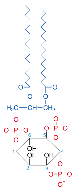
Phosphatidylinositol 4,5-bisphosphate or PtdIns(4,5)P2, also known simply as PIP2 or PI(4,5)P2, is a minor phospholipid component of cell membranes. PtdIns(4,5)P2 is enriched at the plasma membrane where it is a substrate for a number of important signaling proteins. PIP2 also forms lipid clusters that sort proteins.

<span class="mw-page-title-main">Lipid signaling</span> Biological signaling using lipid molecules

Lipid signaling, broadly defined, refers to any biological cell signaling event involving a lipid messenger that binds a protein target, such as a receptor, kinase or phosphatase, which in turn mediate the effects of these lipids on specific cellular responses. Lipid signaling is thought to be qualitatively different from other classical signaling paradigms because lipids can freely diffuse through membranes. One consequence of this is that lipid messengers cannot be stored in vesicles prior to release and so are often biosynthesized "on demand" at their intended site of action. As such, many lipid signaling molecules cannot circulate freely in solution but, rather, exist bound to special carrier proteins in serum.

Phospholipase D (EC 3.1.4.4, lipophosphodiesterase II, lecithinase D, choline phosphatase, PLD; systematic name phosphatidylcholine phosphatidohydrolase) is an enzyme of the phospholipase superfamily that catalyses the following reaction

<span class="mw-page-title-main">Chemokine receptor</span> Cytokine receptor

Chemokine receptors are cytokine receptors found on the surface of certain cells that interact with a type of cytokine called a chemokine. There have been 20 distinct chemokine receptors discovered in humans. Each has a rhodopsin-like 7-transmembrane (7TM) structure and couples to G-protein for signal transduction within a cell, making them members of a large protein family of G protein-coupled receptors. Following interaction with their specific chemokine ligands, chemokine receptors trigger a flux in intracellular calcium (Ca2+) ions (calcium signaling). This causes cell responses, including the onset of a process known as chemotaxis that traffics the cell to a desired location within the organism. Chemokine receptors are divided into different families, CXC chemokine receptors, CC chemokine receptors, CX3C chemokine receptors and XC chemokine receptors that correspond to the 4 distinct subfamilies of chemokines they bind. The four subfamilies of chemokines differ in the spacing of structurally important cysteine residues near the N-terminal of the chemokine.

Prostaglandin receptors or prostanoid receptors represent a sub-class of cell surface membrane receptors that are regarded as the primary receptors for one or more of the classical, naturally occurring prostanoids viz., prostaglandin D2,, PGE2, PGF2alpha, prostacyclin (PGI2), thromboxane A2 (TXA2), and PGH2. They are named based on the prostanoid to which they preferentially bind and respond, e.g. the receptor responsive to PGI2 at lower concentrations than any other prostanoid is named the Prostacyclin receptor (IP). One exception to this rule is the receptor for thromboxane A2 (TP) which binds and responds to PGH2 and TXA2 equally well.

Gq protein alpha subunit is a family of heterotrimeric G protein alpha subunits. This family is also commonly called the Gq/11 (Gq/G11) family or Gq/11/14/15 family to include closely related family members. G alpha subunits may be referred to as Gq alpha, Gαq, or Gqα. Gq proteins couple to G protein-coupled receptors to activate beta-type phospholipase C (PLC-β) enzymes. PLC-β in turn hydrolyzes phosphatidylinositol 4,5-bisphosphate (PIP2) to diacyl glycerol (DAG) and inositol trisphosphate (IP3). IP3 acts as a second messenger to release stored calcium into the cytoplasm, while DAG acts as a second messenger that activates protein kinase C (PKC).

<span class="mw-page-title-main">PLCE1</span> Protein-coding gene in the species Homo sapiens

Phospholipase C epsilon 1 (PLCE1) is an enzyme that in humans is encoded by the PLCE1 gene. This gene encodes a phospholipase enzyme (PLCE1) that catalyzes the hydrolysis of phosphatidylinositol-4,5-bisphosphate to generate two second messengers: inositol 1,4,5-triphosphate (IP3) and diacylglycerol (DAG). Mutations in this gene cause early-onset nephrotic syndrome and have been associated with respiratory chain deficiency with diffuse mesangial sclerosis.

<span class="mw-page-title-main">DLC1</span> Protein-coding gene in the species Homo sapiens

Deleted in Liver Cancer 1 also known as DLC1 and StAR-related lipid transfer protein 12 (STARD12) is a protein which in humans is encoded by the DLC1 gene.

<span class="mw-page-title-main">PLCD3</span> Protein-coding gene in the species Homo sapiens

1-Phosphatidylinositol-4,5-bisphosphate phosphodiesterase delta-3 is an enzyme that in humans is encoded by the PLCD3 gene.

Oocyteactivation is a series of processes that occur in the oocyte during fertilization.

The insulin transduction pathway is a biochemical pathway by which insulin increases the uptake of glucose into fat and muscle cells and reduces the synthesis of glucose in the liver and hence is involved in maintaining glucose homeostasis. This pathway is also influenced by fed versus fasting states, stress levels, and a variety of other hormones.

<span class="mw-page-title-main">Diglyceride</span> Type of fat derived from glycerol and two fatty acids

A diglyceride, or diacylglycerol (DAG), is a glyceride consisting of two fatty acid chains covalently bonded to a glycerol molecule through ester linkages. Two possible forms exist, 1,2-diacylglycerols and 1,3-diacylglycerols. Diglycerides are natural components of food fats, though minor in comparison to triglycerides. DAGs can act as surfactants and are commonly used as emulsifiers in processed foods. DAG-enriched oil has been investigated extensively as a fat substitute due to its ability to suppress the accumulation of body fat; with total annual sales of approximately USD 200 million in Japan since its introduction in the late 1990s till 2009.

Substrate presentation is a biological process that activates a protein. The protein is sequestered away from its substrate and then activated by release and exposure of the protein to its substrate. A substrate is typically the substance on which an enzyme acts but can also be a protein surface to which a ligand binds. The substrate is the material acted upon. In the case of an interaction with an enzyme, the protein or organic substrate typically changes chemical form. Substrate presentation differs from allosteric regulation in that the enzyme need not change its conformation to begin catalysis. Substrate presentation is best described for nanoscopic distances (<100 nm).

References

  1. 1 2 3 Kadamur G, Ross EM (2013). "Mammalian phospholipase C". Annual Review of Physiology. 75: 127–54. doi:10.1146/annurev-physiol-030212-183750. PMID   23140367.
  2. Suh, PG; Park, JI; Manzoli, L; Cocco, L; Peak, JC; Katan, M; Fukami, K; Kataoka, T; Yun, S; Ryu, SH (2008). "Multiple roles of phosphoinositide-specific phospholipase C isozymes". BMB Reports. 41 (6): 415–34. doi: 10.5483/bmbrep.2008.41.6.415 . hdl: 11585/62661 . PMID   18593525.
  3. Titball, RW (1993). "Bacterial phospholipases C." Microbiological Reviews. 57 (2): 347–66. doi:10.1128/MMBR.57.2.347-366.1993. PMC   372913 . PMID   8336671.
  4. Singaravelu, Gunasekaran; Singson, Andrew (January 2013). "Calcium signaling surrounding fertilization in the nematode Caenorhabditis elegans". Cell Calcium. 53 (1): 2–9. doi:10.1016/j.ceca.2012.11.009. PMC   3566351 .
  5. Hicks SN, Jezyk MR, Gershburg S, Seifert JP, Harden TK, Sondek J (August 2008). "General and versatile autoinhibition of PLC isozymes". Molecular Cell. 31 (3): 383–94. doi:10.1016/j.molcel.2008.06.018. PMC   2702322 . PMID   18691970.
  6. 1 2 Sakurai J, Nagahama M, Oda M (November 2004). "Clostridium perfringens alpha-toxin: characterization and mode of action". Journal of Biochemistry. 136 (5): 569–74. doi:10.1093/jb/mvh161. PMID   15632295.
  7. Essen LO, Perisic O, Katan M, Wu Y, Roberts MF, Williams RL (February 1997). "Structural mapping of the catalytic mechanism for a mammalian phosphoinositide-specific phospholipase C". Biochemistry. 36 (7): 1704–18. doi:10.1021/bi962512p. PMID   9048554.
  8. Ellis, MV; James, SR; Perisic, O; Downes, PC; Williams, RL; Katan, M (1998). "Catalytic Domain of Phosphoinositide-specific Phospholipase C (PLC): mutation analysis of residues within the active site of hydrophobic ridge of PLCD1". The Journal of Biological Chemistry. 273 (19): 11650–9. doi: 10.1074/jbc.273.19.11650 . PMID   9565585.
  9. 1 2 Walter F. Boron (2003). Medical Physiology: A Cellular And Molecular Approaoch. Elsevier/Saunders. p. 1300. ISBN   978-1-4160-2328-9. Page 104
  10. GeneGlobe -> GHRH Signaling [ permanent dead link ] Retrieved on May 31, 2009
  11. Bleasdale JE, Thakur NR, Gremban RS, Bundy GL, Fitzpatrick FA, Smith RJ, Bunting S (November 1990). "Selective inhibition of receptor-coupled phospholipase C-dependent processes in human platelets and polymorphonuclear neutrophils". The Journal of Pharmacology and Experimental Therapeutics. 255 (2): 756–68. PMID   2147038.
  12. Macmillan D, McCarron JG (July 2010). "The phospholipase C inhibitor U-73122 inhibits Ca(2+) release from the intracellular sarcoplasmic reticulum Ca(2+) store by inhibiting Ca(2+) pumps in smooth muscle". British Journal of Pharmacology. 160 (6): 1295–301. doi:10.1111/j.1476-5381.2010.00771.x. PMC   2938802 . PMID   20590621.
  13. Huang W, Barrett M, Hajicek N, Hicks S, Harden TK, Sondek J, Zhang Q (February 2013). "Small molecule inhibitors of phospholipase C from a novel high-throughput screen". The Journal of Biological Chemistry. 288 (8): 5840–8. doi: 10.1074/jbc.M112.422501 . PMC   3581404 . PMID   23297405.
  14. Klein RR, Bourdon DM, Costales CL, Wagner CD, White WL, Williams JD, Hicks SN, Sondek J, Thakker DR (April 2011). "Direct activation of human phospholipase C by its well known inhibitor u73122". The Journal of Biological Chemistry. 286 (14): 12407–16. doi: 10.1074/jbc.M110.191783 . PMC   3069444 . PMID   21266572.
  15. Horowitz LF, Hirdes W, Suh BC, Hilgemann DW, Mackie K, Hille B (September 2005). "Phospholipase C in living cells: activation, inhibition, Ca2+ requirement, and regulation of M current". The Journal of General Physiology. 126 (3): 243–62. doi:10.1085/jgp.200509309. PMC   2266577 . PMID   16129772.
  16. Rees, Shaun W. P.; Leung, Euphemia; Reynisson, Jóhannes; Barker, David; Pilkington, Lisa I. (2021-09-01). "Development of 2-Morpholino-N-hydroxybenzamides as anti-proliferative PC-PLC inhibitors". Bioorganic Chemistry. 114: 105152. doi:10.1016/j.bioorg.2021.105152. ISSN   0045-2068. PMID   34328856.
  17. Eurtivong, C.; Pilkington, L. I.; van Rensburg, M.; White, R. M.; Kaur Brar, H.; Rees, S.; Paulin, E. K.; Xu, C. S.; Sharma, N.; Leung, I. K. H.; Leung, E.; Barker, D.; Reynisson, J. (1 February 2020). "Discovery of novel phosphatidylcholine-specific phospholipase C drug-like inhibitors as potential anticancer agents". European Journal of Medicinal Chemistry. 187: 111919. doi:10.1016/j.ejmech.2019.111919. PMID   31810783. S2CID   208813280.
  18. Pilkington, L. I.; Sparrow, K.; Rees, S. W. P.; Paulin, E. K.; van Rensburg, M.; Xu, C. S.; Langley, R. J.; Leung, I. K. H.; Reynisson, J.; Leung, E.; Barker, D. (2020). "Development, Synthesis and Biological Investigation of a Novel Class of Potent PC-PLC Inhibitors". European Journal of Medicinal Chemistry. 191: 112162. doi:10.1016/j.ejmech.2020.112162. PMID   32101781. S2CID   211536972.
  19. Little C, Otnåss AB (June 1975). "The metal ion dependence of phospholipase C from Bacillus cereus". Biochimica et Biophysica Acta (BBA) - Enzymology. 391 (2): 326–33. doi:10.1016/0005-2744(75)90256-9. PMID   807246.
  20. "Phospholipase C, Phosphatidylinositol-specific from Bacillus cereus" (PDF). Product Information. Sigma Aldrich.
  21. Hilgemann DW (October 2007). "Local PIP(2) signals: when, where, and how?". Pflügers Archiv. 455 (1): 55–67. doi:10.1007/s00424-007-0280-9. PMID   17534652. S2CID   29839094.
  22. Hansen (1 May 2015). "Lipid agonism: The PIP2 paradigm of ligand-gated ion channels". Biochimica et Biophysica Acta (BBA) - Molecular and Cell Biology of Lipids. 1851 (5): 620–628. doi:10.1016/j.bbalip.2015.01.011. PMC   4540326 . PMID   25633344.
  23. Falkenburger BH, Jensen JB, Dickson EJ, Suh BC, Hille B (September 2010). "Phosphoinositides: lipid regulators of membrane proteins". The Journal of Physiology. 588 (Pt 17): 3179–85. doi:10.1113/jphysiol.2010.192153. PMC   2976013 . PMID   20519312.
  24. 1 2 3 Alberts B, Lewis J, Raff M, Roberts K, Walter P (2002). Molecular biology of the cell (4th ed.). New York: Garland Science. ISBN   978-0-8153-3218-3.
  25. Li Z, Jiang H, Xie W, Zhang Z, Smrcka AV, Wu D (February 2000). "Roles of PLC-beta2 and -beta3 and PI3Kgamma in chemoattractant-mediated signal transduction". Science. 287 (5455): 1046–9. Bibcode:2000Sci...287.1046L. doi:10.1126/science.287.5455.1046. PMID   10669417.
  26. Gresset A, Sondek J, Harden TK (2012). "The phospholipase C isozymes and their regulation". Phosphoinositides I: Enzymes of Synthesis and Degradation. Subcellular Biochemistry. Vol. 58. pp. 61–94. doi:10.1007/978-94-007-3012-0_3. ISBN   978-94-007-3011-3. PMC   3638883 . PMID   22403074.
  27. Piomelli, Daniele (1993-04-01). "Arachidonic acid in cell signaling" (PDF). Current Opinion in Cell Biology. 5 (2): 274–280. doi:10.1016/0955-0674(93)90116-8. PMID   7685181.
  28. Fujii Y, Sakurai J (May 1989). "Contraction of the rat isolated aorta caused by Clostridium perfringens alpha toxin (phospholipase C): evidence for the involvement of arachidonic acid metabolism". British Journal of Pharmacology. 97 (1): 119–24. doi:10.1111/j.1476-5381.1989.tb11931.x. PMC   1854495 . PMID   2497921.