Endonuclease

Last updated

In molecular biology, endonucleases are enzymes that cleave the phosphodiester bond within a polynucleotide chain (namely DNA or RNA). Some, such as deoxyribonuclease I, cut DNA relatively nonspecifically (without regard to sequence), while many, typically called restriction endonucleases or restriction enzymes, cleave only at very specific nucleotide sequences. Endonucleases differ from exonucleases, which cleave the ends of recognition sequences instead of the middle (endo) portion. Some enzymes known as "exo-endonucleases", however, are not limited to either nuclease function, displaying qualities that are both endo- and exo-like. [1] Evidence suggests that endonuclease activity experiences a lag compared to exonuclease activity. [2]

Contents

Restriction enzymes are endonucleases from eubacteria and archaea that recognize a specific DNA sequence. [3] The nucleotide sequence recognized for cleavage by a restriction enzyme is called the restriction site. Typically, a restriction site will be a palindromic sequence about four to six nucleotides long. Most restriction endonucleases cleave the DNA strand unevenly, leaving complementary single-stranded ends. These ends can reconnect through hybridization and are termed "sticky ends". Once paired, the phosphodiester bonds of the fragments can be joined by DNA ligase. There are hundreds of restriction endonucleases known, each attacking a different restriction site. The DNA fragments cleaved by the same endonuclease can be joined regardless of the origin of the DNA. Such DNA is called recombinant DNA; DNA formed by the joining of genes into new combinations. [4] Restriction endonucleases (restriction enzymes) are divided into three categories, Type I, Type II, and Type III, according to their mechanism of action. These enzymes are often used in genetic engineering to make recombinant DNA for introduction into bacterial, plant, or animal cells, as well as in synthetic biology. [5] One of the more famous endonucleases is Cas9.

Categories

Ultimately, there are three categories of restriction endonucleases that relatively contribute to the cleavage of specific sequences. The types I and III are large multisubunit complexes that include both the endonucleases and methylase activities. Type I can cleave at random sites of about 1000 base pairs or more from the recognition sequence and it requires ATP as source of energy. Type II behaves slightly differently and was first isolated by Hamilton Smith in 1970. They are simpler versions of the endonucleases and require no ATP in their degradation processes. Some examples of type II restriction endonucleases include BamHI, EcoRI, EcoRV, HindIII, and HaeIII. Type III, however, cleaves the DNA at about 25 base pairs from the recognition sequence and also requires ATP in the process. [4]

Notations

The commonly used notation for restriction endonucleases [6] is of the form "VwxyZ", where "Vwx" are, in italics, the first letter of the genus and the first two letters of the species where this restriction endonuclease may be found, for example, Escherichia coli, Eco, and Haemophilus influenzae, Hin. This is followed by the optional, non-italicized symbol "y", which indicates the type or strain identification, for example, EcoR for E. coli strains bearing the drug resistance transfer factor RTF-1, [6] EcoB for E. coli strain B, [7] and Hind for H. influenzae strain d. [6] Finally, when a particular type or strain has several different restriction endonucleases, these are identified by Roman numerals, thus, the restriction endonucleases from H. influenzae strain d are named HindI, HindII, HindIII, etc. Another example: "HaeII" and "HaeIII" refer to bacterium Haemophilus aegyptius (strain not specified), restriction endonucleases number II and number III, respectively. [4] :64–64 The restriction enzymes used in molecular biology usually recognize short target sequences of about 4 – 8 base pairs. For instance, the EcoRI enzyme recognizes and cleaves the sequence 5' – GAATTC – 3'. [8]

Restriction enzyme Eco RI Restriction enzyme Eco RI.JPG
Restriction enzyme Eco RI

Restriction endonucleases come in several types. A restriction endonuclease typically requires a recognition site and a cleavage pattern (typically of nucleotide bases: A, C, G, T). If the recognition site is outside the region of the cleavage pattern, then the restriction endonuclease is referred to as Type I. If the recognition sequence overlaps with the cleavage sequence, then the restriction endonuclease restriction enzyme is Type II.[ citation needed ]

Processes involved with endonucleases

Endonucleases play a role in many aspects of biological life. Below are a couple examples of processes where endonucleases play a crucial role.

DNA repair

Endonucleases play a role in DNA repair. AP endonuclease, specifically, catalyzes the incision of DNA exclusively at AP sites, and therefore prepares DNA for subsequent excision, repair synthesis and DNA ligation. For example, when depurination occurs, this lesion leaves a deoxyribose sugar with a missing base. [9] The AP endonuclease recognizes this sugar and essentially cuts the DNA at this site and then allows for DNA repair to continue. [10] E. coli cells contain two AP endonucleases: endonuclease IV (endoIV) and exonuclease III (exoIII) while in eukaryotes, there is only one AP endonuclease. [11]

APEndonucleasecartoon.gif

Repair of DNA in which the two complementary strands are joined by an interstrand covalent crosslink requires multiple incisions in order to disengage the strands and remove the damage. Incisions are required on both sides of the crosslink and on both strands of the duplex DNA. In mouse embryonic stem cells, an intermediate stage of crosslink repair involves production of double-strand breaks. [12] MUS81/EME1 is a structure specific endonuclease involved in converting interstrand crosslinks to double-strand breaks in a DNA replication-dependent manner. [12] After introduction of a double-strand break, further steps are required to complete the repair process. If a crosslink is not properly repaired it can block DNA replication.[ citation needed ]

Thymine dimer repair

Exposure of bacteriophage (phage) T4 to ultraviolet irradiation induces thymine dimers in the phage DNA. The phage T4 denV gene encodes endonuclease V that catalyzes the initial steps in the repair of these UV-induced thymine dimers. [13] Endonuclease V first cleaves the glycosylic bond on the 5’ side of a pyrimidine dimer and then catalyzes cleavage of the DNA phospodiester bond that originally linked the two nucleotides of the dimer. Subsequent steps in the repair process involve removal of the dimer remnants and repair synthesis to fill in the resulting single-strand gap using the undamaged strand as template.[ citation needed ]

Apoptosis

During apoptosis, Apoptotic endonuclease DFF40 is activated to initiate controlled cellular disassembly. This disintegration is characterized by the cleavage of genomic DNA into specific fragments. The precise role of endonucleases in this context is to cleave the DNA at specific sites, generating fragments with defined lengths. These fragments are then packaged into apoptotic bodies, ensuring a neat and efficient removal of the dying cell without causing inflammation or damage to neighboring cells. [14]

DNA Replication

Flap endonuclease 1 (FEN1) and Dna2 endonuclease are integral to DNA replication on the lagging strand, participating in crucial processes such as primer removal and Okazaki fragment processing. Endonucleases are actively involved in processing these fragments by cleaving the phosphodiester bonds between them. This process is integral to the seamless synthesis and joining of Okazaki fragments, contributing to the overall continuity of the newly replicated DNA strand. [15] [16]

RNA Processing

Endonucleases, more specifically endoribonuclease, play a crucial role in RNA processing, a fundamental step in gene expression. This process involves the precise cleavage of precursor RNA molecules, guided by endonucleases, to generate functional RNAs essential for various cellular functions. Endonucleases selectively cleave precursor RNAs at specific sites, defining the boundaries of functional RNA segments during RNA processing. The outcome of RNA processing is the production of functional RNA molecules, such as transfer RNAs (tRNAs) and ribosomal RNAs (rRNAs). Endonucleases contribute to the precision of this process, ensuring the formation of mature and functional RNA species.

Endonucleases like RNase P and tRNase Z (ELAC2), shape precursor tRNAs into mature, functional tRNAs, crucial for accurate translation during protein synthesis. [17] In ribosome biogenesis, endonucleases from the RNase III family, like DROSHA, play a role in processing precursor rRNAs, contributing to the assembly of functional ribosomes. [18]

DICER and DROSHA also from the RNase III family play a role in the processing pre-miRNA to functional miRNA. [19]

Maturation of Nails and Hairs

The endonuclease DNase1L2 also contribute prominently to the removal of DNA during the formation of hair and nails. This process is essential for the maturation of hair and nail structures and is crucial for the transformation of cells into durable and keratinized structures, ensuring the strength and integrity of hair and nails. [20]

Further discussion

Restriction endonucleases may be found that cleave standard dsDNA (double-stranded DNA), or ssDNA (single-stranded DNA), or even RNA.[ citation needed ] This discussion is restricted to dsDNA; however, the discussion can be extended to the following:

  1. Holliday junctions
  2. Triple-stranded DNA, quadruple-stranded DNA (G-quadruplex)
  3. Double-stranded hybrids of DNA and RNA (one strand is DNA, the other strand is RNA) [4] :72–73
  4. Synthetic or artificial DNA (for example, containing bases other than A, C, G, T, refer to the work of Eric T. Kool). Research with synthetic codons, refer to the research by S. Benner, and enlarging the amino acid set in polypeptides, thus enlarging the proteome or proteomics, see the research by P. Schultz. [4] :chapter 3

In addition, research is now underway to construct synthetic or artificial restriction endonucleases, especially with recognition sites that are unique within a genome.[ citation needed ]

Restriction endonucleases or restriction enzymes typically cleave in two ways: blunt-ended or sticky-ended patterns. An example of a Type I restriction endonuclease. [4] :64

Furthermore, there exist DNA/RNA non-specific endonucleases, such as those that are found in Serratia marcescens , which act on dsDNA, ssDNA, and RNA.[ citation needed ]

Common endonucleases

Below are tables of common prokaryotic and eukaryotic endonucleases. [21]

Prokaryotic EnzymeSourceComments
RecBCD enonucleaseE. coliPartially ATP dependent; also an exonuclease; functions in recombination and repair
T7 endonuclease ( P00641 )phage T7 (gene 3)Essential for replication; preference for single stranded over double stranded DNA
T4 endonuclease II ( P07059 )phage T4 (denA)Splits -TpC- sequence to yield 5'-dCMP- terminated oligonucleotides; chain length of product varies with conditions
Bal 31 endonuclease P. espejiana Also an exonuclease; nibbles away 3' and 5' ends of duplex DNA. A mixture of at least two nucleases, fast and slow. [22]
Endonuclease I (endo I; P25736 )E. coli (endA)Periplasmic location; average chain length of product is 7; inhibited by tRNA; produces double stranded DNA break; produces nick when complexed with tRNA; endo I mutants grow normally
Micrococcal nuclease ( P00644 )StaphylococcusProduces 3'-P termini; requires Ca2+; also acts on RNA; prefers single stranded DNA and AT-rich regions
Endonuclease II (endo VI, exo III; P09030 )E. coli (xthA)Cleavage next to AP site; also a 3'→5' exonuclease; phosphomonoesterase on 3'-P termini
Eukaryotic EnzymeSourceComments
Neurospora endonuclease [23] Neurospora crassa, mitochondriaAlso acts on RNA.
S1 nuclease ( P24021 )Aspergillus oryzaeAlso acts on RNA
P1-nuclease ( P24289 )Penicillium citrinumAlso acts on RNA
Mung bean nuclease Imung bean sproutsAlso acts on RNA
Ustilago nuclease (Dnase I) [24] Ustilago maydisAlso acts on RNA
Dnase I ( P00639 )Bovine pancreasAverage chain length of product is 4; produces double strand break in presence of Mn2+
AP endonuclease Nucleus, mitochondriaInvolved in DNA Base Excision Repair pathway
Endo R [25] HeLa cells Specific for GC sites
FLAP1 NucleusResponsible for processing Okazaki fragments during DNA replication

Mutations

Xeroderma pigmentosa is a rare, autosomal recessive disease caused by a defective UV-specific endonuclease. Patients with mutations are unable to repair DNA damage caused by sunlight. [26]

Sickle Cell anemia is a disease caused by a point mutation. The sequence altered by the mutation eliminates the recognition site for the restriction endonuclease MstII that recognizes the nucleotide sequence. [27]

tRNA splicing endonuclease mutations cause pontocerebellar hypoplasia. Pontocerebellar hypoplasias (PCH) represent a group of neurodegenerative autosomal recessive disorders that is caused by mutations in three of the four different subunits of the tRNA-splicing endonuclease complex. [28]

See also

Related Research Articles

<span class="mw-page-title-main">Primer (molecular biology)</span> Short strand of RNA or DNA that serves as a starting point for DNA synthesis

A primer is a short single-stranded nucleic acid used by all living organisms in the initiation of DNA synthesis. A synthetic primer may also be referred to as an oligo, short for oligonucleotide. DNA polymerase enzymes are only capable of adding nucleotides to the 3’-end of an existing nucleic acid, requiring a primer be bound to the template before DNA polymerase can begin a complementary strand. DNA polymerase adds nucleotides after binding to the RNA primer and synthesizes the whole strand. Later, the RNA strands must be removed accurately and replace them with DNA nucleotides forming a gap region known as a nick that is filled in using an enzyme called ligase. The removal process of the RNA primer requires several enzymes, such as Fen1, Lig1, and others that work in coordination with DNA polymerase, to ensure the removal of the RNA nucleotides and the addition of DNA nucleotides. Living organisms use solely RNA primers, while laboratory techniques in biochemistry and molecular biology that require in vitro DNA synthesis usually use DNA primers, since they are more temperature stable. Primers can be designed in laboratory for specific reactions such as polymerase chain reaction (PCR). When designing PCR primers, there are specific measures that must be taken into consideration, like the melting temperature of the primers and the annealing temperature of the reaction itself. Moreover, the DNA binding sequence of the primer in vitro has to be specifically chosen, which is done using a method called basic local alignment search tool (BLAST) that scans the DNA and finds specific and unique regions for the primer to bind.

A restriction enzyme, restriction endonuclease, REase, ENase orrestrictase is an enzyme that cleaves DNA into fragments at or near specific recognition sites within molecules known as restriction sites. Restriction enzymes are one class of the broader endonuclease group of enzymes. Restriction enzymes are commonly classified into five types, which differ in their structure and whether they cut their DNA substrate at their recognition site, or if the recognition and cleavage sites are separate from one another. To cut DNA, all restriction enzymes make two incisions, once through each sugar-phosphate backbone of the DNA double helix.

The restriction modification system is found in bacteria and other prokaryotic organisms, and provides a defense against foreign DNA, such as that borne by bacteriophages.

Gene knockdown is an experimental technique by which the expression of one or more of an organism's genes is reduced. The reduction can occur either through genetic modification or by treatment with a reagent such as a short DNA or RNA oligonucleotide that has a sequence complementary to either gene or an mRNA transcript.

<span class="mw-page-title-main">Ribonuclease</span> Class of enzyme that catalyzes the degradation of RNA

Ribonuclease is a type of nuclease that catalyzes the degradation of RNA into smaller components. Ribonucleases can be divided into endoribonucleases and exoribonucleases, and comprise several sub-classes within the EC 2.7 and 3.1 classes of enzymes.

<span class="mw-page-title-main">Nuclease</span> Class of enzymes which cleave nucleic acids

In biochemistry, a nuclease is an enzyme capable of cleaving the phosphodiester bonds between nucleotides of nucleic acids. Nucleases variously effect single and double stranded breaks in their target molecules. In living organisms, they are essential machinery for many aspects of DNA repair. Defects in certain nucleases can cause genetic instability or immunodeficiency. Nucleases are also extensively used in molecular cloning.

<span class="mw-page-title-main">Okazaki fragments</span> Transient components of lagging strand of DNA

Okazaki fragments are short sequences of DNA nucleotides which are synthesized discontinuously and later linked together by the enzyme DNA ligase to create the lagging strand during DNA replication. They were discovered in the 1960s by the Japanese molecular biologists Reiji and Tsuneko Okazaki, along with the help of some of their colleagues.

A cDNA library is a combination of cloned cDNA fragments inserted into a collection of host cells, which constitute some portion of the transcriptome of the organism and are stored as a "library". cDNA is produced from fully transcribed mRNA found in the nucleus and therefore contains only the expressed genes of an organism. Similarly, tissue-specific cDNA libraries can be produced. In eukaryotic cells the mature mRNA is already spliced, hence the cDNA produced lacks introns and can be readily expressed in a bacterial cell. While information in cDNA libraries is a powerful and useful tool since gene products are easily identified, the libraries lack information about enhancers, introns, and other regulatory elements found in a genomic DNA library.

<span class="mw-page-title-main">Dicer</span> Enzyme that cleaves double-stranded RNA (dsRNA) into short dsRNA fragments

Dicer, also known as endoribonuclease Dicer or helicase with RNase motif, is an enzyme that in humans is encoded by the DICER1 gene. Being part of the RNase III family, Dicer cleaves double-stranded RNA (dsRNA) and pre-microRNA (pre-miRNA) into short double-stranded RNA fragments called small interfering RNA and microRNA, respectively. These fragments are approximately 20–25 base pairs long with a two-base overhang on the 3′-end. Dicer facilitates the activation of the RNA-induced silencing complex (RISC), which is essential for RNA interference. RISC has a catalytic component Argonaute, which is an endonuclease capable of degrading messenger RNA (mRNA).

<span class="mw-page-title-main">Exonuclease</span> Class of enzymes; type of nuclease

Exonucleases are enzymes that work by cleaving nucleotides one at a time from the end (exo) of a polynucleotide chain. A hydrolyzing reaction that breaks phosphodiester bonds at either the 3′ or the 5′ end occurs. Its close relative is the endonuclease, which cleaves phosphodiester bonds in the middle (endo) of a polynucleotide chain. Eukaryotes and prokaryotes have three types of exonucleases involved in the normal turnover of mRNA: 5′ to 3′ exonuclease (Xrn1), which is a dependent decapping protein; 3′ to 5′ exonuclease, an independent protein; and poly(A)-specific 3′ to 5′ exonuclease.

Mung bean nuclease is a nuclease derived from sprouts of the mung bean that removes nucleotides in a step-wise manner from single-stranded DNA molecules (ssDNA) and is used in biotechnological applications to remove such ssDNA from a mixture also containing double-stranded DNA (dsDNA). This enzyme is useful for transcript mapping, removal of single-stranded regions in DNA hybrids or single-stranded overhangs produced by restriction enzymes, etc. It has an activity similar to Nuclease S1, but it has higher specificity for single-stranded molecules.

<span class="mw-page-title-main">Ribonuclease III</span> Class of enzymes

Ribonuclease III (RNase III or RNase C)(BRENDA 3.1.26.3) is a type of ribonuclease that recognizes dsRNA and cleaves it at specific targeted locations to transform them into mature RNAs. These enzymes are a group of endoribonucleases that are characterized by their ribonuclease domain, which is labelled the RNase III domain. They are ubiquitous compounds in the cell and play a major role in pathways such as RNA precursor synthesis, RNA Silencing, and the pnp autoregulatory mechanism.

<i>Fok</i>I Restriction enzyme

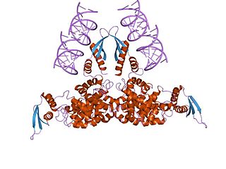
The restriction endonuclease Fok1, naturally found in Flavobacterium okeanokoites, is a bacterial type IIS restriction endonuclease consisting of an N-terminal DNA-binding domain and a non sequence-specific DNA cleavage domain at the C-terminal. Once the protein is bound to duplex DNA via its DNA-binding domain at the 5'-GGATG-3' recognition site, the DNA cleavage domain is activated and cleaves the DNA at two locations, regardless of the nucleotide sequence at the cut site. The DNA is cut 9 nucleotides downstream of the motif on the forward strand, and 13 nucleotides downstream of the motif on the reverse strand, producing two sticky ends with 4-bp overhangs.

<span class="mw-page-title-main">Argonaute</span> Protein that plays a role in RNA silencing process

The Argonaute protein family, first discovered for its evolutionarily conserved stem cell function, plays a central role in RNA silencing processes as essential components of the RNA-induced silencing complex (RISC). RISC is responsible for the gene silencing phenomenon known as RNA interference (RNAi). Argonaute proteins bind different classes of small non-coding RNAs, including microRNAs (miRNAs), small interfering RNAs (siRNAs) and Piwi-interacting RNAs (piRNAs). Small RNAs guide Argonaute proteins to their specific targets through sequence complementarity, which then leads to mRNA cleavage, translation inhibition, and/or the initiation of mRNA decay.

<i>Hae</i>III Enzyme

HaeIII is one of many restriction enzymes (endonucleases) a type of prokaryotic DNA that protects organisms from unknown, foreign DNA. It is a restriction enzyme used in molecular biology laboratories. It was the third endonuclease to be isolated from the Haemophilus aegyptius bacteria. The enzyme's recognition site—the place where it cuts DNA molecules—is the GGCC nucleotide sequence which means it cleaves DNA at the site 5′-GG/CC-3. The recognition site is usually around 4-8 bps.This enzyme's gene has been sequenced and cloned. This is done to make DNA fragments in blunt ends. HaeIII is not effective for single stranded DNA cleavage.

<span class="mw-page-title-main">Nuclease S1</span> Class of enzymes

Nuclease S1 is an endonuclease enzyme that splits single-stranded DNA (ssDNA) and RNA into oligo- or mononucleotides. This enzyme catalyses the following chemical reaction

Deoxyribonuclease IV (phage-T4-induced) is catalyzes the degradation nucleotides in DsDNA by attacking the 5'-terminal end.

<span class="mw-page-title-main">Cas9</span> Microbial protein found in Streptococcus pyogenes M1 GAS

Cas9 is a 160 kilodalton protein which plays a vital role in the immunological defense of certain bacteria against DNA viruses and plasmids, and is heavily utilized in genetic engineering applications. Its main function is to cut DNA and thereby alter a cell's genome. The CRISPR-Cas9 genome editing technique was a significant contributor to the Nobel Prize in Chemistry in 2020 being awarded to Emmanuelle Charpentier and Jennifer Doudna.

<span class="mw-page-title-main">Ribonuclease T</span> Class of enzymes

Ribonuclease T is a ribonuclease enzyme involved in the maturation of transfer RNA and ribosomal RNA in bacteria, as well as in DNA repair pathways. It is a member of the DnaQ family of exonucleases and non-processively acts on the 3' end of single-stranded nucleic acids. RNase T is capable of cleaving both DNA and RNA, with extreme sequence specificity discriminating against cytosine at the 3' end of the substrate.

<i>Eco</i>RI Restriction enzyme

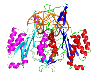
EcoRI is a restriction endonuclease enzyme isolated from species E. coli. It is a restriction enzyme that cleaves DNA double helices into fragments at specific sites, and is also a part of the restriction modification system. The Eco part of the enzyme's name originates from the species from which it was isolated - "E" denotes generic name which is "Escherichia" and "co" denotes species name, "coli" - while the R represents the particular strain, in this case RY13, and the I denotes that it was the first enzyme isolated from this strain.

References

  1. "Properties of Exonucleases and Endonucleases". New England BioLabs. 2017. Retrieved May 21, 2017.
  2. Slor, Hanoch (April 14, 1975). "Differentiation between exonucleases and endonucleases and between haplotomic and diplotomic endonucleases using 3-h-dna-coated wells of plastic depression plates as substrate". Nucleic Acids Research. 2 (6): 897–903. doi:10.1093/nar/2.6.897. PMC   343476 . PMID   167356.
  3. Stephen T. Kilpatrick; Jocelyn E. Krebs; Lewin, Benjamin; Goldstein, Elliott (2011). Lewin's genes X . Boston: Jones and Bartlett. ISBN   978-0-7637-6632-0.
  4. 1 2 3 4 5 6 Cox M, Nelson DR, Lehninger AL (2005). Lehninger principles of biochemistry . San Francisco: W.H. Freeman. pp.  952. ISBN   978-0-7167-4339-2.
  5. Simon M (2010). Emergent computation: Emphasizing Bioinformatics. New York: Springer. p. 437. ISBN   978-1441919632.
  6. 1 2 3 Smith, HO; Nathans, D (15 December 1973). "A suggested nomenclature for bacterial host modification and restriction systems and their enzymes". Journal of Molecular Biology. 81 (3): 419–23. doi:10.1016/0022-2836(73)90152-6. PMID   4588280.
  7. Rubin, RA; Modrich, P (25 October 1977). "EcoRI methylase". The Journal of Biological Chemistry. 252 (20): 7265–72. doi: 10.1016/S0021-9258(19)66964-4 . PMID   332688.
  8. Losick R, Watson JD, Baker TA, Bell S, Gann S, Levine MW (2008). Molecular biology of the gene. San Francisco: Pearson/Benjamin Cummings. ISBN   978-0-8053-9592-1.
  9. Ellenberger T, Friedberg EC, Walker GS, Wolfram S, Wood RJ, Schultz R (2006). DNA repair and mutagenesis. Washington, D.C: ASM Press. ISBN   978-1-55581-319-2.
  10. Alberts B (2002). Molecular biology of the cell. New York: Garland Science. ISBN   978-0-8153-3218-3.
  11. Nishino T, Morikawa K (December 2002). "Structure and function of nucleases in DNA repair: shape, grip and blade of the DNA scissors". Oncogene. 21 (58): 9022–32. doi: 10.1038/sj.onc.1206135 . PMID   12483517.
  12. 1 2 Hanada, K.; Budzowska, M.; Modesti, M.; Maas, A.; Wyman, C.; Essers, J.; Kanaar, R. (2006). "The structure-specific endonuclease Mus81-Eme1 promotes conversion of interstrand DNA crosslinks into double-strands breaks". The EMBO Journal. 25 (20): 4921–4932. doi:10.1038/sj.emboj.7601344. PMC   1618088 . PMID   17036055.
  13. Bernstein, C. (1981). "Deoxyribonucleic acid repair in bacteriophage". Microbiological Reviews. 45 (1): 72–98. doi:10.1128/mr.45.1.72-98.1981. PMC   281499 . PMID   6261109.
  14. Yoshida, Akira; Pommier, Yves; Ueda, Takanori (2006-02-01). "Endonuclease Activation and Chromosomal DNA Fragmentation during Apoptosis in Leukemia Cells". International Journal of Hematology. 84 (1): 31–37. doi:10.1007/BF03342699. ISSN   1865-3774. PMID   16867899. S2CID   25475000.
  15. Jin, Yong Hwan; Obert, Robyn; Burgers, Peter M. J.; Kunkel, Thomas A.; Resnick, Michael A.; Gordenin, Dmitry A. (2001-04-24). "The 3′→5′ exonuclease of DNA polymerase δ can substitute for the 5′ flap endonuclease Rad27/Fen1 in processing Okazaki fragments and preventing genome instability". Proceedings of the National Academy of Sciences of the United States of America. 98 (9): 5122–5127. doi: 10.1073/pnas.091095198 . ISSN   0027-8424. PMC   33174 . PMID   11309502.
  16. Liu, Yuan; Kao, Hui-I; Bambara, Robert A. (June 2004). "Flap Endonuclease 1: A Central Component of DNA Metabolism". Annual Review of Biochemistry. 73 (1): 589–615. doi:10.1146/annurev.biochem.73.012803.092453. ISSN   0066-4154. PMID   15189154.
  17. Hartmann, Roland K.; Gössringer, Markus; Späth, Bettina; Fischer, Susan; Marchfelder, Anita (2009). "The making of tRNAs and more - RNase P and tRNase Z". Progress in Molecular Biology and Translational Science. 85: 319–368. doi:10.1016/S0079-6603(08)00808-8. ISSN   1877-1173. PMID   19215776.
  18. Lejars, Maxence; Kobayashi, Asaki; Hajnsdorf, Eliane (December 2021). "RNase III, Ribosome Biogenesis and Beyond". Microorganisms. 9 (12): 2608. doi: 10.3390/microorganisms9122608 . PMC   8708148 . PMID   34946208.
  19. Kuehbacher, Angelika; Urbich, Carmen; Zeiher, Andreas M.; Dimmeler, Stefanie (2007-07-06). "Role of Dicer and Drosha for Endothelial MicroRNA Expression and Angiogenesis". Circulation Research. 101 (1): 59–68. doi:10.1161/CIRCRESAHA.107.153916. ISSN   0009-7330. PMID   17540974.
  20. Fischer, Heinz; Szabo, Sandra; Scherz, Jennifer; Jaeger, Karin; Rossiter, Heidemarie; Buchberger, Maria; Ghannadan, Minoo; Hermann, Marcela; Theussl, Hans-Christian; Tobin, Desmond J.; Wagner, Erwin F.; Tschachler, Erwin; Eckhart, Leopold (June 2011). "Essential role of the keratinocyte-specific endonuclease DNase1L2 in the removal of nuclear DNA from hair and nails". The Journal of Investigative Dermatology. 131 (6): 1208–1215. doi:10.1038/jid.2011.13. ISSN   0022-202X. PMC   3185332 . PMID   21307874.
  21. Tania A. Baker; Kornberg, Arthur (2005). DNA replication. University Science. ISBN   978-1-891389-44-3.
  22. Wei, CF; Alianell, GA; Bencen, GH; Gray HB, Jr (25 November 1983). "Isolation and comparison of two molecular species of the BAL 31 nuclease from Alteromonas espejiana with distinct kinetic properties". The Journal of Biological Chemistry. 258 (22): 13506–12. doi: 10.1016/S0021-9258(17)43942-1 . PMID   6643438.
  23. Linn, S; Lehman, IR (10 June 1966). "An endonuclease from mitochondria of Neurospora crassa". The Journal of Biological Chemistry. 241 (11): 2694–9. doi: 10.1016/S0021-9258(18)96595-6 . PMID   4287861.
  24. Holloman, WK; Holliday, R (10 December 1973). "Studies on a nuclease from Ustilago maydis. I. Purification, properties, and implication in recombination of the enzyme". The Journal of Biological Chemistry. 248 (23): 8107–13. doi: 10.1016/S0021-9258(19)43199-2 . PMID   4201782.
  25. Gottlieb, J; Muzyczka, N (5 July 1990). "Purification and characterization of HeLa endonuclease R. A G-specific mammalian endonuclease". The Journal of Biological Chemistry. 265 (19): 10836–41. doi: 10.1016/S0021-9258(19)38522-9 . PMID   2358441.
  26. Medical Biochemistry at a Glance. New York: Wiley. 2012. ISBN   978-0-470-65451-4.
  27. Ferrier DR, Champe PC, Harvey RP (2008). Biochemistry. Philadelphia: Wolters Kluwer/Lippincott Williams & Wilkins. ISBN   978-0-7817-6960-0.
  28. Budde BS, Namavar Y, Barth PG, Poll-The BT, Nürnberg G, Becker C, van Ruissen F, Weterman MA, Fluiter K, te Beek ET, Aronica E, van der Knaap MS, Höhne W, Toliat MR, Crow YJ, Steinling M, Voit T, Roelenso F, Brussel W, Brockmann K, Kyllerman M, Boltshauser E, Hammersen G, Willemsen M, Basel-Vanagaite L, Krägeloh-Mann I, de Vries LS, Sztriha L, Muntoni F, Ferrie CD, Battini R, Hennekam RC, Grillo E, Beemer FA, Stoets LM, Wollnik B, Nürnberg P, Baas F (September 2008). "tRNA splicing endonuclease mutations cause pontocerebellar hypoplasia". Nat. Genet. 40 (9): 1113–8. doi:10.1038/ng.204. PMID   18711368. S2CID   205345070.