Dentate gyrus

Last updated

Dentate gyrus
HippocampalRegions.jpg
Diagram of hippocampal regions. DG: Dentate gyrus.
Gray717.png
Coronal section of brain immediately in front of pons. (Label for "Gyrus dentatus" is at bottom center.)
Details
Part of Temporal lobe
Artery Posterior cerebral
Anterior choroidal
Identifiers
Latin gyrus dentatus
MeSH D018891
NeuroNames 179
NeuroLex ID birnlex_1178
TA98 A14.1.09.237
A14.1.09.339
TA2 5521
FMA 61922
Anatomical terms of neuroanatomy

The dentate gyrus (DG) is part of the hippocampal formation in the temporal lobe of the brain, which also includes the hippocampus and the subiculum. The dentate gyrus is part of the hippocampal trisynaptic circuit and is thought to contribute to the formation of new episodic memories, [1] [2] the spontaneous exploration of novel environments [2] and other functions. [3]

Contents

It is notable as being one of a select few brain structures which may have significant rates of adult neurogenesis in many species of mammals, from rodents to primates. [4] Other sites of adult neurogenesis may include the subventricular zone, the striatum [5] and the cerebellum. [6] However, whether significant neurogenesis exists in the adult human dentate gyrus has been a matter of debate. [7] [8] [9] [10] [11]

Structure

Location of the dentate gyrus and relations to other structures. Cerebral Gyri - Medial Surface2.png
Location of the dentate gyrus and relations to other structures.

The dentate gyrus, like the hippocampus, consists of three distinct layers: an outer molecular layer, a middle granule cell layer, and an inner polymorphic layer. [12] (In the hippocampus the outer layer is the molecular layer, the middle layer is the pyramidal layer, and the inner layer the stratum oriens). The polymorphic layer is also the hilus of the dentate gyrus (CA4, the junction of the hippocampus and dentate gyrus). [13] [14]

The granule layer is between the overlying molecular layer and the underlying hilus (polymorphic layer). [10] The granule cells of the granule layer project their axons known as mossy fibers to make excitatory synapses on the dendrites of CA3 pyramidal neurons. The granule cells are tightly packed together in a laminated manner that dampens the excitability of neurons. [15]

Some of the basal dendrites of the granule cells curve up into the molecular layer. Most basal dendrites enter the hilus. These hilar dendrites are shorter and thinner, and have fewer side branches. [16]

A second excitatory cell type in the hilus is the mossy cell, [13] which projects its axons widely along the septotemporal axis (running from the septal area to the temporal lobe) with the ipsilateral projection skipping the first 1–2 mm near the cell bodies, [17] an unusual configuration, hypothesized to prepare a set of cell assemblies in CA3 for a data retrieval role, by randomizing their cell distribution. [18]

Between the hilus and the granule cell layer is a region called the subgranular zone which is a site of adult neurogenesis. [10]

The anteromedial continuation of the dentate gyrus is called the tail of the dentate gyrus, or the band of Giacomini. Most of the dentate gyrus is not exposed on the surface of the brain but the band of Giacomini is visible, and makes an important landmark of the inferior surface of the uncus. [19]

Trisynaptic circuit

The trisynaptic circuit consists of excitatory cells (mostly stellate cells) in layer II of the entorhinal cortex, projecting to the granule cell layer of the dentate gyrus via the perforant path. [20] [21] The dentate gyrus receives no direct inputs from other cortical structures. [22] The perforant path is divided into the medial and lateral perforant paths, generated, respectively, at the medial and lateral portions of the entorhinal cortex. The medial perforant path synapses onto the proximal dendritic area of the granule cells, whereas the lateral perforant path does so onto their distal dendrites. Most lateral views of the dentate gyrus may appear to suggest a structure consisting of just one entity, but medial movement may provide evidence of the ventral and dorsal parts of the dentate gyrus. [23] The axons of the granule cells called mossy fibres, make excitatory synaptic connections with the pyramidal cells of CA3 and CA1. [21]

Development

The granule cells in the dentate gyrus are distinguished by their late time of formation during brain development. In rats, approximately 85% of the granule cells are generated after birth. [24] In humans, it is estimated that granule cells begin to be generated during gestation weeks 10.5 to 11, and continue being generated during the second and third trimesters, after birth and all the way into adulthood. [25] [26] The germinal sources of granule cells and their migration pathways [27] have been studied during rat brain development. The oldest granule cells are generated in a specific region of the hippocampal neuroepithelium and migrate into the primordial dentate gyrus around embryonic days (E) 17/18, and then settle as the outermost cells in the forming granular layer. Next, dentate precursor cells move out of this same area of the hippocampal neuroepithelium and, retaining their mitotic capacity, invade the hilus (core) of the forming dentate gyrus. This dispersed germinal matrix is the source of granule cells from that point on. The newly generated granule cells accumulate under the older cells that began to settle in the granular layer. As more granule cells are produced, the layer thickens and the cells are stacked up according to age—the oldest being the most superficial and the youngest being deeper. [28] The granule cell precursors remain in a subgranular zone that becomes progressively thinner as the dentate gyrus grows, but these precursor cells are retained in adult rats. These sparsely scattered cells constantly generate granule cell neurons, [29] [30] which add to the total population. There are a variety of other differences in the rat, monkey and human dentate gyrus. The granule cells only have apical dendrites in the rat. But in the monkey and human, many granule cells also have basal dendrites. [1]

Function

The subgranular zone (in rat brain). (A) Regions of the dentate gyrus: the hilus, subgranular zone (sgz), granule cell layer (GCL), and molecular layer (ML). Cells were stained for doublecortin (DCX). (B) Closeup of subgranular zone, located between the hilus and GCL, a site of adult neurogenesis. Doublecortin expression-2.png
The subgranular zone (in rat brain). (A) Regions of the dentate gyrus: the hilus, subgranular zone (sgz), granule cell layer (GCL), and molecular layer (ML). Cells were stained for doublecortin (DCX). (B) Closeup of subgranular zone, located between the hilus and GCL, a site of adult neurogenesis.
Phenotypes of proliferating cells in the dentate gyrus. A fragment of an illustration from Faiz et al., 2005. Proliferating cells in the dentate gyrus (crop).jpg
Phenotypes of proliferating cells in the dentate gyrus. A fragment of an illustration from Faiz et al., 2005.

The dentate gyrus is thought to contribute to the formation of memories, and to play a role in depression.

The role of the hippocampus in learning and memory has been studied for many decades particularly since the late 1950s, following the results of surgery, in an American male, to remove most of the hippocampus. [33] It remains unclear how the hippocampus enables new memory formation, but one process, called long term potentiation (LTP), occurs in this brain region. [34] LTP involves long-lasting strengthening of synaptic connections after repeated stimulation. [20] While the dentate gyrus shows LTP, it is also one of the few regions of the mammalian brain where adult neurogenesis (the formation of new neurons) takes place. Some studies hypothesize that new memories could preferentially use newly formed granule cells of the dentate gyrus, providing a potential mechanism for distinguishing multiple instances of similar events or multiple visits to the same location. [35] Correspondingly, it has been proposed that the immature, newborn granule cells are receptive to form new synaptic connections with the axons arriving from the layer II of the entorhinal cortex, this way a particular new constellation of events is remembered as an episodic memory by first associating the events in the young granule cells that have the appropriate, permissive age. [36] This concept is reinforced by the fact that increased neurogenesis is associated with improved spatial memory in rodents, as seen through performance in a maze. [37]

The dentate gyrus is known to serve as a pre-processing unit. While the CA3 subfield is involved in encoding, storage, and retrieval of memory, the dentate gyrus is important in pattern separation. [21] When information enters via the perforant path, the dentate gyrus separates very similar information into distinct and unique details. [38] [39] This ensures that new memories are encoded separately without input from previously stored memories of similar feature, [10] and prepares the relevant data for storage in the CA3 region. [38] Pattern separation gives the ability to differentiate one memory from other stored memories. [40] Pattern separation begins in the dentate gyrus. Granule cells in the dentate gyrus process sensory information using competitive learning, and relay a preliminary representation to form place fields. [41] Place fields are extremely specific, as they are capable of remapping and adjusting firing rates in response to subtle sensory signal changes. This specificity is critical for pattern separation, as it distinguishes memories from one another. [40]

The dentate gyrus shows a specific form of neural plasticity resulting from the ongoing integration of newly formed excitatory granule cells. [10]

Clinical significance

Memory

One of the most prominent early cases of anterograde amnesia (inability to form new memories) linking the hippocampus to memory formation was the case of Henry Molaison (anonymously known as Patient H.M. until his death in 2008). [34] His epilepsy was treated with surgical removal of his hippocampi (left and right hemispheres each have their own hippocampus) as well as some surrounding tissue. This targeted brain tissue removal left Mr. Molaison with an inability to form new memories, and the hippocampus has been thought critical to memory formation since that time, though the processes involved are unclear. [34]

Stress and depression

The dentate gyrus may also have a functional role in stress and depression. For instance, in the rat, neurogenesis has been found to increase in response to chronic treatment with antidepressants. [42] The physiological effects of stress, often characterized by release of glucocorticoids such as cortisol, as well as activation of the sympathetic nervous system (a division of the autonomic nervous system), have been shown to inhibit the process of neurogenesis in primates. [43] Both endogenous and exogenous glucocorticoids are known to cause psychosis and depression, [44] implying that neurogenesis in the dentate gyrus may play an important role in modulating symptoms of stress and depression. [45]

Blood sugar

Studies by researchers at Columbia University Medical Center indicate that poor glucose control can lead to deleterious effects on the dentate gyrus, resulting in memory decline. [46]

Other

Some evidence seen in the mouse suggests that neurogenesis in the dentate gyrus increases in response to aerobic exercise. [47] Several experiments have shown neurogenesis (the development of nerve tissues) often increases in the dentate gyrus of adult rodents when they are exposed to an enriched environment. [48] [49]

Spatial behavior

Studies have shown that after having about 90% of their dentate gyrus cells destroyed, rats had extreme difficulty in maneuvering through a maze they had previously navigated. When being tested a number of times to see whether they could learn a maze, the results showed that the rats did not improve at all, indicating that their working memories were severely impaired. Rats had trouble with place strategies because they could not consolidate learned information about a maze into their working memory, and, thus, could not remember it when maneuvering through the same maze in a later trial. Every time a rat entered the maze, the rat behaved as if it was seeing the maze for the first time. [50]

Related Research Articles

<span class="mw-page-title-main">Hippocampus</span> Vertebrate brain region involved in memory consolidation

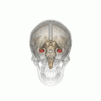
The hippocampus is a major component of the brain of humans and other vertebrates. Humans and other mammals have two hippocampi, one in each side of the brain. The hippocampus is part of the limbic system, and plays important roles in the consolidation of information from short-term memory to long-term memory, and in spatial memory that enables navigation. The hippocampus is located in the allocortex, with neural projections into the neocortex, in humans as well as other primates. The hippocampus, as the medial pallium, is a structure found in all vertebrates. In humans, it contains two main interlocking parts: the hippocampus proper, and the dentate gyrus.

<span class="mw-page-title-main">Olfactory bulb</span> Neural structure

The olfactory bulb is a neural structure of the vertebrate forebrain involved in olfaction, the sense of smell. It sends olfactory information to be further processed in the amygdala, the orbitofrontal cortex (OFC) and the hippocampus where it plays a role in emotion, memory and learning. The bulb is divided into two distinct structures: the main olfactory bulb and the accessory olfactory bulb. The main olfactory bulb connects to the amygdala via the piriform cortex of the primary olfactory cortex and directly projects from the main olfactory bulb to specific amygdala areas. The accessory olfactory bulb resides on the dorsal-posterior region of the main olfactory bulb and forms a parallel pathway. Destruction of the olfactory bulb results in ipsilateral anosmia, while irritative lesions of the uncus can result in olfactory and gustatory hallucinations.

<span class="mw-page-title-main">Adult neurogenesis</span> Generating of neurons from neural stem cells in adults

Adult neurogenesis is the process in which neurons are generated from neural stem cells in the adult. This process differs from prenatal neurogenesis.

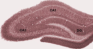
Schaffer collaterals are axon collaterals given off by CA3 pyramidal cells in the hippocampus. These collaterals project to area CA1 of the hippocampus and are an integral part of memory formation and the emotional network of the Papez circuit, and of the hippocampal trisynaptic loop. It is one of the most studied synapses in the world and named after the Hungarian anatomist-neurologist Károly Schaffer.

<span class="mw-page-title-main">Perforant path</span>

In the brain, the perforant path or perforant pathway provides a connectional route from the entorhinal cortex to all fields of the hippocampal formation, including the dentate gyrus, all CA fields, and the subiculum.

Elizabeth Gould is an American neuroscientist and the Dorman T. Warren Professor of Psychology at Princeton University. She was an early investigator of adult neurogenesis in the hippocampus, a research area that continues to be controversial. In November 2002, Discover magazine listed her as one of the 50 most important women scientists.

The stratum lucidum of the hippocampus is a layer of the hippocampus between the stratum pyramidale and the stratum radiatum. It is the tract of the mossy fiber projections, both inhibitory and excitatory from the granule cells of the dentate gyrus. One mossy fiber may make up to 37 connections to a single pyramidal cell, and innervate around 12 pyramidal cells on top of that. Any given pyramidal cell in the stratum lucidum may get input from as many as 50 granule cells.

<span class="mw-page-title-main">Mossy fiber (hippocampus)</span> Pathway in the hippocampus

In the hippocampus, the mossy fiber pathway consists of unmyelinated axons projecting from granule cells in the dentate gyrus that terminate on modulatory hilar mossy cells and in Cornu Ammonis area 3 (CA3), a region involved in encoding short-term memory. These axons were first described as mossy fibers by Santiago Ramón y Cajal as they displayed varicosities along their lengths that gave them a mossy appearance. The axons that make up the pathway emerge from the basal portions of the granule cells and pass through the hilus of the dentate gyrus before entering the stratum lucidum of CA3. Granule cell synapses tend to be glutamatergic, though immunohistological data has indicated that some synapses contain neuropeptidergic elements including opiate peptides such as dynorphin and enkephalin. There is also evidence for co-localization of both GABAergic and glutamatergic neurotransmitters within mossy fiber terminals. GABAergic and glutamatergic co-localization in mossy fiber boutons has been observed primarily in the developing hippocampus, but in adulthood, evidence suggests that mossy fiber synapses may alternate which neurotransmitter is released through activity-dependent regulation.

<span class="mw-page-title-main">Subgranular zone</span>

The subgranular zone (SGZ) is a brain region in the hippocampus where adult neurogenesis occurs. The other major site of adult neurogenesis is the subventricular zone (SVZ) in the brain.

The trisynaptic circuit, or trisynaptic loop is a relay of synaptic transmission in the hippocampus. The circuit was initially described by the neuroanatomist Santiago Ramon y Cajal, in the early twentieth century, using the Golgi staining method. After the discovery of the trisynaptic circuit, a series of research has been conducted to determine the mechanisms driving this circuit. Today, research is focused on how this loop interacts with other parts of the brain, and how it influences human physiology and behaviour. For example, it has been shown that disruptions within the trisynaptic circuit lead to behavioural changes in rodent and feline models.

<span class="mw-page-title-main">Hippocampus anatomy</span>

Hippocampus anatomy describes the physical aspects and properties of the hippocampus, a neural structure in the medial temporal lobe of the brain. It has a distinctive, curved shape that has been likened to the sea-horse monster of Greek mythology and the ram's horns of Amun in Egyptian mythology. This general layout holds across the full range of mammalian species, from hedgehog to human, although the details vary. For example, in the rat, the two hippocampi look similar to a pair of bananas, joined at the stems. In primate brains, including humans, the portion of the hippocampus near the base of the temporal lobe is much broader than the part at the top. Due to the three-dimensional curvature of this structure, two-dimensional sections such as shown are commonly seen. Neuroimaging pictures can show a number of different shapes, depending on the angle and location of the cut.

<span class="mw-page-title-main">Fascia dentata</span>

The fascia dentata is the earliest stage of the hippocampal circuit. Its primary input is the perforant path from the superficial layers of entorhinal cortex. Its principal neurons are tiny granule cells which give rise to unmyelinated axons called the mossy fibers which project to the hilus and CA3. The fascia dentata of the rat contains approximately 1,000,000 granule cells. It receives feedback connections from mossy cells in the hilus at distant levels in the septal and temporal directions. The fascia dentata and the hilus together make up the dentate gyrus. As with all regions of the hippocampus, the dentate gyrus also receives GABAergic and cholinergic input from the medial septum and the diagonal band of Broca.

<span class="mw-page-title-main">Granule cell</span> Type of neuron with a very small cell body

The name granule cell has been used for a number of different types of neurons whose only common feature is that they all have very small cell bodies. Granule cells are found within the granular layer of the cerebellum, the dentate gyrus of the hippocampus, the superficial layer of the dorsal cochlear nucleus, the olfactory bulb, and the cerebral cortex.

Endogenous regeneration in the brain is the ability of cells to engage in the repair and regeneration process. While the brain has a limited capacity for regeneration, endogenous neural stem cells, as well as numerous pro-regenerative molecules, can participate in replacing and repairing damaged or diseased neurons and glial cells. Another benefit that can be achieved by using endogenous regeneration could be avoiding an immune response from the host.

<span class="mw-page-title-main">Glucocorticoids in hippocampal development</span> HippoCampus

The hippocampus is an area of the brain integral to learning and memory. Removal of this structure can result in the inability to form new memories as most famously demonstrated in a patient referred to as HM. The unique morphology of the hippocampus can be appreciated without the use of special stains and this distinct circuitry has helped further the understanding of neuronal signal potentiation. The following will provide an introduction to hippocampal development with particular focus on the role of glucocorticoid signaling.

Neurogenesis is the process by which nervous system cells, the neurons, are produced by neural stem cells (NSCs). In short, it is brain growth in relation to its organization. This occurs in all species of animals except the porifera (sponges) and placozoans. Types of NSCs include neuroepithelial cells (NECs), radial glial cells (RGCs), basal progenitors (BPs), intermediate neuronal precursors (INPs), subventricular zone astrocytes, and subgranular zone radial astrocytes, among others.

Attila Losonczy is a Hungarian neuroscientist, Professor of Neuroscience at Columbia University Medical Center. Losonczy's main area of research is on the relationship between neural networks and behavior, specifically with regard to learning in the hippocampus.

<span class="mw-page-title-main">Hippocampus proper</span> Part of the brain of mammals

The hippocampus proper refers to the actual structure of the hippocampus which is made up of three regions or subfields. The subfields CA1, CA2, and CA3 use the initials of cornu Ammonis, an earlier name of the hippocampus.

The supramammillary nucleus (SuM), or supramammillary area, is a thin layer of cells in the brain that lies above the mammillary bodies. It can be considered part of the hypothalamus and diencephalon. The nucleus can be divided into medial and lateral sections. The medial SuM, or SuMM, is made of smaller cells which release dopamine and give input to the lateral septal nucleus. The lateral SuM, or SuML, is made of larger cells that project to the hippocampus.

<span class="mw-page-title-main">Neurogenesis hypothesis of depression</span> Theory of depression

Adult neurogenesis is the process by which functional, mature neurons are produced from neural stem cells (NSCs) in the adult brain. In most mammals, including humans, it only occurs in the subgranular zone of the hippocampus, and in the olfactory bulb. The neurogenesis hypothesis of depression proposes that major depressive disorder is caused, at least partly, by impaired neurogenesis in the subgranular zone of the hippocampus.

References

  1. 1 2 Amaral, David G.; Scharfman, Helen E.; Lavenex, Pierre (2007). "The dentate gyrus: Fundamental neuroanatomical organization (Dentate gyrus for dummies)". The Dentate Gyrus: A Comprehensive Guide to Structure, Function, and Clinical Implications. Progress in Brain Research. Vol. 163. pp. 3–790. doi:10.1016/S0079-6123(07)63001-5. ISBN   9780444530158. PMC   2492885 . PMID   17765709.
  2. 1 2 Saab BJ, Georgiou J, Nath A, Lee FJ, Wang M, Michalon A, Liu F, Mansuy IM, Roder JC (2009). "NCS-1 in the dentate gyrus promotes exploration, synaptic plasticity, and rapid acquisition of spatial memory". Neuron. 63 (5): 643–56. doi: 10.1016/j.neuron.2009.08.014 . PMID   19755107. S2CID   5321020.
  3. Scharfman, Helen E., ed. (2011). The Dentate Gyrus: A Comprehensive Guide to Structure, Function, and Clinical Implications. Elsevier. ISBN   978-0-08-055175-3.[ page needed ]
  4. Cameron HA, McKay RD (July 2001). "Adult neurogenesis produces a large pool of new granule cells in the dentate gyrus". J. Comp. Neurol. 435 (4): 406–17. doi:10.1002/cne.1040. PMID   11406822. S2CID   15254735.
  5. Ernst, A; Alkass, K; Bernard, S; Salehpour, M; Perl, S; Tisdale, J; Possnert, G; Druid, H; Frisén, J (27 February 2014). "Neurogenesis in the striatum of the adult human brain". Cell. 156 (5): 1072–83. doi: 10.1016/j.cell.2014.01.044 . PMID   24561062.
  6. Ponti G, Peretto P, Bonfanti L (2008). "Genesis of neuronal and glial progenitors in the cerebellar cortex of peripuberal and adult rabbits". PLOS ONE. 3 (6): e2366. Bibcode:2008PLoSO...3.2366P. doi: 10.1371/journal.pone.0002366 . PMC   2396292 . PMID   18523645.
  7. Sorrells SF, Paredes MF, Cebrian-Silla A, Sandoval K, Qi D, Kelley KW, et al. (March 2018). "Human hippocampal neurogenesis drops sharply in children to undetectable levels in adults". Nature. 555 (7696): 377–381. Bibcode:2018Natur.555..377S. doi:10.1038/nature25975. PMC   6179355 . PMID   29513649.
  8. Boldrini M, Fulmore CA, Tartt AN, Simeon LR, Pavlova I, Poposka V, et al. (April 2018). "Human Hippocampal Neurogenesis Persists throughout Aging". Cell Stem Cell. 22 (4): 589–599.e5. doi:10.1016/j.stem.2018.03.015. PMC   5957089 . PMID   29625071.
  9. Abbott, Louise C.; Nigussie, Fikru (January 2020). "Adult neurogenesis in the mammalian dentate gyrus". Anatomia, Histologia, Embryologia. 49 (1): 3–16. doi:10.1111/ahe.12496. PMID   31568602. S2CID   203622372.
  10. 1 2 3 4 5 Tuncdemir, Sebnem Nur; Lacefield, Clay Orion; Hen, Rene (November 2019). "Contributions of adult neurogenesis to dentate gyrus network activity and computations". Behavioural Brain Research. 374: 112112. doi:10.1016/j.bbr.2019.112112. PMC   6724741 . PMID   31377252.
  11. Zhou, Yi; Su, Yijing; Li, Shiying; Kennedy, Benjamin C.; Zhang, Daniel Y.; Bond, Allison M.; Sun, Yusha; Jacob, Fadi; Lu, Lu; Hu, Peng; Viaene, Angela N.; Helbig, Ingo; Kessler, Sudha K.; Lucas, Timothy; Salinas, Ryan D. (July 2022). "Molecular landscapes of human hippocampal immature neurons across lifespan". Nature. 607 (7919): 527–533. doi:10.1038/s41586-022-04912-w. ISSN   1476-4687. PMC   9316413 . PMID   35794479.
  12. Treves, A.; Tashiro, A.; Witter, M.P.; Moser, E.I. (July 2008). "What is the mammalian dentate gyrus good for?". Neuroscience. 154 (4): 1155–1172. doi:10.1016/j.neuroscience.2008.04.073. PMID   18554812. S2CID   14710031.
  13. 1 2 Scharfman, Helen E. (September 2016). "The enigmatic mossy cell of the dentate gyrus". Nature Reviews Neuroscience. 17 (9): 562–575. doi:10.1038/nrn.2016.87. PMC   5369357 . PMID   27466143.
  14. Haines, D; Mihailoff, G (2018). Fundamental neuroscience for basic and clinical applications (Fifth ed.). Elsevier. p. 461. ISBN   9780323396325.
  15. Nadler, J. Victor (2003). "The recurrent mossy fiber pathway of the epileptic brain". Neurochemical Research. 28 (11): 1649–1658. doi:10.1023/a:1026004904199. PMID   14584819. S2CID   2566342.
  16. Seress, László; Mrzljak, Ladislav (March 1987). "Basal dendrites of granule cells are normal features of the fetal and adult dentate gyrus of both monkey and human hippocampal formations". Brain Research. 405 (1): 169–174. doi:10.1016/0006-8993(87)91003-1. PMID   3567591. S2CID   23358962.
  17. Amaral DG, Witter MP (1989). "The three-dimensional organization of the hippocampal formation: a review of anatomical data". Neuroscience. 31 (3): 571–591. doi:10.1016/0306-4522(89)90424-7. PMID   2687721. S2CID   28430607.
  18. Legéndy CR (2017). "On the 'data stirring' role of the dentate gyrus of the hippocampus". Reviews in the Neurosciences. 28 (6): 599–615. doi:10.1515/revneuro-2016-0080. PMID   28593904. S2CID   3716652.
  19. Elgendy, Azza. "Band of Giacomini | Radiology Reference Article | Radiopaedia.org". Radiopaedia. Retrieved 17 October 2019.
  20. 1 2 Blumenfeld, Hal (2010). Neuroanatomy through clinical cases (2nd ed.). Sunderland, Mass.: Sinauer Associates. ISBN   978-0878936137.
  21. 1 2 3 Senzai, Yuta (March 2019). "Function of local circuits in the hippocampal dentate gyrus-CA3 system". Neuroscience Research. 140: 43–52. doi:10.1016/j.neures.2018.11.003. PMID   30408501. S2CID   53220907.
  22. Nolte, John (2002). The Human Brain: An Introduction to Its Functional Neuroanatomy (fifth ed.). pp. 570–573.
  23. Rachel A. Dalley; Lydia L. Ng; Angela L. Guillozet-Bongaarts (2008). "Dentate Gyrus". Nature Precedings. doi: 10.1038/npre.2008.2095.1 .
  24. Bayer SA, Altman J (November 1974). "Hippocampal development in the rat: cytogenesis and morphogenesis examined with autoradiography and low-level X-irradiation". J. Comp. Neurol. 158 (1): 55–79. doi:10.1002/cne.901580105. PMID   4430737. S2CID   17968282.
  25. Bayer SA, Altman J (2008). The Human Brain During The Early First Trimester. Vol. 5 Atlas of Human Central Nervous System Development. Appendix, p. 497.
  26. Eriksson PS, Perfilieva E, Björk-Eriksson T, et al. (November 1998). "Neurogenesis in the adult human hippocampus". Nat. Med. 4 (11): 1313–7. doi: 10.1038/3305 . PMID   9809557.
  27. Altman J, Bayer SA (November 1990). "Migration and distribution of two populations of hippocampal granule cell precursors during the perinatal and postnatal periods". J. Comp. Neurol. 301 (3): 365–81. doi:10.1002/cne.903010304. PMID   2262596. S2CID   7425653.
  28. Angevine JB (October 1965). "Time of neuron origin in the hippocampal region. An autoradiographic study in the mouse". Exp Neurol Suppl (Suppl 2): Suppl 2:1–70. PMID   5838955.
  29. Bayer SA, Yackel JW, Puri PS (May 1982). "Neurons in the rat dentate gyrus granular layer substantially increase during juvenile and adult life". Science. 216 (4548): 890–2. Bibcode:1982Sci...216..890B. doi:10.1126/science.7079742. PMID   7079742.
  30. Bayer SA (1982). "Changes in the total number of dentate granule cells in juvenile and adult rats: a correlated volumetric and 3H-thymidine autoradiographic study". Exp Brain Res. 46 (3): 315–23. doi:10.1007/bf00238626. PMID   7095040. S2CID   18663323.
  31. Oomen, Charlotte A.; Girardi, Carlos E. N.; Cahyadi, Rudy; Verbeek, Eva C.; Krugers, Harm; Joëls, Marian; Lucassen, Paul J. (29 January 2009). "Opposite Effects of Early Maternal Deprivation on Neurogenesis in Male versus Female Rats". PLOS ONE. 4 (1): e3675. Bibcode:2009PLoSO...4.3675O. doi: 10.1371/journal.pone.0003675 . PMC   2629844 . PMID   19180242.
  32. Faiz M, Acarin L, Castellano B, Gonzalez B (2005). "Proliferation dynamics of germinative zone cells in the intact and excitotoxically lesioned postnatal rat brain". BMC Neurosci . 6 (1): 26. doi: 10.1186/1471-2202-6-26 . PMC   1087489 . PMID   15826306.
  33. Benedict Carey (4 December 2008). "H. M., an Unforgettable Amnesiac, Dies at 82". The New York Times . Retrieved 5 December 2008. In 1953, he underwent an experimental brain operation in Hartford to correct a seizure disorder, only to emerge from it fundamentally and irreparably changed. He developed a syndrome neurologists call profound amnesia. He had lost the ability to form new declarative memories.
  34. 1 2 3 Kandel ER, Schwartz J, Jessell T, Siegelbaum S, Hudspeth AJ (2013). Principles of neural science (5th ed.). McGraw Hill Professional. ISBN   978-0-07-139011-8.
  35. Nakashiba T, Cushman JD, Pelkey KA, Renaudineau S, Buhl DL, McHugh TJ, et al. (March 2012). "Young dentate granule cells mediate pattern separation, whereas old granule cells facilitate pattern completion". Cell. 149 (1): 188–201. doi:10.1016/j.cell.2012.01.046. PMC   3319279 . PMID   22365813.
  36. Kovács KA (September 2020). "Episodic Memories: How do the Hippocampus and the Entorhinal Ring Attractors Cooperate to Create Them?". Frontiers in Systems Neuroscience. 14: 68. doi: 10.3389/fnsys.2020.559186 . PMC   7511719 . PMID   33013334.
  37. Bliss RM (August 2007). ""Food and the Aging Mind". First in a Series: Nutrition and Brain Function". USDA. USDA.gov. Retrieved 27 February 2010.
  38. 1 2 Jonas, Peter; Lisman, John (10 September 2014). "Structure, function, and plasticity of hippocampal dentate gyrus microcircuits". Frontiers in Neural Circuits. 8: 107. doi: 10.3389/fncir.2014.00107 . PMC   4159971 . PMID   25309334.
  39. Lamothe-Molina, Paul J.; Franzelin, Andreas; Auksutat, Lea; Laprell, Laura; Alhbeck, Joachim; Kneussel, Matthias; Engel, Andreas K.; Morellini, Fabio; Oertner, Thomas G. (31 August 2020). "cFos ensembles in the dentate gyrus rapidly segregate over time and do not form a stable map of space" (PDF). doi:10.1101/2020.08.29.273391. S2CID   221510080.{{cite journal}}: Cite journal requires |journal= (help)
  40. 1 2 Moser, Edvard I.; Kropff, Emilio; Moser, May-Britt (1 July 2008). "Place Cells, Grid Cells, and the Brain's Spatial Representation System". Annual Review of Neuroscience. 31 (1): 69–89. doi:10.1146/annurev.neuro.31.061307.090723. PMID   18284371.
  41. Rolls, Edmund T. (2013). "The mechanisms for pattern completion and pattern separation in the hippocampus". Frontiers in Systems Neuroscience. 7: 74. doi: 10.3389/fnsys.2013.00074 . PMC   3812781 . PMID   24198767.
  42. Malberg JE, Eisch AJ, Nestler EJ, Duman RS (December 2000). "Chronic antidepressant treatment increases neurogenesis in adult rat hippocampus". J. Neurosci. 20 (24): 9104–10. doi:10.1523/JNEUROSCI.20-24-09104.2000. PMC   6773038 . PMID   11124987.
  43. Gould E, Tanapat P, McEwen BS, Flügge G, Fuchs E (March 1998). "Proliferation of granule cell precursors in the dentate gyrus of adult monkeys is diminished by stress". Proc. Natl. Acad. Sci. U.S.A. 95 (6): 3168–71. Bibcode:1998PNAS...95.3168G. doi: 10.1073/pnas.95.6.3168 . PMC   19713 . PMID   9501234.
  44. Jacobs BL, van Praag H, Gage FH (May 2000). "Adult brain neurogenesis and psychiatry: a novel theory of depression". Mol. Psychiatry. 5 (3): 262–9. doi: 10.1038/sj.mp.4000712 . PMID   10889528.
  45. Surget A, Tanti A, Leonardo ED, Laugeray A, Rainer Q, Touma C, Palme R, Griebel G, Ibarguen-Vargas Y, Hen R, Belzung C (December 2011). "Antidepressants recruit new neurons to improve stress response regulation". Mol. Psychiatry. 16 (12): 1177–88. doi:10.1038/mp.2011.48. PMC   3223314 . PMID   21537331.
  46. "Blood Sugar Control Linked to Memory Decline, Study Says". The New York Times. 1 January 2009. Retrieved 13 March 2011.
  47. Praag, H (1999). "Running increases cell proliferation and neurogenesis in the adult mouse dentate gyrus". Nature Neuroscience. 2 (3): 266–270. doi:10.1038/6368. PMID   10195220. S2CID   7170664.
  48. Kempermann G, Kuhn HG, Gage FH (April 1997). "More hippocampal neurons in adult mice living in an enriched environment". Nature. 386 (6624): 493–5. Bibcode:1997Natur.386..493K. doi:10.1038/386493a0. PMID   9087407. S2CID   4281128.
  49. Eadie BD, Redila VA, Christie BR (May 2005). "Voluntary exercise alters the cytoarchitecture of the adult dentate gyrus by increasing cellular proliferation, dendritic complexity, and spine density". J. Comp. Neurol. 486 (1): 39–47. doi:10.1002/cne.20493. hdl: 2429/15467 . PMID   15834963. S2CID   8386870.
  50. Xavier GF, Costa VC (August 2009). "Dentate gyrus and spatial behaviour". Prog. Neuropsychopharmacol. Biol. Psychiatry. 33 (5): 762–73. doi: 10.1016/j.pnpbp.2009.03.036 . PMID   19375476. S2CID   1115081.