The abducens nerve is the sixth cranial nerve (CNVI), in humans, that controls the movement of the lateral rectus muscle, responsible for outward gaze. It is a somatic efferent nerve.

The oculomotor nerve is the third cranial nerve. It enters the orbit via the superior orbital fissure and innervates extrinsic eye muscles that enable most movements of the eye and that raise the eyelid. The nerve also contains fibers that innervate the intrinsic eye muscles that enable pupillary constriction and accommodation. The oculomotor nerve is derived from the basal plate of the embryonic midbrain. Cranial nerves IV and VI also participate in control of eye movement.

The omohyoid muscle is a muscle that depresses the hyoid. It is located in the front of the neck and consists of two bellies separated by an intermediate tendon. Its superior belly serves as the most lateral member of the infrahyoid muscles, located lateral to both the sternothyroid and thyrohyoid muscles. Its name derives from the Greek "omos" meaning shoulder, giving one of its attachments, and "hyoid", giving the other attachment – the hyoid bone.

In anatomy, the olivary bodies or simply olives are a pair of prominent oval structures in the medulla oblongata, the lower portion of the brainstem. They contain the olivary nuclei.

The spinocerebellar tract is a nerve tract originating in the spinal cord and terminating in the same side (ipsilateral) of the cerebellum.

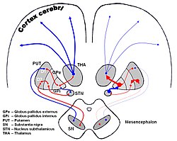
The subthalamus or prethalamus is a part of the diencephalon. Its most prominent structure is the subthalamic nucleus. The subthalamus connects to the globus pallidus, a basal nucleus of the telencephalon.

The trigeminal ganglion is a sensory ganglion of the trigeminal nerve that occupies a cavity in the dura mater, covering the trigeminal impression near the apex of the petrous part of the temporal bone.

The apex of the posterior grey column, one of the three grey columns of the spinal cord, is capped by a V-shaped or crescentic mass of translucent, gelatinous neuroglia, termed the substantia gelatinosa of Rolando, which contains both neuroglia cells, and small nerve cells. The gelatinous appearance is due to a very low concentration of myelinated fibers. It extends the entire length of the spinal cord and into the medulla oblongata where it becomes the spinal nucleus of the trigeminal nerve.

The posterior commissure is a rounded band of white fibers crossing the middle line on the dorsal aspect of the rostral end of the cerebral aqueduct. It is important in the bilateral pupillary light reflex.

The substantia innominata also innominate substance, or substantia innominata of Meynert is a series of layers in the human brain consisting partly of gray and partly of white matter, which lies below the anterior part of the thalamus and lentiform nucleus. It is included as part of the anterior perforated substance. It is part of the basal forebrain structures and includes the nucleus basalis. A portion of the substantia innominata, below the globus pallidus is considered as part of the extended amygdala.

The anterior external arcuate fibers vary as to their prominence: in some cases they form an almost continuous layer covering the medullary pyramids and olivary body, while in other cases they are barely visible on the surface.
The mammillothalamic tract arises from cells in both the medial and lateral nuclei of the mammillary body and by fibers that are directly continued from the fornix.

The posterolateral tract is a small strand situated in relation to the tip of the posterior column close to the entrance of the posterior nerve roots. It is present throughout the spinal cord, and is most developed in the upper cervical regions.

The olivospinal fasciculus (Helweg) was thought to arise in the vicinity of the inferior olivary nucleus in the medulla oblongata, and was thought to be seen only in the cervical region of the medulla spinalis, where it forms a small triangular area at the periphery, close to the most lateral of the anterior nerve roots. Its existence is now strongly doubted.
The paramedian reticular nucleus sends its connections to the spinal cord in a mostly ipsilateral manner, although there is some decussation.

The thalamic fasciculus is a component of the subthalamus. It is synonymous with field H1 of Forel. Nerve fibres form a tract containing cerebellothalamic (crossed) and pallidothalamic (uncrossed) fibres, that is insinuated between the thalamus and the zona incerta.
The anterolateral central arteries are a group of small arteries arising from the anterior part of the circle of Willis and supply the basal ganglia. They arise at the commencement of the middle cerebral artery and are arranged in two sets:
- Internal striate: passes upward through the inner segments of the lentiform nucleus and supplies the lentiform nucleus, caudate nucleus, and internal capsule;
- External striate: ascends through the outer segment of the lentiform nucleus and supplies the caudate nucleus.

The salivatory nuclei are the superior salivatory nucleus, and the inferior salivatory nucleus that innervate the salivary glands. They are located in the pontine tegmentum in the brainstem. They both are examples of cranial nerve nuclei.
The internal globus pallidus and the external globus pallidus (GPe) make up the globus pallidus. The GPi is one of the output nuclei of the basal ganglia. The GABAergic neurons send their axons to the ventral anterior nucleus (VA) and the ventral lateral nucleus (VL) in the dorsal thalamus, to the centromedian complex, and to the pedunculopontine complex.
The fields of Forel are areas in a deep part of the brain known as the diencephalon. They are below the thalamus and consist of three defined, white matter areas of the subthalamus. These three regions are also named "H fields":