Facial muscles

Last updated
Facial muscles
Gray378.png
Head
Lateral head anatomy.jpg
Lateral head anatomy
Details
Nerve facial nerve
Identifiers
Latin musculi faciei
MeSH D005152
TA98 A04.1.03.001
TA2 2060
FMA 71288
Anatomical terms of muscle

The facial muscles are a group of striated skeletal muscles supplied by the facial nerve (cranial nerve VII) that, among other things, control facial expression. These muscles are also called mimetic muscles. They are only found in mammals, although they derive from neural crest cells found in all vertebrates. They are the only muscles that attach to the dermis. [1]

Contents

Structure

The facial muscles are just under the skin (subcutaneous) muscles that control facial expression. They generally originate from the surface of the skull bone (rarely the fascia), and insert on the skin of the face. When they contract, the skin moves. These muscles also cause wrinkles at right angles to the muscles’ action line. [2]

Nerve supply

The facial muscles are supplied by the facial nerve (cranial nerve VII), with each nerve serving one side of the face. [2] In contrast, the nearby masticatory muscles are supplied by the mandibular nerve, a branch of the trigeminal nerve (cranial nerve V).

List of muscles

The facial muscles include: [3]

The platysma is supplied by the facial nerve. Although it is mostly in the neck and can be grouped with the neck muscles by location, it can be considered a muscle of facial expression due to its common nerve supply.

The stylohyoid muscle, stapedius and posterior belly of the digastric muscle are also supplied by the facial nerve, but are not considered muscles of facial expression.

Facial muscles by type of movement

Movement [4] TargetTarget motion or directionPrime moverOriginInsertion
Raising eyebrows (e.g., showing surprise)Skin of scalpAnterior Occipitofrontalis frontal bellyEpicraneal aponeurosisUnderneath skin of forehead
Tensing and retracting scalpSkin of scalpPosterior Occipitofrontalis, occipital bellyOccipital bone; mastoid process (temporal bone)Epicraneal aponeurosis
Lowering eyebrows (e.g., scowling, frowning)Skin underneath eyebrowsInferior Corrugator supercilii Frontal boneSkin underneath eyebrow
Flaring nostrils Nasal cartilage (pushes nostrils open when cartilage is compressed)Inferior compression; posterior compressionNasalis Maxilla Nasal bone
Raising upper lipUpper lipElevation Levator labii superioris Maxilla Underneath skin at corners of the mouth; orbicularis oris
Lowering lower lipLower lipDepression Depressor labii inferioris Mandible Underneath skin of lower lip
Opening mouth and sliding lower jaw left and rightLower jawDepression, lateral Depressor angulus oris Mandible Underneath skin at corners of mouth
SmilingCorners of mouthLateral elevation Zygomaticus major Zygomatic bone Underneath skin at corners of mouth (dimple area), orbicularis oris
Shaping of lips (as during speech)LipsMultiple Orbicularis oris Tissue surrounding lipsUnderneath skin at corners of the mouth
Lateral movement of cheeks (e.g., sucking on a straw; also used to compress air in mouth while blowing)CheeksLateral Buccinator Maxilla, mandible; sphenoid bone (via pterygomandibular raphae) Orbicularis oris
Pursing of lips by straightening them laterallyCorners of mouthLateral Risorius Fascia of parotid salivary glandUnderneath the skin at corners of the mouth
Protrusion of lower lip (e.g., pouting expression)Lower lip and skin of chinProtraction Mentalis Mandible Underneath skin of chin

Development

The facial muscles are derived from the second branchial/pharyngeal arch. They, like the branchial arches, originally derive from neural crest cells. In humans, they typically begin forming around the eighth week of embryonic development. [1]

Clinical significance

An inability to form facial expressions on one side of the face may be the first sign of damage to the nerve of these muscles. Damage to the facial nerve results in facial paralysis of the muscles of facial expression on the involved side. Paralysis is the loss of voluntary muscle action; the facial nerve has become damaged permanently or temporarily. This damage can occur with a stroke, Bell palsy, or parotid salivary gland cancer (malignant neoplasm) because the facial nerve travels through the gland. The parotid gland can also be damaged permanently by surgery or temporarily by trauma. These situations of paralysis not only inhibit facial expression but also seriously impair the patient’s ability to speak, either permanently or temporarily. [2]

See also

Related Research Articles

<span class="mw-page-title-main">Facial nerve</span> Cranial nerve VII, for the face and tasting

The facial nerve, also known as the seventh cranial nerve, cranial nerve VII, or simply CN VII, is a cranial nerve that emerges from the pons of the brainstem, controls the muscles of facial expression, and functions in the conveyance of taste sensations from the anterior two-thirds of the tongue. The nerve typically travels from the pons through the facial canal in the temporal bone and exits the skull at the stylomastoid foramen. It arises from the brainstem from an area posterior to the cranial nerve VI and anterior to cranial nerve VIII.

Articles related to anatomy include:

<span class="mw-page-title-main">Orbicularis oris muscle</span> Kissing muscles

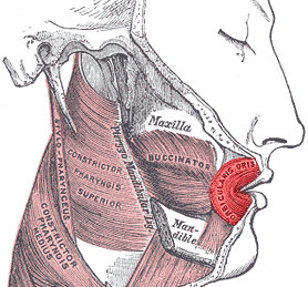
In human anatomy, the orbicularis oris muscle is a complex of muscles in the lips that encircles the mouth. It is not a true sphincter, as was once thought, as it is actually composed of four independent quadrants that interlace and give only an appearance of circularity.

<span class="mw-page-title-main">Lip</span> Visible body parts at the mouth

The lips are a horizontal pair of soft appendages attached to the jaws and are the most visible part of the mouth of many animals, including humans. Vertebrate lips are soft, movable and serve to facilitate the ingestion of food and the articulation of sound and speech. Human lips are also a somatosensory organ, and can be an erogenous zone when used in kissing and other acts of intimacy.

<span class="mw-page-title-main">Platysma muscle</span> Human neck muscle

The platysma muscle is a superficial muscle of the human neck that overlaps the sternocleidomastoid. It covers the anterior surface of the neck superficially. When it contracts, it produces a slight wrinkling of the neck, and a "bowstring" effect on either side of the neck.

<span class="mw-page-title-main">Depressor labii inferioris muscle</span> Facial muscle that helps to lower the bottom lip

The depressor labii inferioris is a facial muscle. It helps to lower the bottom lip.

<span class="mw-page-title-main">Zygomaticus major muscle</span> Facial muscle that raises the corner of the mouth and moves it posteriorly during smiling

The zygomaticus major muscle is a muscle of the face. It arises from either zygomatic arch (cheekbone); it inserts at the corner of the mouth. It is innervated by branches of the facial nerve.

<span class="mw-page-title-main">Zygomaticus minor muscle</span> Facial muscle that draws the upper lip upwards and backwards during smiling

The zygomaticus minor muscle is a muscle of facial expression. It originates from the zygomatic bone, lateral to the rest of the levator labii superioris muscle, and inserts into the outer part of the upper lip. It draws the upper lip backward, upward, and outward and is used in smiling. It is innervated by the facial nerve (VII).

<span class="mw-page-title-main">Levator labii superioris</span> Muscle of the face

The levator labii superioris is a muscle of the human body used in facial expression. It is a broad sheet, the origin of which extends from the side of the nose to the zygomatic bone.

<span class="mw-page-title-main">Levator anguli oris</span> Muscle of the mouth

The levator anguli oris (caninus) is a facial muscle of the mouth arising from the canine fossa, immediately below the infraorbital foramen. It elevates angle of mouth medially. Its fibers are inserted into the angle of the mouth, intermingling with those of the zygomaticus, triangularis, and orbicularis oris. Specifically, the levator anguli oris is innervated by the buccal branches of the facial nerve.

<span class="mw-page-title-main">Depressor anguli oris muscle</span> Facial muscle that depresses the corner of the mouth during frowning

The depressor anguli oris muscle is a facial muscle. It originates from the mandible and inserts into the angle of the mouth. It is associated with frowning, as it depresses the corner of the mouth.

<span class="mw-page-title-main">Facial artery</span>

The facial artery is a branch of the external carotid artery that supplies structures of the superficial face.

<span class="mw-page-title-main">Modiolus (face)</span> Chiasma of facial muscles

In facial anatomy, the modiolus is a dense, compact, mobile, fibromuscular tissue mass of facial muscles formed by the interlacing of a number of muscles just lateral to the angle of the mouth opposite the second upper premolar tooth.

<span class="mw-page-title-main">Infraorbital artery</span>

The infraorbital artery is a small artery in the head that arises from the maxillary artery and passes through the inferior orbital fissure to enter the orbit, then passes forward along the floor of the orbit, finally exiting the orbit through the infraorbital foramen to reach the face.

<span class="mw-page-title-main">Marginal mandibular branch of the facial nerve</span>

The marginal mandibular branch of the facial nerve arises from the facial nerve in the parotid gland at the parotid plexus. It passes anterior-ward deep to the platysma and depressor anguli oris muscles. It provides motor innervation to muscles of the lower lip and chin: the depressor labii inferioris muscle, depressor anguli oris muscle, and mentalis muscle. It communicates with the mental branch of the inferior alveolar nerve.

<span class="mw-page-title-main">Buccal branches of the facial nerve</span>

The buccal branches of the facial nerve, are of larger size than the rest of the branches, pass horizontally forward to be distributed below the orbit and around the mouth.

<span class="mw-page-title-main">Buccal fat pad</span>

The buccal fat pad is one of several encapsulated fat masses in the cheek. It is a deep fat pad located on either side of the face between the buccinator muscle and several more superficial muscles. The inferior portion of the buccal fat pad is contained within the buccal space. It should not be confused with the malar fat pad, which is directly below the skin of the cheek. It should also not be confused with jowl fat pads. It is implicated in the formation of hollow cheeks and the nasolabial fold, but not in the formation of jowls.

<span class="mw-page-title-main">Outline of human anatomy</span> Overview of and topical guide to human anatomy

The following outline is provided as an overview of and topical guide to human anatomy:

Levator muscle can refer to:

<span class="mw-page-title-main">Canine space</span>

The canine space, is a fascial space of the head and neck. It is a thin potential space on the face, and is paired on either side. It is located between the levator anguli oris muscle inferiorly and the levator labii superioris muscle superiorly. The term is derived from the fact that the space is in the region of the canine fossa, and that infections originating from the maxillary canine tooth may spread to involve the space. Infra-orbital is derived from infra- meaning below and orbit which refers to the eye socket.

References

  1. 1 2 Wilkins, Adam S. (2017). "History of the Face I". Making Faces. Belknap Press. p. 169. ISBN   9780674725522.
  2. 1 2 3 Illustrated Anatomy of the Head and Neck, Fehrenbach and Herring, Elsevier, 2012, page 89
  3. Kyung Won, PhD. Chung (2005). Gross Anatomy (Board Review). Hagerstown, MD: Lippincott Williams & Wilkins. p. 364. ISBN   0-7817-5309-0.
  4. Creative Commons by small.svg  This article incorporates text available under the CC BY 4.0 license.Betts, J Gordon; Desaix, Peter; Johnson, Eddie; Johnson, Jody E; Korol, Oksana; Kruse, Dean; Poe, Brandon; Wise, James; Womble, Mark D; Young, Kelly A (June 8, 2023). Anatomy & Physiology. Houston: OpenStax CNX. 11.3 Axial muscles of the head, neck and back. ISBN   978-1-947172-04-3.