Orbicularis oris muscle

Last updated
Orbicularis oris
Orbicularis oris.jpg
Details
Origin Maxilla and mandible
Insertion Skin around the lips
Artery Inferior labial artery and superior labial artery.
Nerve Cranial nerve VII, buccal branch
Actions It is sometimes known as the kissing muscle [1] because it is used to pucker the lips.
Identifiers
Latin musculus orbicularis oris
TA98 A04.1.03.023
TA2 2073
FMA 46841
Anatomical terms of muscle

In human anatomy, the orbicularis oris muscle is a complex of muscles in the lips that encircles the mouth. [2] It is not a true sphincter, as was once thought, as it is actually composed of four independent quadrants that interlace and give only an appearance of circularity. [3]

Contents

It is also one of the muscles used in the playing of all brass instruments and some woodwind instruments. This muscle closes the mouth and puckers the lips when it contracts.

Structure

The orbicularis oris is not a simple sphincter muscle like the orbicularis oculi; it consists of numerous strata of muscular fibers surrounding the orifice of the mouth, but having different direction. It consists partly of fibers derived from the other facial muscles which are inserted into the lips, and partly of fibers proper to the lips. Of the former, a considerable number are derived from the buccinator and form the deeper stratum of the orbicularis.

Some of the buccinator fibers—namely, those near the middle of the muscle—decussate at the angle of the mouth, those arising from the maxilla passing to the lower lip, and those from the mandible to the upper lip. The uppermost and lowermost fibers of the buccinator pass across the lips from side to side without decussation.

Superficial to this stratum is a second, formed on either side by the caninus and triangularis, which cross each other at the angle of the mouth; those from the caninus passing to the lower lip, and those from the triangularis to the upper lip, along which they run, to be inserted into the skin near the median line. In addition to these, fibers from the quadratus labii superioris, the zygomaticus, and the quadratus labii inferioris intermingle with the transverse fibers above described, and have principally an oblique direction. The proper fibers of the lips are oblique, and pass from the under surface of the skin to the mucous membrane, through the thickness of the lip.

Finally, fibers occur by which the muscle is connected with the maxilla and the septum of the nose above and with the mandible below. In the upper lip, these consist of two bands, lateral and medial, on either side of the middle line; the lateral band m. incisivus labii superioris arises from the alveolar border of the maxilla, opposite the lateral incisor tooth, and arching lateralward is continuous with the other muscles at the angle of the mouth; the medial band m. nasolabialis connects the upper lip to the back of the septum of the nose.

The interval between the two medial bands corresponds with the depression, called the philtrum, seen on the lip beneath the septum of the nose. The additional fibers for the lower lip constitute a slip m. incisivus labii inferioris on either side of the middle line; this arises from the mandible, lateral to the Mentalis, and intermingles with the other muscles at the angle of the mouth.

Clinical significance

Babies are occasionally born without one or both sides of this particular muscle, resulting in a slight droop to the affected side of the face.

Additional images

See also

Related Research Articles

Articles related to anatomy include:

<span class="mw-page-title-main">Buccinator muscle</span> Muscle

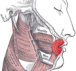
The buccinator is a thin quadrilateral muscle occupying the interval between the maxilla and the mandible at the side of the face. It forms the anterior part of the cheek or the lateral wall of the oral cavity.

<span class="mw-page-title-main">Lip</span> Visible body parts at the mouth

The lips are a horizontal pair of soft appendages attached to the jaws and are the most visible part of the mouth of many animals, including humans. Vertebrate lips are soft, movable and serve to facilitate the ingestion of food and the articulation of sound and speech. Human lips are also a somatosensory organ, and can be an erogenous zone when used in kissing and other acts of intimacy.

<span class="mw-page-title-main">Platysma muscle</span> Human neck muscle

The platysma muscle is a superficial muscle of the human neck that overlaps the sternocleidomastoid. It covers the anterior surface of the neck superficially. When it contracts, it produces a slight wrinkling of the neck, and a "bowstring" effect on either side of the neck.

<span class="mw-page-title-main">Depressor labii inferioris muscle</span> Facial muscle that helps to lower the bottom lip

The depressor labii inferioris is a facial muscle. It helps to lower the bottom lip.

<span class="mw-page-title-main">Orbicularis oculi muscle</span> Facial muscle that closes the eyelids

The orbicularis oculi is a muscle in the face that closes the eyelids. It arises from the nasal part of the frontal bone, from the frontal process of the maxilla in front of the lacrimal groove, and from the anterior surface and borders of a short fibrous band, the medial palpebral ligament.

<span class="mw-page-title-main">Levator labii superioris</span> Muscle of the face

The levator labii superioris is a muscle of the human body used in facial expression. It is a broad sheet, the origin of which extends from the side of the nose to the zygomatic bone.

<span class="mw-page-title-main">Levator anguli oris</span> Muscle of the mouth

The levator anguli oris (caninus) is a facial muscle of the mouth arising from the canine fossa, immediately below the infraorbital foramen. It elevates angle of mouth medially. Its fibers are inserted into the angle of the mouth, intermingling with those of the zygomaticus, triangularis, and orbicularis oris. Specifically, the levator anguli oris is innervated by the buccal branches of the facial nerve.

<span class="mw-page-title-main">Depressor anguli oris muscle</span> Facial muscle that depresses the corner of the mouth during frowning

The depressor anguli oris muscle is a facial muscle. It originates from the mandible and inserts into the angle of the mouth. It is associated with frowning, as it depresses the corner of the mouth.

<span class="mw-page-title-main">Facial artery</span> Blood vessel supplying blood to the superficial face

The facial artery is a branch of the external carotid artery that supplies structures of the superficial face.

<span class="mw-page-title-main">Modiolus (face)</span> Chiasma of facial muscles

In facial anatomy, the modiolus is a dense, compact, mobile, fibromuscular tissue mass of facial muscles formed by the interlacing of a number of muscles just lateral to the angle of the mouth opposite the second upper premolar tooth.

<span class="mw-page-title-main">Frontal process of maxilla</span> Plate which forms part of the lateral boundary of the nose

The frontal process of the maxilla is a strong plate, which projects upward, medialward, and backward from the maxilla, forming part of the lateral boundary of the nose.

<span class="mw-page-title-main">Angular artery</span> Artery of the face

The angular artery is an artery of the face. It is the terminal part of the facial artery. It ascends to the medial angle of the eye's orbit. It is accompanied by the angular vein. It ends by anastomosing with the dorsal nasal branch of the ophthalmic artery. It supplies the lacrimal sac, the orbicularis oculi muscle, and the outer side of the nose.

<span class="mw-page-title-main">Buccal branches of the facial nerve</span>

The buccal branches of the facial nerve, are of larger size than the rest of the branches, pass horizontally forward to be distributed below the orbit and around the mouth.

<span class="mw-page-title-main">Buccal space</span>

The buccal space is a fascial space of the head and neck. It is a potential space in the cheek, and is paired on each side. The buccal space is superficial to the buccinator muscle and deep to the platysma muscle and the skin. The buccal space is part of the subcutaneous space, which is continuous from head to toe.

<span class="mw-page-title-main">Facial muscles</span> Muscles of the face

The facial muscles are a group of striated skeletal muscles supplied by the facial nerve that, among other things, control facial expression. These muscles are also called mimetic muscles. They are only found in mammals, although they derive from neural crest cells found in all vertebrates. They are the only muscles that attach to the dermis.

<span class="mw-page-title-main">Human nose</span> Feature of the human face

The human nose is the first organ of the respiratory system. It is also the principal organ in the olfactory system. The shape of the nose is determined by the nasal bones and the nasal cartilages, including the nasal septum, which separates the nostrils and divides the nasal cavity into two.

<span class="mw-page-title-main">Outline of human anatomy</span> Overview of and topical guide to human anatomy

The following outline is provided as an overview of and topical guide to human anatomy:

<span class="mw-page-title-main">Canine space</span>

The canine space, is a fascial space of the head and neck. It is a thin potential space on the face, and is paired on either side. It is located between the levator anguli oris muscle inferiorly and the levator labii superioris muscle superiorly. The term is derived from the fact that the space is in the region of the canine fossa, and that infections originating from the maxillary canine tooth may spread to involve the space. Infra-orbital is derived from infra- meaning below and orbit which refers to the eye socket.

References

PD-icon.svgThis article incorporates text in the public domain from page 384 of the 20th edition of Gray's Anatomy (1918)

  1. "Muscles - Facial". BBC : Science & Nature : Human Body & Mind. Retrieved 2010-10-03.
  2. "orbicularis oris muscle". TheFreeDictionary : Mosby's Dental Dictionary, 2nd edition. 2008. Retrieved 2010-10-03.
  3. Saladin, "Anatomy & Physiology: The Unity of Form and Function". 5th edition. McGraw Hill. Page 330