Time-lapse microscopy

Last updated
Time-lapse microscope
Olympus FluoView FV1000 Confocal Microscope - NCMIR.jpg
A time-lapse microscope. The transparent cell incubator is necessary to keep cells alive during observation.
Other names(Time-lapse) microcinematograph, (Time-lapse) video microscope, Time-lapse cinemicrograph
UsesObservation of slow microscopic processes
InventorJean Comandon and other contemporaries
Related items Time-lapse photography, Live-cell imaging

Time-lapse microscopy is time-lapse photography applied to microscopy. Microscope image sequences are recorded and then viewed at a greater speed to give an accelerated view of the microscopic process.

Contents

Before the introduction of the video tape recorder in the 1960s, time-lapse microscopy recordings were made on photographic film. During this period, time-lapse microscopy was referred to as microcinematography. With the increasing use of video recorders, the term time-lapse video microscopy was gradually adopted. Today, the term video is increasingly dropped, reflecting that a digital still camera is used to record the individual image frames, instead of a video recorder.

Applications

TimeLapseMicroscopyCancerCells.gif
A time-lapse movie of dividing cancer cells, created by using a phase-contrast microscope.
Time-lapse video of dividing cells.gif
Cell division over 42 hours. The time-lapse movie was created by using a phase microscope.
Jean Comandon, pioneer of microcinematography, recorded this time-lapse film in c. 1910, using an ultramicroscope. The film show living spiral shaped syphilis bacteria moving among red blood cells of frog. Notice the back-and-forth movement, characterizing the disease-causing form.

Time-lapse microscopy can be used to observe any microscopic object over time. However, its main use is within cell biology to observe artificially cultured cells. Depending on the cell culture, different microscopy techniques can be applied to enhance characteristics of the cells as most cells are transparent. [1]

To enhance observations further, cells have therefore traditionally been stained before observation. Unfortunately, the staining process kills the cells. The development of less destructive staining methods and methods to observe unstained cells has led to that cell biologists increasingly observe living cells. This is known as live-cell imaging. A few tools have been developed to identify and analyze single cells during live-cell imaging. [2] [3] [4]

Time-lapse microscopy is the method that extends live-cell imaging from a single observation in time to the observation of cellular dynamics over long periods of time. [5] [6] Time-lapse microscopy is primarily used in research, but is clinically used in IVF clinics as studies has proven it to increase pregnancy rates, lower abortion rates and predict aneuploidy [7] [8]

Modern approaches are further extending time-lapse microscopy observations beyond making movies of cellular dynamics. Traditionally, cells have been observed in a microscope and measured in a cytometer. Increasingly this boundary is blurred as cytometric techniques are being integrated with imaging techniques for monitoring and measuring dynamic activities of cells and subcellular structures. [5]

History

One of the microcinematographs used at the Marey Institute during the late 19th century Marey's micro-cinematograph.png
One of the microcinematographs used at the Marey Institute during the late 19th century

The Cheese Mites by Martin Duncan from 1903 is one of the earliest microcinematographic films. [9] However, the early development of scientific microcinematography took place in Paris. The first reported time-lapse microscope was assembled in the late 1890s at the Marey Institute, founded by the pioneer of chronophotography, Étienne-Jules Marey. [10] [11] [12] It was, however, Jean Comandon who made the first significant scientific contributions around 1910. [13] [14]

Comandon was a trained microbiologist specializing in syphilis research. Inspired by Victor Henri's microcinematic work on Brownian motion, [15] [16] [17] he used the newly invented ultramicroscope to study the movements of the syphilis bacteria. [18] At the time, the ultramicroscope was the only microscope in which the thin spiral shaped bacteria was visible. Using an enormous cinema camera bolted to the fragile microscope, he demonstrated visually that the movement of the disease-causing bacteria is uniquely different from the non-disease-causing form. Comandon's films proved instrumental in teaching doctors how to distinguish the two forms. [19] [20]

Comandon's extensive pioneering work inspired others to adopt microcinematography. Heniz Rosenberger builds a microcinematograph in the mid 1920s. In collerboration with Alexis Carrel, they used the device to further develop Carrel's cell culturing techniques. [21] Similar work was conducted by Warren Lewis. [22]

During World War II, Carl Zeiss AG released the first phase-contrast microscope on the market. With this new microscope, cellular details could for the first time be observed without using lethal stains. [1] By setting up some of the first time-lapse experiments with chicken fibroblasts and a phase-contrast microscope, Michael Abercrombie described the basis of our current understanding of cell migration in 1953. [23] [24]

With the broad introduction of the digital camera at the beginning of this century, time-lapse microscopy has been made dramatically more accessible and is currently experiencing an unrepresented raise in scientific publications. [5]

See also

Related Research Articles

<span class="mw-page-title-main">Electron microscope</span> Type of microscope with electrons as a source of illumination

An electron microscope is a microscope that uses a beam of electrons as a source of illumination. They use electron optics that are analogous to the glass lenses of an optical light microscope to control the electron beam, for instance focusing them to produce magnified images or electron diffraction patterns. As the wavelength of an electron can be up to 100,000 times smaller than that of visible light, electron microscopes have a much higher resolution of about 0.1 nm, which compares to about 200 nm for light microscopes. Electron microscope may refer to:

<span class="mw-page-title-main">Histology</span> Study of the microscopic anatomy of cells and tissues of plants and animals

Histology, also known as microscopic anatomy or microanatomy, is the branch of biology that studies the microscopic anatomy of biological tissues. Histology is the microscopic counterpart to gross anatomy, which looks at larger structures visible without a microscope. Although one may divide microscopic anatomy into organology, the study of organs, histology, the study of tissues, and cytology, the study of cells, modern usage places all of these topics under the field of histology. In medicine, histopathology is the branch of histology that includes the microscopic identification and study of diseased tissue. In the field of paleontology, the term paleohistology refers to the histology of fossil organisms.

<span class="mw-page-title-main">Microscopy</span> Viewing of objects which are too small to be seen with the naked eye

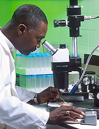
Microscopy is the technical field of using microscopes to view objects and areas of objects that cannot be seen with the naked eye. There are three well-known branches of microscopy: optical, electron, and scanning probe microscopy, along with the emerging field of X-ray microscopy.

<span class="mw-page-title-main">Optical microscope</span> Microscope that uses visible light

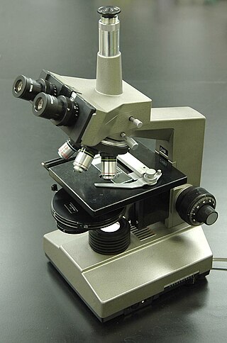
The optical microscope, also referred to as a light microscope, is a type of microscope that commonly uses visible light and a system of lenses to generate magnified images of small objects. Optical microscopes are the oldest design of microscope and were possibly invented in their present compound form in the 17th century. Basic optical microscopes can be very simple, although many complex designs aim to improve resolution and sample contrast.

An ultramicroscope is a microscope with a system that lights the object in a way that allows viewing of tiny particles via light scattering, and not light reflection or absorption. When the diameter of a particle is below or near the wavelength of visible light, the particle cannot be seen in a light microscope with the usual methods of illumination. The ultra- in ultramicroscope refers to the ability to see objects whose diameter is shorter than the wavelength of visible light, on the model of the ultra- in ultraviolet.

<span class="mw-page-title-main">Fluorescence microscope</span> Optical microscope that uses fluorescence and phosphorescence

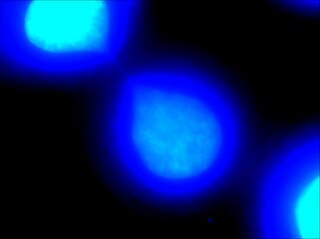
A fluorescence microscope is an optical microscope that uses fluorescence instead of, or in addition to, scattering, reflection, and attenuation or absorption, to study the properties of organic or inorganic substances. "Fluorescence microscope" refers to any microscope that uses fluorescence to generate an image, whether it is a simple set up like an epifluorescence microscope or a more complicated design such as a confocal microscope, which uses optical sectioning to get better resolution of the fluorescence image.

<span class="mw-page-title-main">Confocal microscopy</span> Optical imaging technique

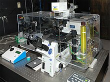
Confocal microscopy, most frequently confocal laser scanning microscopy (CLSM) or laser scanning confocal microscopy (LSCM), is an optical imaging technique for increasing optical resolution and contrast of a micrograph by means of using a spatial pinhole to block out-of-focus light in image formation. Capturing multiple two-dimensional images at different depths in a sample enables the reconstruction of three-dimensional structures within an object. This technique is used extensively in the scientific and industrial communities and typical applications are in life sciences, semiconductor inspection and materials science.

<span class="mw-page-title-main">Two-photon excitation microscopy</span>

Two-photon excitation microscopy is a fluorescence imaging technique that is particularly well-suited to image scattering living tissue of up to about one millimeter in thickness. Unlike traditional fluorescence microscopy, where the excitation wavelength is shorter than the emission wavelength, two-photon excitation requires simultaneous excitation by two photons with longer wavelength than the emitted light. The laser is focused onto a specific location in the tissue and scanned across the sample to sequentially produce the image. Due to the non-linearity of two-photon excitation, mainly fluorophores in the micrometer-sized focus of the laser beam are excited, which results in the spatial resolution of the image. This contrasts with confocal microscopy, where the spatial resolution is produced by the interaction of excitation focus and the confined detection with a pinhole.

<span class="mw-page-title-main">Phase-contrast microscopy</span> Optical microscopy technique

Phase-contrast microscopy (PCM) is an optical microscopy technique that converts phase shifts in light passing through a transparent specimen to brightness changes in the image. Phase shifts themselves are invisible, but become visible when shown as brightness variations.

<span class="mw-page-title-main">Medical microbiology</span> Branch of medical science

Medical microbiology, the large subset of microbiology that is applied to medicine, is a branch of medical science concerned with the prevention, diagnosis and treatment of infectious diseases. In addition, this field of science studies various clinical applications of microbes for the improvement of health. There are four kinds of microorganisms that cause infectious disease: bacteria, fungi, parasites and viruses, and one type of infectious protein called prion.

<span class="mw-page-title-main">Immunolabeling</span> Procedure for detection and localization of an antigen

Immunolabeling is a biochemical process that enables the detection and localization of an antigen to a particular site within a cell, tissue, or organ. Antigens are organic molecules, usually proteins, capable of binding to an antibody. These antigens can be visualized using a combination of antigen-specific antibody as well as a means of detection, called a tag, that is covalently linked to the antibody. If the immunolabeling process is meant to reveal information about a cell or its substructures, the process is called immunocytochemistry. Immunolabeling of larger structures is called immunohistochemistry.

<span class="mw-page-title-main">Intravital microscopy</span> Imaging of cells in living animals

Intravital microscopy is a form of microscopy that allows observing biological processes in live animals at a high resolution that makes distinguishing between individual cells of a tissue possible.

<span class="mw-page-title-main">Cytometry</span> Measurement of number and characteristics of cells

Cytometry is the measurement of number and characteristics of cells. Variables that can be measured by cytometric methods include cell size, cell count, cell morphology, cell cycle phase, DNA content, and the existence or absence of specific proteins on the cell surface or in the cytoplasm. Cytometry is used to characterize and count blood cells in common blood tests such as the complete blood count. In a similar fashion, cytometry is also used in cell biology research and in medical diagnostics to characterize cells in a wide range of applications associated with diseases such as cancer and AIDS.

Super-resolution microscopy is a series of techniques in optical microscopy that allow such images to have resolutions higher than those imposed by the diffraction limit, which is due to the diffraction of light. Super-resolution imaging techniques rely on the near-field or on the far-field. Among techniques that rely on the latter are those that improve the resolution only modestly beyond the diffraction-limit, such as confocal microscopy with closed pinhole or aided by computational methods such as deconvolution or detector-based pixel reassignment, the 4Pi microscope, and structured-illumination microscopy technologies such as SIM and SMI.

<span class="mw-page-title-main">Chromatin bridge</span> Medical condition

Chromatin bridge is a mitotic occurrence that forms when telomeres of sister chromatids fuse together and fail to completely segregate into their respective daughter cells. Because this event is most prevalent during anaphase, the term anaphase bridge is often used as a substitute. After the formation of individual daughter cells, the DNA bridge connecting homologous chromosomes remains fixed. As the daughter cells exit mitosis and re-enter interphase, the chromatin bridge becomes known as an interphase bridge. These phenomena are usually visualized using the laboratory techniques of staining and fluorescence microscopy.

<span class="mw-page-title-main">Quantitative phase-contrast microscopy</span>

Quantitative phase contrast microscopy or quantitative phase imaging are the collective names for a group of microscopy methods that quantify the phase shift that occurs when light waves pass through a more optically dense object.

<span class="mw-page-title-main">Light sheet fluorescence microscopy</span> Fluorescence microscopy technique

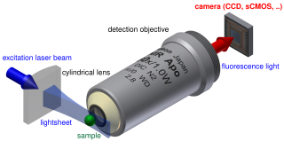
Light sheet fluorescence microscopy (LSFM) is a fluorescence microscopy technique with an intermediate-to-high optical resolution, but good optical sectioning capabilities and high speed. In contrast to epifluorescence microscopy only a thin slice of the sample is illuminated perpendicularly to the direction of observation. For illumination, a laser light-sheet is used, i.e. a laser beam which is focused only in one direction. A second method uses a circular beam scanned in one direction to create the lightsheet. As only the actually observed section is illuminated, this method reduces the photodamage and stress induced on a living sample. Also the good optical sectioning capability reduces the background signal and thus creates images with higher contrast, comparable to confocal microscopy. Because light sheet fluorescence microscopy scans samples by using a plane of light instead of a point, it can acquire images at speeds 100 to 1,000 times faster than those offered by point-scanning methods.

<span class="mw-page-title-main">Live-cell imaging</span> Study of living cells using time-lapse microscopy

Live-cell imaging is the study of living cells using time-lapse microscopy. It is used by scientists to obtain a better understanding of biological function through the study of cellular dynamics. Live-cell imaging was pioneered in the first decade of the 21st century. One of the first time-lapse microcinematographic films of cells ever made was made by Julius Ries, showing the fertilization and development of the sea urchin egg. Since then, several microscopy methods have been developed to study living cells in greater detail with less effort. A newer type of imaging using quantum dots have been used, as they are shown to be more stable. The development of holotomographic microscopy has disregarded phototoxicity and other staining-derived disadvantages by implementing digital staining based on cells’ refractive index.

<span class="mw-page-title-main">Cell unroofing</span> Methods to isolate and expose cell membranes

Cell unroofing is any of various methods to isolate and expose the cell membrane of cells. Differently from the more common membrane extraction protocols performed with multiple steps of centrifugation, in cell unroofing the aim is to tear and preserve patches of the plasma membrane in order to perform in situ experiments using.

Jean Comandon was a French microbiologist and filmmaker. He was one of the leading figures in the development of microcinematography in Paris and its use in science research and education.

References

  1. 1 2 "The Phase Contrast Microscope". Nobel Media AB.
  2. Stylianidou, Stella; Brennan, Connor; Nissen, Silas B.; Kuwada, Nathan J.; Wiggins, Paul A. (August 29, 2016). "SuperSegger: robust image segmentation, analysis and lineage tracking of bacterial cells" (PDF). Molecular Microbiology. 102 (4): 690–700. doi:10.1111/mmi.13486. PMID   27569113. S2CID   10684951.
  3. Young, Jonathan W.; Locke, James C. W.; Altinok, Alphan; Rosenfeld, Nitzan; Bacarian, Tigran; Swain, Peter S.; Mjolsness, Eric; Elowitz, Michael B. (December 15, 2011). "Measuring single-cell gene expression dynamics in bacteria using fluorescence time-lapse microscopy". Nature Protocols. 7 (1): 80–88. doi:10.1038/nprot.2011.432. PMC   4161363 . PMID   22179594.
  4. Merouane, Amine; Rey-Villamizar, Nicolas; Lu, Yanbin; Liadi, Ivan; Romain, Gabrielle; Lu, Jennifer; Singh, Harjeet; Cooper, Laurence J. N.; Varadarajan, Navin; Roysam, Badrinath (2015-10-01). "Automated profiling of individual cell–cell interactions from high-throughput time-lapse imaging microscopy in nanowell grids (TIMING)". Bioinformatics. 31 (19): 3189–3197. doi:10.1093/bioinformatics/btv355. ISSN   1367-4803. PMC   4693004 . PMID   26059718.
  5. 1 2 3 Coutu, D. L.; Schroeder, T. (2013). "Probing cellular processes by long-term live imaging - historic problems and current solutions". Journal of Cell Science. 126 (Pt 17): 3805–15. doi: 10.1242/jcs.118349 . PMID   23943879.
  6. Landecker, H. (2009). "Seeing things: From microcinematography to live cell imaging". Nature Methods. 6 (10): 707–709. doi:10.1038/nmeth1009-707. PMID   19953685. S2CID   6521488.
  7. Meseguer, M.; Rubio, I.; Cruz, M.; Basile, N.; Marcos, J.; Requena, A. (2012). "Embryo incubation and selection in a time-lapse monitoring system improves pregnancy outcome compared with a standard incubator: A retrospective cohort study". Fertility and Sterility. 98 (6): 1481–1489.e10. doi: 10.1016/j.fertnstert.2012.08.016 . PMID   22975113.
  8. Campbell, A.; Fishel, S.; Bowman, N.; Duffy, S.; Sedler, M.; Hickman, C. F. L. (2013). "Modelling a risk classification of aneuploidy in human embryos using non-invasive morphokinetics". Reproductive BioMedicine Online. 26 (5): 477–485. doi: 10.1016/j.rbmo.2013.02.006 . PMID   23518033.
  9. Rohrer, Finlo. "Cheese mites and other wonders". BBC News Magazine. Retrieved 2011-04-24.
  10. Talbot, Frederick A. (1913). Practical cinematography and its applications. W. Heinemann. OL   7220960M.
  11. "Le cinéma au service de la science". Institut national de l'audiovisuel. Retrieved 2013-01-09.
  12. Landecker, Hannah (2006). "Microcinematography and the History of Science and Film". Isis. 97: 121–132. doi:10.1086/501105. S2CID   144554305.
  13. "Jean Comandon (1877-1970)". Institut Pasteur. Archived from the original on 2014-12-05.
  14. "MICROBES CAUGHT IN ACTION.; Moving Pictures of Them a Great Aid In Medical Research". The New York Times. October 31, 1909.
  15. Bigg, Charlotte (2011). "Chapter 6: A visual history of Jean Perrin's Brownian motion curves" (PDF). In Daston, Lorraine; Lunbeck, Elizabeth (eds.). Histories of Scientific Observation. The University of Chicago Press.[ permanent dead link ]
  16. Bigg, Charlotte (2008). "Evident atoms: visuality in Jean Perrin's Brownian motion research" (PDF). Studies in History and Philosophy of Science Part A. 39 (3): 312–322. doi:10.1016/j.shpsa.2008.06.003.[ permanent dead link ]
  17. Henri, Victor (1908). "Étude cinématographique des mouvements browniens". Comptes Rendus Hebdomadaires des Séances de l'Académie des Sciences (146): 1024–1026.
  18. Landecker, Hannah (2005). "Cellular Features: Microcinematography and Film Theory". Critical Inquiry. 31 (4): 903–937. doi:10.1086/444519. S2CID   162894152.
  19. Bayly, H. W. (1910). "Demonstration by the Ultra-microscope of living Treponema pallidum and various Spirochaetes". Proceedings of the Royal Society of Medicine. 3 (Clin Sect): 3–6. doi:10.1177/003591571000300202. PMC   1961544 . PMID   19974144.
  20. Roux, P.; Münter, S.; Frischknecht, F.; Herbomel, P.; Shorte, S. L. (2004). "Focusing light on infection in four dimensions". Cellular Microbiology. 6 (4): 333–343. doi: 10.1111/j.1462-5822.2004.00374.x . PMID   15009025. S2CID   12228598.
  21. Rosenberger, Heinz (1929). "Micro-Cinematography in Medical Research". J Dent Res. 9 (3): 343–352. doi:10.1177/00220345290090030501. S2CID   71952151.
  22. "Warren H. (Warren Harmon) Lewis papers, ca. 1913-1964". American Philosophical Society. Retrieved 2011-04-24.
  23. Hoyos-Flight, Monica. "Milestone 2: Nature Milestones in Cytoskeleton". Nature Publishing Group.
  24. Abercrombie, M.; Heaysman, J. E. (1953). "Observations on the social behaviour of cells in tissue culture. I. Speed of movement of chick heart fibroblasts in relation to their mutual contacts". Experimental Cell Research. 5 (1): 111–131. doi:10.1016/0014-4827(53)90098-6. PMID   13083622.

Historic time-lapse microscopy films