Gyrification

Last updated
Gyrification in the human brain PretermSurfaces HiRes.png
Gyrification in the human brain

Gyrification is the process of forming the characteristic folds of the cerebral cortex. [1]

Contents

The peak of such a fold is called a gyrus (pl. gyri), and its trough is called a sulcus (pl. sulci). The neurons of the cerebral cortex reside in a thin layer of gray matter, only 2–4 mm thick, at the surface of the brain. [2] Much of the interior volume is occupied by white matter, which consists of long axonal projections to and from the cortical neurons residing near the surface. Gyrification allows a larger cortical surface area and hence greater cognitive functionality to fit inside a smaller cranium.

In most mammals, gyrification begins during fetal development. Primates, cetaceans, and ungulates have extensive cortical gyri, with a few species exceptions, while rodents generally have none. Gyrification in some animals, for example the ferret, continues well into postnatal life. [3]

Gyrification during human brain development

Human cortical development. Human Cortical Development.png
Human cortical development.

As fetal development proceeds, gyri and sulci begin to take shape with the emergence of deepening indentations on the surface of the cortex. Not all gyri begin to develop at the same time. Instead, the primary cortical gyri form first (beginning as early as gestational week 10 in humans), followed by secondary and tertiary gyri later in development. [4] One of the first and most prominent sulci is the lateral sulcus (also known as the lateral fissure or Sylvian fissure), followed by others such as the central sulcus, which separates the motor cortex (precentral gyrus) from somatosensory cortex (postcentral gyrus). [5] Most cortical gyri and sulci begin to take shape between weeks 24 and 38 of gestation, and continue to enlarge and mature after birth.

Evolutionary advantages

One advantage of gyrification is thought to be increased speed of brain cell communication, since cortical folds allow for cells to be closer to one other, requiring less time and energy to transmit neuronal electrical impulses, termed action potentials. [6] There is evidence to suggest a positive relationship between gyrification and cognitive information processing speed, as well as better verbal working memory. [7] Additionally, because a large cranium requires a larger pelvis during childbirth, with implied difficulty in bipedalism, a smaller cranium is more easily delivered. [8] [9]

Theories on causality in gyrification

Mechanical buckling

The mechanisms of cortical gyrification are not well understood, and several hypotheses are debated in the scientific literature. A popular hypothesis dating back to the time of Retzius in the late 19th century asserts that mechanical buckling forces due to the expanding brain tissue cause the cortical surface to fold. [10] Many theories since have been loosely tied to this hypothesis.

An external growth constraint of the cranium is not thought to cause gyrification. This is primarily because the primordium of the cranium during the period of fetal brain development is not yet ossified (hardened into the bone through calcification). The tissue covering the embryonic cerebral cortex is several thin layers of ectoderm (future skin) and mesenchyme (future muscle and connective tissue, including the future cranium). These thin layers grow easily along with cortical expansion but eventually, the cranial mesenchyme differentiates into cartilage; ossification of the cranial plates does not occur until later in development. The human cranium continues to grow substantially along with the brain after birth until the cranial plates finally fuse after several years. Experimental studies in animals have furthermore shown that cortical folding can occur without external constraints. [11] Cranial growth is thus thought to be driven by brain growth; mechanical and genetic factors intrinsic to the brain are now thought to be the primary drivers of gyrification. [6] The only observed role that the cranium may play in gyrification is in flattening of gyri as the brain matures after the cranial plates fuse. [11]

Axonal tension

An alternative theory suggests that axonal tension forces between highly interconnected cortical areas pull local cortical areas towards each other, inducing folds. [12] This model has been criticised: A numerical computer simulation could not produce a biologically realistic folding pattern. [13] One study showed that gyrification can be experimentally induced in the embryonic mouse, but at early stages in the absence of axonal connections. [14]

Differential tangential expansion

More recently, the theory of differential tangential expansion has been proposed, stating that folding patterns of the brain are a result of different tangential expansion rates between different cortical areas. [15] This is proposed to be due to areal differences in early progenitor division rates.

Mechanical factors

Cortical thickness

Early conditions of the brain have a strong influence on its final level of gyrification. In particular, there is an inverse relationship between cortical thickness and gyrification. Areas of the brain with low values of thickness are found to have higher levels of gyrification. The reverse is also true, that areas of the brain with high values of thickness are found to have lower levels of gyrification. [6]

Growth speed

There is some dispute over the growth rates through which cortical and subcortical layers of the brain develop. Purely isotropic growth suggests that the grey (outer shell) and white matter (inner core) layers each grow at separate rates, that are uniform in all dimensions. Tangential growth suggests that the grey matter grows at a faster rate than the inner white matter and that the growth rate of the grey matter determines the growth rate of the white matter. Though both methods are differential, with the cortex growing more rapidly than the subcortex, tangential growth has been suggested as a more plausible model. [6]

Creases on the brain's surface are formed as a result of instability, and tangential growth models reach levels of instability that cause creasing more frequently than isotropic models. This level is called a critical point, at which, the models prefer to release potential energy by destabilizing and forming creases to become more stable. [6]

Genetic factors

The pattern of cortical gyri and sulci is not random; most of the major convolutions are conserved between individuals and are also found across species. This reproducibility may suggest that genetic mechanisms can specify the location of major gyri. Studies of monozygotic and dizygotic twins of the late 1990s support this idea, [16] particularly with regards to primary gyri and sulci, whereas there is more variability among secondary and tertiary gyri. [17] Therefore, one may hypothesize that secondary and tertiary folds could be more sensitive to genetic and environmental factors. [18] The first gene reported to influence gyrification was Trnp1. [19] Local expression levels of Trnp1, can determine the future position of developing folds/gyri in human brains. [19] [20] [21] Genes that influence cortical progenitor dynamics, neurogenesis and neuronal migration, as well as genes that influence the development of cortical circuits and axonal projections may all contribute to gyrification. Trnp1 is a DNA-binding factor that has been shown to regulate other genes that regulate the proliferation of cortical progenitor cells thereby serving as a master gene-regulator. [6] [19] In addition, the fibroblast growth factor (FGF)- and sonic hedgehog (SHH)-signaling pathways have recently been reported to be able to induce cortical folds, with a full complement of cortical layers, in mice that live to adulthood. [14] [22] These FGF and Shh factors regulate cortical stem cell proliferation and neurogenesis dynamics. Roles for beta-catenin (part of the Wnt pathway) and appropriate levels of cell death of cortical progenitors have also been found. [23] [24]

Cell biological determinants

Cortical stem cells, known as radial glial cells (RGC)s, reside in the ventricular zone and generate the excitatory glutamatergic neurons of the cerebral cortex. [25] [26] These cells rapidly proliferate through self-renewal at early developmental stages, expanding the progenitor pool and increasing cortical surface area. At this stage, the pattern of cortical areas is genetically programmed by a system of signaling centers through the process of cortical patterning, and the primordial map of cortical functional areas at this stage is called a 'protomap'. [27] Cortical neurogenesis begins to deplete the pool of progenitor cells, subject to the influences of many genetic cues such as fibroblast growth factors (FGF)s and Notch. [28] RGCs generate intermediate neuronal precursors that divide further in the subventricular zone (SVZ), amplifying the number of cortical neurons being produced. [29] The long fibers of RGCs project all the way through the developing cortex to the pial surface of the brain, and these fibers serve as physical guides for neuronal migration. [30] A second class of RGC, termed basal RGCs (bRGC)s, forms a third progenitor pool in the outer SVZ. [31] Basal RGCs are generally much more abundant in higher mammals. Both classic RGCs and the recently described bRGCs represent guiding cues that lead newborn neurons to their destination in the cortex. Increased numbers of bRGCs increase the density of guiding fibers in an otherwise fanning out array which would lose fiber density. [32] The scientific literature points to differences in the dynamics of proliferation and neuronal differentiation in each of these progenitor zones across mammalian species, and such differences may account for the large differences in cortical size and gyrification among mammals. One hypothesis suggests that certain progenitor cells generate abundant neurons destined for the outer cortical layers, causing greater surface area increase in the outer layers compared with the inner cortical layers. [32] It remains unclear how this may work without further mechanistic elements. [33] [34]

Variation across species

A 'gyrification index' (GI) has been used as a measure of the magnitude of cortical convolutions on the surface of the mammalian brain. [6] [35] Reptile and bird brains do not show gyrification. Mammals with a high GI are generally larger than those with a low GI; for example the pilot whale and bottlenose dolphin show the highest GI values. The human brain, while larger than that of a horse, shows a similar GI. Rodents generally show the lowest GIs. Nonetheless, some rodents show gyrencephaly and a few primate species are quite lissencephalic. [36]

A linear relation between mammals expressed in gyrification terms has been found by Mota & Herculano-Houzel, 2015. [37] They suggest a model that combines morphometric measurements (Cortical Thickness, Exposed Area, and Total Area) which could be a way to describe gyrification.

Neurological disorders of gyrification

Lissencephaly

A cerebral cortex lacking surface convolutions is said to be lissencephalic, meaning 'smooth-brained'. [38] During embryonic development, all mammalian brains begin as lissencephalic structures derived from the neural tube. Some, like mouse brains, remain lissencephalic throughout adulthood. It has been shown that lissencephalic species possess many of the molecular cues needed to achieve gyrencephaly, but a large variety of genes are involved in the regulation of the neural progenitor proliferation and neurogenic processes that underlie gyrification. It is hypothesized that spatiotemporal differences in these molecular pathways, including FGF, Shh, and Trnp1 and likely many others, determine the timing and extent of gyrification in various species. [19] [22]

Lissencephaly is a human disease state. For humans with lissencephaly, a large number of neurons fail to reach the outer cortex during neuronal migration, and remain under the cortical plate. [39] This displacement results in not only defects in cortical connections, but also a thickened cortex, consistent with the idea that a brain with a thicker cortex will have a lesser degree of gyrification. [40]

Polymicrogyria

Polymicrogyria is a condition in which the brain has an overly convoluted cortex. Though at the surface, the brain appears smooth with a few sulci, looking at the interior of the brain reveals a convoluted structure with a large number of secondary and tertiary folds. [40] Brain imaging with MRI reveals a brain with polymicrogyria to have a thin cortex, consistent with the idea that a brain with a thin cortex will have a high level of gyrification. [39] [40] A wide array of genes when mutated have been shown to cause Polymicrogyria in humans, ranging from mTORopathies (e.g. AKT3) to channelopathies (sodium channels, "SCN3A"). [41]

Autism

Patients with autism have overall higher levels of cortical gyrification, [42] but only in the temporal, parietal, and occipital lobes, as well as part of the cingulate cortex. [43] The higher levels of gyrification are found to relate to greater local connectivity in autistic brains, suggesting hyperconnectivity.

Trnp1, which was able to induce gyrification in animal models, has been hypothesized to be associated with disorders of gyrification in some cases of autism, but a review in 2012 found only one reported case of a mutation, in a patient with Rett syndrome (not ASD). [44]

The folds of autistic human brains are found to experience slight shifts in location, early in brain development. Specifically, different patterns appear in the superior frontal sulcus, Sylvian fissure, inferior frontal gyrus, superior temporal gyrus, and olfactory sulci. [45] These areas relate to working memory, emotional processing, language, and eye gaze, [46] and their difference in location and level of gyrification when compared to a neurotypical human brain could explain some altered behaviors in autistic patients.

Schizophrenia

A more prevalent condition, schizophrenia, has also been associated with structural abnormalities in the brain. A reduced cortical thickness and increased gyrification is seen similar to the changes shown in those with autism. [39] [47]

Zika virus malformations

Cortical malformations induced by the Zika virus are due to infection during pregnancy, and are generally classified as microcephaly, or 'small-brain'. Due to the large reduction in volume of the cerebral cortex in microcephaly, changes in gyrification are not unexpected. Studies of the mechanism of Zika malformations indicate that the principal defect is due to infection of radial glial cells and subsequent cell death. [48] [49] Death of cortical stem cells causes the loss of all expected daughter cells, and the scope of the malformation thus depends on the timing of infection as well as its severity during the schedule of neural stem cell proliferation and neurogenesis. Earlier infections would generally be expected to produce a more severe malformation. [50] [51] The microcephaly and gyrification malformations are permanent and there are no known treatments.

Measurements of gyrification

Cortical Gyrification can be measured in terms of the Gyrification Index (GI), [52] Fractal Dimensionality [53] and a combination of morphometric terms (Area, Thickness, Volume). [37] The GI is defined as the ratio between the Total Area and the Exposed Area ("perimeter of the brain delineated on two-dimensional coronal sections" [54] ). FreeSurfer, a surface reconstruction Software is one of the tools available to measure the GI. [55]

Additional images

See also

Related Research Articles

<span class="mw-page-title-main">Cerebral cortex</span> Outer layer of the cerebrum of the mammalian brain

The cerebral cortex, also known as the cerebral mantle, is the outer layer of neural tissue of the cerebrum of the brain in humans and other mammals. The cerebral cortex mostly consists of the six-layered neocortex, with just 10% consisting of the allocortex. It is separated into two cortices, by the longitudinal fissure that divides the cerebrum into the left and right cerebral hemispheres. The two hemispheres are joined beneath the cortex by the corpus callosum. The cerebral cortex is the largest site of neural integration in the central nervous system. It plays a key role in attention, perception, awareness, thought, memory, language, and consciousness. The cerebral cortex is part of the brain responsible for cognition.

The development of the nervous system, or neural development (neurodevelopment), refers to the processes that generate, shape, and reshape the nervous system of animals, from the earliest stages of embryonic development to adulthood. The field of neural development draws on both neuroscience and developmental biology to describe and provide insight into the cellular and molecular mechanisms by which complex nervous systems develop, from nematodes and fruit flies to mammals.

<span class="mw-page-title-main">Lissencephaly</span> Medical condition

Lissencephaly is a set of rare brain disorders whereby the whole or parts of the surface of the brain appear smooth. It is caused by defective neuronal migration during the 12th to 24th weeks of gestation resulting in a lack of development of brain folds (gyri) and grooves (sulci). It is a form of cephalic disorder. Terms such as agyria and pachygyria are used to describe the appearance of the surface of the brain.

<span class="mw-page-title-main">Neocortex</span> Mammalian structure involved in higher-order brain functions

The neocortex, also called the neopallium, isocortex, or the six-layered cortex, is a set of layers of the mammalian cerebral cortex involved in higher-order brain functions such as sensory perception, cognition, generation of motor commands, spatial reasoning and language. The neocortex is further subdivided into the true isocortex and the proisocortex.

<span class="mw-page-title-main">Polymicrogyria</span> Medical condition

Polymicrogyria (PMG) is a condition that affects the development of the human brain by multiple small gyri (microgyri) creating excessive folding of the brain leading to an abnormally thick cortex. This abnormality can affect either one region of the brain or multiple regions.

<span class="mw-page-title-main">Gyrus</span> Ridge on the cerebral cortex of the brain

In neuroanatomy, a gyrus is a ridge on the cerebral cortex. It is generally surrounded by one or more sulci. Gyri and sulci create the folded appearance of the brain in humans and other mammals.

Pachygyria is a congenital malformation of the cerebral hemisphere. It results in unusually thick convolutions of the cerebral cortex. Typically, children have developmental delay and seizures, the onset and severity depending on the severity of the cortical malformation. Infantile spasms are common in affected children, as is intractable epilepsy.

<span class="mw-page-title-main">Sulcus (neuroanatomy)</span> Fold in the surface of the brain

In neuroanatomy, a sulcus is a depression or groove in the cerebral cortex. It surrounds a gyrus, creating the characteristic folded appearance of the brain in humans and other mammals. The larger sulci are usually called fissures.

<span class="mw-page-title-main">Radial glial cell</span> Bipolar-shaped progenitor cells of all neurons in the cerebral cortex and some glia

Radial glial cells, or radial glial progenitor cells (RGPs), are bipolar-shaped progenitor cells that are responsible for producing all of the neurons in the cerebral cortex. RGPs also produce certain lineages of glia, including astrocytes and oligodendrocytes. Their cell bodies (somata) reside in the embryonic ventricular zone, which lies next to the developing ventricular system.

<span class="mw-page-title-main">Evolution of the brain</span> Overview of the evolution of the brain

There is much to be discovered about the evolution of the brain and the principles that govern it. While much has been discovered, not everything currently known is well understood. The evolution of the brain has appeared to exhibit diverging adaptations within taxonomic classes such as Mammalia and more vastly diverse adaptations across other taxonomic classes. Brain to body size scales allometrically. This means as body size changes, so do other physiological, anatomical, and biochemical constructs connecting the brain to the body. Small bodied mammals have relatively large brains compared to their bodies whereas large mammals have a smaller brain to body ratios. If brain weight is plotted against body weight for primates, the regression line of the sample points can indicate the brain power of a primate species. Lemurs for example fall below this line which means that for a primate of equivalent size, we would expect a larger brain size. Humans lie well above the line indicating that humans are more encephalized than lemurs. In fact, humans are more encephalized compared to all other primates. This means that human brains have exhibited a larger evolutionary increase in its complexity relative to its size. Some of these evolutionary changes have been found to be linked to multiple genetic factors, such as proteins and other organelles.

<span class="mw-page-title-main">Ganglionic eminence</span>

The ganglionic eminence (GE) is a transitory structure in the development of the nervous system that guides cell and axon migration. It is present in the embryonic and fetal stages of neural development found between the thalamus and caudate nucleus.

The Protomap is a primordial molecular map of the functional areas of the mammalian cerebral cortex during early embryonic development, at a stage when neural stem cells are still the dominant cell type. The protomap is a feature of the ventricular zone, which contains the principal cortical progenitor cells, known as radial glial cells. Through a process called 'cortical patterning', the protomap is patterned by a system of signaling centers in the embryo, which provide positional information and cell fate instructions. These early genetic instructions set in motion a development and maturation process that gives rise to the mature functional areas of the cortex, for example the visual, somatosensory, and motor areas. The term protomap was coined by Pasko Rakic. The protomap hypothesis was opposed by the protocortex hypothesis, which proposes that cortical proto-areas initially have the same potential, and that regionalization in large part is controlled by external influences, such as axonal inputs from the thalamus to the cortex. However, a series of papers in the year 2000 and in 2001 provided strong evidence against the protocortex hypothesis, and the protomap hypothesis has been well accepted since then. The protomap hypothesis, together with the related radial unit hypothesis, forms our core understanding of the embryonic development of the cerebral cortex. Once the basic structure is present and cortical neurons have migrated to their final destinations, many other processes contribute to the maturation of functional cortical circuits.

<span class="mw-page-title-main">Eomesodermin</span> Protein-coding gene in the species Homo sapiens

Eomesodermin also known as T-box brain protein 2 (Tbr2) is a protein that in humans is encoded by the EOMES gene.

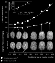
The development of the cerebral cortex, known as corticogenesis is the process during which the cerebral cortex of the brain is formed as part of the development of the nervous system of mammals including its development in humans. The cortex is the outer layer of the brain and is composed of up to six layers. Neurons formed in the ventricular zone migrate to their final locations in one of the six layers of the cortex. The process occurs from embryonic day 10 to 17 in mice and between gestational weeks seven to 18 in humans.

<span class="mw-page-title-main">Ventricular zone</span> Transient embryonic layer of tissue containing neural stem cells

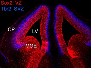
In vertebrates, the ventricular zone (VZ) is a transient embryonic layer of tissue containing neural stem cells, principally radial glial cells, of the central nervous system (CNS). The VZ is so named because it lines the ventricular system, which contains cerebrospinal fluid (CSF). The embryonic ventricular system contains growth factors and other nutrients needed for the proper function of neural stem cells. Neurogenesis, or the generation of neurons, occurs in the VZ during embryonic and fetal development as a function of the Notch pathway, and the newborn neurons must migrate substantial distances to their final destination in the developing brain or spinal cord where they will establish neural circuits. A secondary proliferative zone, the subventricular zone (SVZ), lies adjacent to the VZ. In the embryonic cerebral cortex, the SVZ contains intermediate neuronal progenitors that continue to divide into post-mitotic neurons. Through the process of neurogenesis, the parent neural stem cell pool is depleted and the VZ disappears. The balance between the rates of stem cell proliferation and neurogenesis changes during development, and species from mouse to human show large differences in the number of cell cycles, cell cycle length, and other parameters, which is thought to give rise to the large diversity in brain size and structure.

Neurogenesis is the process by which nervous system cells, the neurons, are produced by neural stem cells (NSCs). In short, it is brain growth in relation to its organization. This occurs in all species of animals except the porifera (sponges) and placozoans. Types of NSCs include neuroepithelial cells (NECs), radial glial cells (RGCs), basal progenitors (BPs), intermediate neuronal precursors (INPs), subventricular zone astrocytes, and subgranular zone radial astrocytes, among others.

<span class="mw-page-title-main">Radial unit hypothesis</span> Conceptual theory of cerebral cortex development

The Radial Unit Hypothesis (RUH) is a conceptual theory of cerebral cortex development, first described by Pasko Rakic. The RUH states that the cerebral cortex develops during embryogenesis as an array of interacting cortical columns, or 'radial units', each of which originates from a transient stem cell layer called the ventricular zone, which contains neural stem cells known as radial glial cells.

<span class="mw-page-title-main">TRNP1</span> Protein-coding gene in the species Homo sapiens

TMF-regulated nuclear protein 1 is a nuclear protein that in humans is encoded by the TRNP1 gene. TRNP1 plays a crucial role in cellular proliferation and brain development. Trnp1 is the first protein that has been shown to play a major role in cortical folding. It represents a major stem cell factor, that is involved in crucial processes during brain but also stem cell development. Trnp1 controls gene expression levels and is sufficient to induce gyri and sulci in the developing brain. Local differences of Trnp1 expression levels in the human brain correlate with cortical folding.

<span class="mw-page-title-main">Microlissencephaly</span> Microcephaly combined with lissencephaly

Microlissencephaly (MLIS) is a rare congenital brain disorder that combines severe microcephaly with lissencephaly. Microlissencephaly is a heterogeneous disorder, i.e. it has many different causes and a variable clinical course. Microlissencephaly is a malformation of cortical development (MCD) that occurs due to failure of neuronal migration between the third and fifth month of gestation as well as stem cell population abnormalities. Numerous genes have been found to be associated with microlissencephaly, however, the pathophysiology is still not completely understood.

Intermediate progenitor cells (IPCs) are a type of progenitor cell in the developing cerebral cortex. They are multipolar cells produced by radial glial cells who have undergone asymmetric division. IPCs can produce neuron cells via neurogenesis and are responsible for ensuring the proper quantity of cortical neurons are produced. In mammals, neural stem cells are the primary progenitors during embryogenesis whereas intermediate progenitor cells are the secondary progenitors.

References

  1. Rakic, P (October 2009). "Evolution of the neocortex: a perspective from developmental biology". Nature Reviews Neuroscience. 10 (10): 724–35. doi:10.1038/nrn2719. PMC   2913577 . PMID   19763105.
  2. Kandel, Eric R.; Schwartz, James H.; Jessell, Thomas M.; Siegelbaum, Steven A.; Hudspeth, A.J., eds. (2012) [1981]. Principles of Neural Science (5th ed.). New York: McGraw Hill. ISBN   978-0-07-139011-8.
  3. Smart, IH; McSherry, GM (June 1986). "Gyrus formation in the cerebral cortex in the ferret. I. Description of the external changes". Journal of Anatomy. 146: 141–52. PMC   1166530 . PMID   3693054.
  4. Rajagopalan, V; Scott, J; Habas, PA; Kim, K; Corbett-Detig, J; Rousseau, F; Barkovich, AJ; Glenn, OA; Studholme, C (23 February 2011). "Local tissue growth patterns underlying normal fetal human brain gyrification quantified in utero". The Journal of Neuroscience. 31 (8): 2878–87. doi:10.1523/jneurosci.5458-10.2011. PMC   3093305 . PMID   21414909.
  5. Bayer, Shirley A; Altman, Joseph (2005). The Human Brain during the Second Trimester. Boca Raton, FL USA: CRC Press. ISBN   978-0-8493-1422-3.
  6. 1 2 3 4 5 6 7 Striedter, Georg F.; Srinivasan, Shyam; Monuki, Edwin S. (2015-01-01). "Cortical Folding: When, Where, How, and Why?". Annual Review of Neuroscience. 38 (1): 291–307. doi: 10.1146/annurev-neuro-071714-034128 . PMID   25897870.
  7. Gautam, Prapti; Anstey, Kaarin J.; Wen, Wei; Sachdev, Perminder S.; Cherbuin, Nicolas (2015-07-01). "Cortical gyrification and its relationships with cortical volume, cortical thickness, and cognitive performance in healthy mid-life adults". Behavioural Brain Research. 287: 331–339. doi:10.1016/j.bbr.2015.03.018. PMID   25804360. S2CID   7476449.
  8. Jordaan, HV (March 1976). "Newborn: adult brain ratios in hominid evolution". American Journal of Physical Anthropology. 44 (2): 271–8. doi:10.1002/ajpa.1330440209. PMID   816206.
  9. Weiner, S; Monge, J; Mann, A (September 2008). "Bipedalism and parturition: an evolutionary imperative for cesarean delivery?". Clinics in Perinatology. 35 (3): 469–78, ix. doi:10.1016/j.clp.2008.06.003. PMID   18952015.
  10. Ronan, L; Voets, N; Rua, C; Alexander-Bloch, A; Hough, M; Mackay, C; Crow, TJ; James, A; Giedd, JN; Fletcher, PC (August 2014). "Differential tangential expansion as a mechanism for cortical gyrification". Cerebral Cortex. 24 (8): 2219–28. doi:10.1093/cercor/bht082. PMC   4089386 . PMID   23542881.
  11. 1 2 Tallinen, Tuomas; Chung, Jun Young; Biggins, John S.; Mahadevan, L. (2014-09-02). "Gyrification from constrained cortical expansion". Proceedings of the National Academy of Sciences. 111 (35): 12667–12672. arXiv: 1503.03853 . Bibcode:2014PNAS..11112667T. doi: 10.1073/pnas.1406015111 . ISSN   0027-8424. PMC   4156754 . PMID   25136099.
  12. Van Essen, DC (23 January 1997). "A tension-based theory of morphogenesis and compact wiring in the central nervous system". Nature. 385 (6614): 313–8. Bibcode:1997Natur.385..313E. doi:10.1038/385313a0. PMID   9002514. S2CID   4355025.
  13. Xu, G; Knutsen, A K; Dikranian, K; Kroenke, C D; Bayly, P V; Taber, L A (2010). "Axons pull on the brain, but tension does not drive cortical folding". Journal of Biomechanical Engineering. 132 (7): 071013. doi:10.1115/1.4001683. PMC   3170872 . PMID   20590291.
  14. 1 2 Rash, BG; Tomasi, S; Lim, HD; Suh, CY; Vaccarino, FM (26 June 2013). "Cortical gyrification induced by fibroblast growth factor 2 in the mouse brain". The Journal of Neuroscience. 33 (26): 10802–14. doi:10.1523/jneurosci.3621-12.2013. PMC   3693057 . PMID   23804101.
  15. Ronan, L; Voets, N; Rua, C; Alexander-Bloch, A; Hough, M; Mackay, C; Crow, TJ; James, A; Giedd, JN; Fletcher, PC (August 2014). "Differential tangential expansion as a mechanism for cortical gyrification". Cerebral Cortex. 24 (8): 2219–28. doi:10.1093/cercor/bht082. PMC   4089386 . PMID   23542881.
  16. Bartley, AJ; Jones, DW; Weinberger, DR (February 1997). "Genetic variability of human brain size and cortical gyral patterns". Brain. 120 (2): 257–69. doi: 10.1093/brain/120.2.257 . PMID   9117373.
  17. White, Tonya; Su, Shu; Schmidt, Marcus; Kao, Chiu-Yen; Sapiro, Guillermo (2010-02-01). "The development of gyrification in childhood and adolescence". Brain and Cognition. Adolescent Brain Development: Current Themes and Future Directions. 72 (1): 36–45. doi:10.1016/j.bandc.2009.10.009. PMC   2815169 . PMID   19942335.
  18. Gómez-Robles, Aida; Hopkins, William D.; Sherwood, Chet C. (2013-06-22). "Increased morphological asymmetry, evolvability and plasticity in human brain evolution". Proc. R. Soc. B. 280 (1761): 20130575. doi:10.1098/rspb.2013.0575. ISSN   0962-8452. PMC   3652445 . PMID   23615289.
  19. 1 2 3 4 Stahl, Ronny; Walcher, Tessa; De Juan Romero, Camino; Pilz, Gregor Alexander; Cappello, Silvia; Irmler, Martin; Sanz-Aquela, José Miguel; Beckers, Johannes; Blum, Robert (2013-04-25). "Trnp1 regulates expansion and folding of the mammalian cerebral cortex by control of radial glial fate". Cell. 153 (3): 535–549. doi: 10.1016/j.cell.2013.03.027 . hdl: 10261/338716 . ISSN   1097-4172. PMID   23622239.
  20. de Juan Romero, Camino; Bruder, Carl; Tomasello, Ugo; Sanz-Anquela, José Miguel; Borrell, Víctor (2015-07-14). "Discrete domains of gene expression in germinal layers distinguish the development of gyrencephaly". The EMBO Journal. 34 (14): 1859–1874. doi:10.15252/embj.201591176. ISSN   1460-2075. PMC   4547892 . PMID   25916825.
  21. Fernández, Virginia; Llinares-Benadero, Cristina; Borrell, Víctor (2016-05-17). "Cerebral cortex expansion and folding: what have we learned?". The EMBO Journal. 35 (10): 1021–1044. doi:10.15252/embj.201593701. ISSN   1460-2075. PMC   4868950 . PMID   27056680.
  22. 1 2 Wang, L; Hou, S; Han, YG (23 May 2016). "Hedgehog signaling promotes basal progenitor expansion and the growth and folding of the neocortex". Nature Neuroscience. 19 (7): 888–96. doi:10.1038/nn.4307. PMC   4925239 . PMID   27214567.
  23. Chenn, A; Walsh, CA (19 July 2002). "Regulation of cerebral cortical size by control of cell cycle exit in neural precursors". Science. 297 (5580): 365–9. Bibcode:2002Sci...297..365C. doi:10.1126/science.1074192. PMID   12130776. S2CID   15145974.
  24. Kuida, K; Haydar, TF; Kuan, CY; Gu, Y; Taya, C; Karasuyama, H; Su, MS; Rakic, P; Flavell, RA (7 August 1998). "Reduced apoptosis and cytochrome c-mediated caspase activation in mice lacking caspase 9". Cell. 94 (3): 325–37. doi: 10.1016/s0092-8674(00)81476-2 . PMID   9708735.
  25. Noctor, SC; Flint, AC; Weissman, TA; Dammerman, RS; Kriegstein, AR (8 February 2001). "Neurons derived from radial glial cells establish radial units in neocortex". Nature. 409 (6821): 714–20. Bibcode:2001Natur.409..714N. doi:10.1038/35055553. PMID   11217860. S2CID   3041502.
  26. Malatesta, P; Hartfuss, E; Götz, M (December 2000). "Isolation of radial glial cells by fluorescent-activated cell sorting reveals a neuronal lineage". Development. 127 (24): 5253–63. doi:10.1242/dev.127.24.5253. PMID   11076748.
  27. Rakic, P (8 July 1988). "Specification of cerebral cortical areas". Science. 241 (4862): 170–6. Bibcode:1988Sci...241..170R. doi:10.1126/science.3291116. PMID   3291116.
  28. Rash, BG; Lim, HD; Breunig, JJ; Vaccarino, FM (26 October 2011). "FGF signaling expands embryonic cortical surface area by regulating Notch-dependent neurogenesis". The Journal of Neuroscience. 31 (43): 15604–17. doi:10.1523/jneurosci.4439-11.2011. PMC   3235689 . PMID   22031906.
  29. Noctor, SC; Martínez-Cerdeño, V; Ivic, L; Kriegstein, AR (February 2004). "Cortical neurons arise in symmetric and asymmetric division zones and migrate through specific phases". Nature Neuroscience. 7 (2): 136–44. doi:10.1038/nn1172. PMID   14703572. S2CID   15946842.
  30. Rakic, P (May 1972). "Mode of cell migration to the superficial layers of fetal monkey neocortex". The Journal of Comparative Neurology. 145 (1): 61–83. doi:10.1002/cne.901450105. PMID   4624784. S2CID   41001390.
  31. LaMonica, BE; Lui, JH; Wang, X; Kriegstein, AR (October 2012). "OSVZ progenitors in the human cortex: an updated perspective on neurodevelopmental disease". Current Opinion in Neurobiology. 22 (5): 747–53. doi:10.1016/j.conb.2012.03.006. PMC   3402619 . PMID   22487088.
  32. 1 2 Hansen, DV; Lui, JH; Parker, PR; Kriegstein, AR (25 March 2010). "Neurogenic radial glia in the outer subventricular zone of human neocortex". Nature. 464 (7288): 554–561. Bibcode:2010Natur.464..554H. doi:10.1038/nature08845. PMID   20154730. S2CID   4412132.
  33. Hevner, RF; Haydar, TF (February 2012). "The (not necessarily) convoluted role of basal radial glia in cortical neurogenesis". Cerebral Cortex. 22 (2): 465–8. doi:10.1093/cercor/bhr336. PMC   3256413 . PMID   22116731.
  34. García-Moreno, F; Vasistha, NA; Trevia, N; Bourne, JA; Molnár, Z (February 2012). "Compartmentalization of cerebral cortical germinal zones in a lissencephalic primate and gyrencephalic rodent". Cerebral Cortex. 22 (2): 482–92. doi: 10.1093/cercor/bhr312 . PMID   22114081.
  35. Zilles, K; Armstrong, E; Moser, KH; Schleicher, A; Stephan, H (1989). "Gyrification in the cerebral cortex of primates". Brain, Behavior and Evolution. 34 (3): 143–50. doi:10.1159/000116500. PMID   2512000.
  36. Hevner, RF; Haydar, TF (February 2012). "The (not necessarily) convoluted role of basal radial glia in cortical neurogenesis". Cerebral Cortex. 22 (2): 465–8. doi:10.1093/cercor/bhr336. PMC   3256413 . PMID   22116731.
  37. 1 2 Mota, Bruno; Herculano-Houzel, Suzana (2015-07-03). "Cortical folding scales universally with surface area and thickness, not number of neurons". Science. 349 (6243): 74–77. Bibcode:2015Sci...349...74M. doi:10.1126/science.aaa9101. ISSN   0036-8075. PMID   26138976. S2CID   24572675.
  38. Armstrong, E; Schleicher, A; Omran, H; Curtis, M; Zilles, K (1991). "The ontogeny of human gyrification". Cerebral Cortex. 5 (1): 56–63. doi:10.1093/cercor/5.1.56. PMID   7719130.
  39. 1 2 3 Budday, Silvia; Raybaud, Charles; Kuhl, Ellen (2014-07-10). "A mechanical model predicts morphological abnormalities in the developing human brain". Scientific Reports. 4: 5644. Bibcode:2014NatSR...4E5644B. doi:10.1038/srep05644. PMC   4090617 . PMID   25008163.
  40. 1 2 3 Ross, M. Elizabeth; Walsh, Christopher A. (2001-01-01). "Human Brain Malformations and Their Lessons for Neuronal Migration". Annual Review of Neuroscience. 24 (1): 1041–1070. doi:10.1146/annurev.neuro.24.1.1041. PMID   11520927. S2CID   18582415.
  41. Smith RS, Kenny CJ, Ganesh V, Jang A, Borges-Monroy R, Partlow JN, et al. (September 2018). "Sodium Channel SCN3A (NaV1.3) Regulation of Human Cerebral Cortical Folding and Oral Motor Development". Neuron. 99 (5): 905–913.e7. doi:10.1016/j.neuron.2018.07.052. PMC   6226006 . PMID   30146301.
  42. Wallace, Gregory L.; Robustelli, Briana; Dankner, Nathan; Kenworthy, Lauren; Giedd, Jay N.; Martin, Alex (2013-06-01). "Increased gyrification, but comparable surface area in adolescents with autism spectrum disorders". Brain. 136 (6): 1956–1967. doi:10.1093/brain/awt106. ISSN   0006-8950. PMC   3673467 . PMID   23715094.
  43. Yang, Daniel Y.-J.; Beam, Danielle; Pelphrey, Kevin A.; Abdullahi, Sebiha; Jou, Roger J. (2016-01-25). "Cortical morphological markers in children with autism: a structural magnetic resonance imaging study of thickness, area, volume, and gyrification". Molecular Autism. 7 (1): 11. doi: 10.1186/s13229-016-0076-x . PMC   4727390 . PMID   26816612.
  44. Stahl, Ronny (2012). Identification and functional analysis of Trnp1: a novel DNA associated protein with a key role in neurogenesis (Text.PhDThesis). Ludwig-Maximilians-Universität München. pp. 86–88.
  45. Chen, Jason A.; Peñagarikano, Olga; Belgard, T. Grant; Swarup, Vivek; Geschwind, Daniel H. (2015-01-01). "The Emerging Picture of Autism Spectrum Disorder: Genetics and Pathology". Annual Review of Pathology: Mechanisms of Disease. 10 (1): 111–144. doi: 10.1146/annurev-pathol-012414-040405 . PMID   25621659.
  46. Levitt, Jennifer G.; Blanton, Rebecca E.; Smalley, Susan; Thompson, P. M.; Guthrie, Donald; McCracken, James T.; Sadoun, Tania; Heinichen, Laura; Toga, Arthur W. (2003-07-01). "Cortical Sulcal Maps in Autism". Cerebral Cortex. 13 (7): 728–735. doi: 10.1093/cercor/13.7.728 . ISSN   1047-3211. PMID   12816888.
  47. Palaniyappan, Lena; Mallikarjun, Pavan; Joseph, Verghese; White, Thomas P.; Liddle, Peter F. (2011). "Folding of the Prefrontal Cortex in Schizophrenia: Regional Differences in Gyrification". Biological Psychiatry. 69 (10): 974–979. doi:10.1016/j.biopsych.2010.12.012. PMID   21257157. S2CID   16645055.
  48. Nowakowski, TJ; Pollen, AA; Di Lullo, E; Sandoval-Espinosa, C; Bershteyn, M; Kriegstein, AR (5 May 2016). "Expression Analysis Highlights AXL as a Candidate Zika Virus Entry Receptor in Neural Stem Cells". Cell Stem Cell. 18 (5): 591–6. doi:10.1016/j.stem.2016.03.012. PMC   4860115 . PMID   27038591.
  49. Li, C; Xu, D; Ye, Q; Hong, S; Jiang, Y; Liu, X; Zhang, N; Shi, L; Qin, CF; Xu, Z (11 May 2016). "Zika Virus Disrupts Neural Progenitor Development and Leads to Microcephaly in Mice". Cell Stem Cell. 19 (1): 120–6. doi: 10.1016/j.stem.2016.04.017 . PMID   27179424.
  50. Wu, Kong-Yan; Zuo, Guo-Long; Li, Xiao-Feng; Ye, Qing; Deng, Yong-Qiang; Huang, Xing-Yao; Cao, Wu-Chun; Qin, Cheng-Feng; Luo, Zhen-Ge (2016-05-13). "Vertical transmission of Zika virus targeting the radial glial cells affects cortex development of offspring mice". Cell Research. 26 (6): 645–654. doi:10.1038/cr.2016.58. ISSN   1748-7838. PMC   4897185 . PMID   27174054.
  51. Tang, Hengli; Hammack, Christy; Ogden, Sarah C.; Wen, Zhexing; Qian, Xuyu; Li, Yujing; Yao, Bing; Shin, Jaehoon; Zhang, Feiran (2016-05-05). "Zika Virus Infects Human Cortical Neural Progenitors and Attenuates Their Growth". Cell Stem Cell. 18 (5): 587–590. doi:10.1016/j.stem.2016.02.016. ISSN   1934-5909. PMC   5299540 . PMID   26952870.
  52. Zilles, Karl; Armstrong, Este; Schleicher, Axel; Kretschmann, Hans-Joachim (1988-11-01). "The human pattern of gyrification in the cerebral cortex". Anatomy and Embryology. 179 (2): 173–179. doi:10.1007/BF00304699. ISSN   0340-2061. PMID   3232854. S2CID   8739203.
  53. Madan, Christopher R.; Kensinger, Elizabeth A. (2016-07-01). "Cortical complexity as a measure of age-related brain atrophy". NeuroImage. 134: 617–629. doi:10.1016/j.neuroimage.2016.04.029. ISSN   1053-8119. PMC   4945358 . PMID   27103141.
  54. "LGI - Free Surfer Wiki". surfer.nmr.mgh.harvard.edu. Retrieved 2018-05-02.
  55. Schaer, M.; Cuadra, M.B.; Tamarit, L.; Lazeyras, F.; Eliez, S.; Thiran, J.-P. (February 2008). "A Surface-Based Approach to Quantify Local Cortical Gyrification". IEEE Transactions on Medical Imaging. 27 (2): 161–170. doi:10.1109/tmi.2007.903576. ISSN   0278-0062. PMID   18334438. S2CID   756173.