Lumbar puncture

Last updated

Lumbar puncture
Thisisspinaltap.jpg
Lumbar puncture in a sitting position. The reddish-brown swirls on the patient's back are tincture of iodine (an antiseptic).
Other namesSpinal tap
ICD-9-CM 03.31
MeSH D013129
eMedicine 80773

Lumbar puncture (LP), also known as a spinal tap, is a medical procedure in which a needle is inserted into the spinal canal, most commonly to collect cerebrospinal fluid (CSF) for diagnostic testing. The main reason for a lumbar puncture is to help diagnose diseases of the central nervous system, including the brain and spine. Examples of these conditions include meningitis and subarachnoid hemorrhage. It may also be used therapeutically in some conditions. Increased intracranial pressure (pressure in the skull) is a contraindication, due to risk of brain matter being compressed and pushed toward the spine. Sometimes, lumbar puncture cannot be performed safely (for example due to a severe bleeding tendency). It is regarded as a safe procedure, but post-dural-puncture headache is a common side effect if a small atraumatic needle is not used. [1]

Contents

The procedure is typically performed under local anesthesia using a sterile technique. A hypodermic needle is used to access the subarachnoid space and collect fluid. Fluid may be sent for biochemical, microbiological, and cytological analysis. Using ultrasound to landmark may increase success. [2]

Lumbar puncture was first introduced in 1891 by the German physician Heinrich Quincke.

Medical uses

The reason for a lumbar puncture may be to make a diagnosis [3] [4] [5] or to treat a disease, as outlined below. [4]

Diagnosis

The chief diagnostic indications of lumbar puncture are for collection of cerebrospinal fluid (CSF). Analysis of CSF may exclude infectious, [4] [6] inflammatory, [4] and neoplastic diseases [4] affecting the central nervous system. The most common purpose is in suspected meningitis, [7] since there is no other reliable tool with which meningitis, a life-threatening but highly treatable condition, can be excluded. A lumbar puncture can also be used to detect whether someone has Stage 1 or Stage 2 Trypanosoma brucei . Young infants commonly require lumbar puncture as a part of the routine workup for fever without a source. [8] This is due to higher rates of meningitis than in older persons. Infants also do not reliably show classic symptoms of meningeal irritation (meningismus) like neck stiffness and headache the way adults do. [7] In any age group, subarachnoid hemorrhage, hydrocephalus, benign intracranial hypertension, and many other diagnoses may be supported or excluded with this test. It may also be used to detect the presence of malignant cells in the CSF, as in carcinomatous meningitis or medulloblastoma. CSF containing less than 10 red blood cells (RBCs)/mm3 constitutes a "negative" tap in the context of a workup for subarachnoid hemorrhage, for example. Taps that are "positive" have an RBC count of 100/mm³ or more. [9]

Treatment

Lumbar punctures may also be done to inject medications into the cerebrospinal fluid ("intrathecally"), particularly for spinal anesthesia [10] or chemotherapy.

Serial lumbar punctures may be useful in temporary treatment of idiopathic intracranial hypertension (IIH). This disease is characterized by increased pressure of CSF which may cause headache and permanent loss of vision. While mainstays of treatment are medication, in some cases lumbar puncture performed multiple times may improve symptoms. It is not recommended as a staple of treatment due to discomfort and risk of the procedure, and the short duration of its efficacy. [11] [12]

Additionally, some people with normal pressure hydrocephalus (characterized by urinary incontinence, a changed ability to walk properly, and dementia) receive some relief of symptoms after removal of CSF. [13]

Contraindications

Lumbar puncture should not be performed in the following situations:

Adverse effects

Headache

Post-dural-puncture headache with nausea is the most common complication; it often responds to pain medications and infusion of fluids. It was long taught that this complication can be prevented by strict maintenance of a supine posture for two hours after the successful puncture; this has not been borne out in modern studies involving large numbers of people. Doing the procedure with the person on their side might decrease the risk. [16] Intravenous caffeine injection is often quite effective in aborting these spinal headaches. A headache that is persistent despite a long period of bedrest and occurs only when sitting up may be indicative of a CSF leak from the lumbar puncture site. It can be treated by more bedrest, or by an epidural blood patch, where the person's own blood is injected back into the site of leakage to cause a clot to form and seal off the leak. [17]

The risk of headache and need for analgesia and blood patch is much reduced if "atraumatic" needles are used. This does not affect the success rate of the procedure in other ways. [18] [19] Although the cost and difficulty are similar, adoption remains low – only 16% ca. 2014. [20]

The headaches may be caused by inadvertent puncture of the dura mater. [21]

Other

Contact between the side of the lumbar puncture needle and a spinal nerve root can result in anomalous sensations (paresthesia) in a leg during the procedure; this is harmless and people can be warned about it in advance to minimize their anxiety if it should occur.

Serious complications of a properly performed lumbar puncture are extremely rare. [4] They include spinal or epidural bleeding, adhesive arachnoiditis and trauma to the spinal cord [10] or spinal nerve roots resulting in weakness or loss of sensation, or even paraplegia. The latter is exceedingly rare, since the level at which the spinal cord ends (normally the inferior border of L1, although it is slightly lower in infants) is several vertebral spaces above the proper location for a lumbar puncture (L3/L4). There are case reports of lumbar puncture resulting in perforation of abnormal dural arterio-venous malformations, resulting in catastrophic epidural hemorrhage; this is exceedingly rare. [10]

The procedure is not recommended when epidural infection is present or suspected, when topical infections or dermatological conditions pose a risk of infection at the puncture site or in patients with severe psychosis or neurosis with back pain. Some authorities believe that withdrawal of fluid when initial pressures are abnormal could result in spinal cord compression or cerebral herniation; others believe that such events are merely coincidental in time, occurring independently as a result of the same pathology that the lumbar puncture was performed to diagnose. In any case, computed tomography of the brain is often performed prior to lumbar puncture if an intracranial mass is suspected. [22]

CSF leaks can result from a lumbar puncture procedure. [23] [24] [25] [26]

Technique

Mechanism

The brain and spinal cord are enveloped by a layer of cerebrospinal fluid, 125–150 mL in total (in adults) which acts as a shock absorber and provides a medium for the transfer of nutrients and waste products. The majority is produced by the choroid plexus in the brain and circulates from there to other areas, before being reabsorbed into the circulation (predominantly by the arachnoid granulations). [27]

The cerebrospinal fluid can be accessed most safely in the lumbar cistern. Below the first or second lumbar vertebrae (L1 or L2) the spinal cord terminates (conus medullaris). Nerves continue down the spine below this, but in a loose bundle of nerve fibers called the cauda equina. There is lower risk with inserting a needle into the spine at the level of the cauda equina because these loose fibers move out of the way of the needle without being damaged. [27] The lumbar cistern extends into the sacrum up to the S2 vertebra. [27]

Procedure

Illustration depicting lumbar puncture (spinal tap) Spinal Tap.png
Illustration depicting lumbar puncture (spinal tap)
Spinal needles used in lumbar puncture Spinal needles.jpg
Spinal needles used in lumbar puncture
Illustration depicting common positions for lumbar puncture procedure Blausen 0617 LumbarPuncture.png
Illustration depicting common positions for lumbar puncture procedure

The person is usually placed on their side (left more commonly than right). The patient bends the neck so the chin is close to the chest, hunches the back, and brings knees toward the chest. This approximates a fetal position as much as possible. Patients may also sit on a stool and bend their head and shoulders forward. The area around the lower back is prepared using aseptic technique. Once the appropriate location is palpated, local anaesthetic is infiltrated under the skin and then injected along the intended path of the spinal needle. A spinal needle is inserted between the lumbar vertebrae L3/L4, L4/L5 [10] or L5/S1 [10] and pushed in until there is a "give" as it enters the lumbar cistern wherein the ligamentum flavum is housed. The needle is again pushed until there is a second 'give' that indicates the needle is now past the dura mater. The arachnoid membrane and the dura mater exist in flush contact with one another in the living person's spine due to fluid pressure from CSF in the subarachnoid space pushing the arachnoid membrane out towards the dura. Therefore, once the needle has pierced the dura mater it has also traversed the thinner arachnoid membrane. The needle is then in the subarachnoid space. The stylet from the spinal needle is then withdrawn and drops of cerebrospinal fluid are collected. The opening pressure of the cerebrospinal fluid may be taken during this collection by using a simple column manometer. The procedure is ended by withdrawing the needle while placing pressure on the puncture site. The spinal level is so selected to avoid spinal injuries. [10] In the past, the patient would lie on their back for at least six hours and be monitored for signs of neurological problems. There is no scientific evidence that this provides any benefit. The technique described is almost identical to that used in spinal anesthesia, except that spinal anesthesia is more often done with the patient in a seated position.[ citation needed ]

The upright seated position is advantageous in that there is less distortion of spinal anatomy which allows for easier withdrawal of fluid. Some practitioners prefer it for lumbar puncture in obese patients, where lying on their side would cause a scoliosis and unreliable anatomical landmarks. However, opening pressures are notoriously unreliable when measured in the seated position. Therefore, patients will ideally lie on their side if practitioners need to measure opening pressure.[ citation needed ]

Reinsertion of the stylet may decrease the rate of post lumbar puncture headaches. [15]

Although not available in all clinical settings, use of ultrasound is helpful for visualizing the interspinous space and assessing the depth of the spine from the skin. Use of ultrasound reduces the number of needle insertions and redirections, and results in higher rates of successful lumbar puncture. [28] If the procedure is difficult, such as in people with spinal deformities such as scoliosis, it can also be performed under fluoroscopy (under continuous X-ray imaging). [29]

Children

In children, a sitting flexed position was as successful as lying on the side with respect to obtaining non-traumatic CSF, CSF for culture, and cell count. There was a higher success rate in obtaining CSF in the first attempt in infants younger than 12 months in the sitting flexed position. [30]

The spine of an infant at the time of birth differs from the adult spine. The conus medullaris (bottom of the spinal cord) terminates at the level of L1 in adults, but may range in term neonates (newly born babies) from L1–L3 levels. [31] It is important to insert the spinal needle below the conus medullaris at the L3/L4 or L4/L5 interspinous levels. [32] With growth of the spine, the conus typically reaches the adult level (L1) by 2 years of age. [31]

The ligamentum flavum and dura mater are not as thick in infants and children as they are in adults. Therefore, it is difficult to assess when the needle passes through them into the subarachnoid space because the characteristic "pop" or "give" may be subtle or nonexistent in the pediatric lumbar puncture. To decrease the chances of inserting the spinal needle too far, some clinicians use the "Cincinnati" method. This method involves removing the stylet of the spinal needle once the needle has advanced through the dermis. After removal of the stylet, the needle is inserted until CSF starts to come out of the needle. Once all of the CSF is collected, the stylet is then reinserted before removal of the needle. [32]

Newborn infants

Lumbar punctures are often used to diagnose or verify an infection in very young babies and can cause quite a bit of pain unless appropriate pain control is used (analgesia). [8] Managing pain is important for infants undergoing this procedure. [8] Approaches for pain control include topical pain medications (anaesthetics such as lidocaine). The most effective approach for pain control in infants who require a lumbar puncture is not clear. [8]

Interpretation

Analysis of the cerebrospinal fluid generally includes a cell count and determination of the glucose and protein concentrations. The other analytical studies of cerebrospinal fluid are conducted according to the diagnostic suspicion. [4]

Pressure determination

Lumbar puncture in a child suspected of having meningitis Spinal tap newborn.JPG
Lumbar puncture in a child suspected of having meningitis

Increased CSF pressure can indicate congestive heart failure, cerebral edema, subarachnoid hemorrhage, hypo-osmolality resulting from hemodialysis, meningeal inflammation, purulent meningitis or tuberculous meningitis, hydrocephalus, or pseudotumor cerebri. [27] In the setting of raised pressure (or normal pressure hydrocephalus, where the pressure is normal but there is excessive CSF), lumbar puncture may be therapeutic. [27]

Decreased CSF pressure can indicate complete subarachnoid blockage, leakage of spinal fluid, severe dehydration, hyperosmolality, or circulatory collapse. Significant changes in pressure during the procedure can indicate tumors or spinal blockage resulting in a large pool of CSF, or hydrocephalus associated with large volumes of CSF. [27]

Cell count

The presence of white blood cells in cerebrospinal fluid is called pleocytosis. A small number of monocytes can be normal; the presence of granulocytes is always an abnormal finding. A large number of granulocytes often heralds bacterial meningitis. White cells can also indicate reaction to repeated lumbar punctures, reactions to prior injections of medicines or dyes, central nervous system hemorrhage, leukemia, recent epileptic seizure, or a metastatic tumor. When peripheral blood contaminates the withdrawn CSF, a common procedural complication, white blood cells will be present along with erythrocytes, and their ratio will be the same as that in the peripheral blood.[ citation needed ]

The finding of erythrophagocytosis, [33] where phagocytosed erythrocytes are observed, signifies haemorrhage into the CSF that preceded the lumbar puncture. Therefore, when erythrocytes are detected in the CSF sample, erythrophagocytosis suggests causes other than a traumatic tap, such as intracranial haemorrhage and haemorrhagic herpetic encephalitis. In which case, further investigations are warranted, including imaging and viral culture.[ citation needed ]

Microbiology

CSF can be sent to the microbiology lab for various types of smears and cultures to diagnose infections.

Chemistry

Several substances found in cerebrospinal fluid are available for diagnostic measurement.

InfectionAppearance [54] WBCs / mm3 [55] Protein (g/L) [55] Glucose [55]
NormalClear<50.15 to 0.45> 2/3 of blood glucose
Bacterial Yellowish, turbid> 1,000 (mostly PMNs)> 1Low
Viral Clear< 200 (mostly lymphocytes)Mild increaseNormal or mildly low
Tuberculosis Yellowish and viscousModest increaseMarkedly IncreasedDecreased
Fungal Yellowish and viscous< 50 (mostly lymphocytes)Initially normal, then increasedNormal or mildly low

History

Lumbar puncture, early 20th century Meningitis - Lumbar puncture.jpg
Lumbar puncture, early 20th century

The first technique for accessing the dural space was described by the London physician Walter Essex Wynter. In 1889 he developed a crude cut down with cannulation in four patients with tuberculous meningitis. The main purpose was the treatment of raised intracranial pressure rather than for diagnosis. [56] The technique for needle lumbar puncture was then introduced by the German physician Heinrich Quincke, who credits Wynter with the earlier discovery; he first reported his experiences at an internal medicine conference in Wiesbaden, Germany, in 1891. [57] He subsequently published a book on the subject. [58] [59]

The lumbar puncture procedure was taken to the United States by Arthur H. Wentworth an assistant professor at the Harvard Medical School, based at Children's Hospital. In 1893 he published a long paper on diagnosing cerebrospinal meningitis by examining spinal fluid. [60] However, he was criticized by antivivisectionists for having obtained spinal fluid from children. He was acquitted, but, nevertheless, he was uninvited from the then forming Johns Hopkins School of Medicine, where he would have been the first professor of pediatrics.[ citation needed ]

Historically lumbar punctures were also employed in the process of performing a pneumoencephalography, a nowadays obsolete X-ray imaging study of the brain that was performed extensively from the 1920s until the advent of modern non-invasive neuroimaging techniques such as MRI and CT in the 1970s. During this quite painful procedure, CSF was replaced with air or some other gas via the lumbar puncture in order to enhance the appearance of certain areas of the brain on plain radiographs.

Related Research Articles

<span class="mw-page-title-main">Cerebrospinal fluid</span> Clear, colorless bodily fluid found in the brain and spinal cord

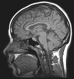
Cerebrospinal fluid (CSF) is a clear, colorless body fluid found within the tissue that surrounds the brain and spinal cord of all vertebrates.

<span class="mw-page-title-main">Idiopathic intracranial hypertension</span> Medical condition

Idiopathic intracranial hypertension (IIH), previously known as pseudotumor cerebri and benign intracranial hypertension, is a condition characterized by increased intracranial pressure without a detectable cause. The main symptoms are headache, vision problems, ringing in the ears, and shoulder pain. Complications may include vision loss.

<span class="mw-page-title-main">Viral meningitis</span> Medical condition

Viral meningitis, also known as aseptic meningitis, is a type of meningitis due to a viral infection. It results in inflammation of the meninges. Symptoms commonly include headache, fever, sensitivity to light and neck stiffness.

<span class="mw-page-title-main">Papilledema</span> Eye disorder

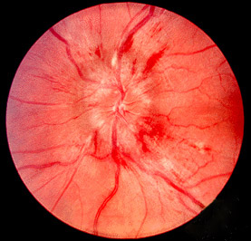
Papilledema or papilloedema is optic disc swelling that is caused by increased intracranial pressure due to any cause. The swelling is usually bilateral and can occur over a period of hours to weeks. Unilateral presentation is extremely rare.

<span class="mw-page-title-main">Intracranial pressure</span> Pressure exerted by fluids inside the skull and on the brain

Intracranial pressure (ICP) is the pressure exerted by fluids such as cerebrospinal fluid (CSF) inside the skull and on the brain tissue. ICP is measured in millimeters of mercury (mmHg) and at rest, is normally 7–15 mmHg for a supine adult. The body has various mechanisms by which it keeps the ICP stable, with CSF pressures varying by about 1 mmHg in normal adults through shifts in production and absorption of CSF.

<span class="mw-page-title-main">Spinal anaesthesia</span> Form of neuraxial regional anaesthesia

Spinal anaesthesia, also called spinal block, subarachnoid block, intradural block and intrathecal block, is a form of neuraxial regional anaesthesia involving the injection of a local anaesthetic or opioid into the subarachnoid space, generally through a fine needle, usually 9 cm (3.5 in) long. It is a safe and effective form of anesthesia usually performed by anesthesiologists that can be used as an alternative to general anesthesia commonly in surgeries involving the lower extremities and surgeries below the umbilicus. The local anesthetic with or without an opioid injected into the cerebrospinal fluid provides locoregional anaesthesia: true analgesia, motor, sensory and autonomic (sympathetic) blockade. Administering analgesics in the cerebrospinal fluid without a local anaesthetic produces locoregional analgesia: markedly reduced pain sensation, some autonomic blockade, but no sensory or motor block. Locoregional analgesia, due to mainly the absence of motor and sympathetic block may be preferred over locoregional anaesthesia in some postoperative care settings. The tip of the spinal needle has a point or small bevel. Recently, pencil point needles have been made available.

<span class="mw-page-title-main">Aseptic meningitis</span> Medical condition

Aseptic meningitis is the inflammation of the meninges, a membrane covering the brain and spinal cord, in patients whose cerebral spinal fluid test result is negative with routine bacterial cultures. Aseptic meningitis is caused by viruses, mycobacteria, spirochetes, fungi, medications, and cancer malignancies. The testing for both meningitis and aseptic meningitis is mostly the same. A cerebrospinal fluid sample is taken by lumbar puncture and is tested for leukocyte levels to determine if there is an infection and goes on to further testing to see what the actual cause is. The symptoms are the same for both meningitis and aseptic meningitis but the severity of the symptoms and the treatment can depend on the certain cause.

<span class="mw-page-title-main">Myelography</span> Medical imaging technique

Myelography is a type of radiographic examination that uses a contrast medium to detect pathology of the spinal cord, including the location of a spinal cord injury, cysts, and tumors. Historically the procedure involved the injection of a radiocontrast agent into the cervical or lumbar spine, followed by several X-ray projections. Today, myelography has largely been replaced by the use of MRI scans, although the technique is still sometimes used under certain circumstances – though now usually in conjunction with CT rather than X-ray projections.

<span class="mw-page-title-main">Tuberculous meningitis</span> Medical condition

Tuberculous meningitis, also known as TB meningitis or tubercular meningitis, is a specific type of bacterial meningitis caused by the Mycobacterium tuberculosis infection of the meninges—the system of membranes which envelop the central nervous system.

<span class="mw-page-title-main">Xanthochromia</span> Medical condition

Xanthochromia, from the Greek xanthos (ξανθός) "yellow" and chroma (χρώμα) "colour", is the yellowish appearance of cerebrospinal fluid that occurs several hours after bleeding into the subarachnoid space caused by certain medical conditions, most commonly subarachnoid hemorrhage. Its presence can be determined by either spectrophotometry or simple visual examination. It is unclear which method is superior.

Cisternography is a medical imaging technique to examine the flow of cerebrospinal fluid (CSF) in the brain, and spinal cord. The gold standard for diagnosis of a cranial cerebrospinal fluid leak is CT cisternography. For the diagnosis of a spinal CSF leak radionuclide cisternography also known as radioisotope cisternography is used. The third type of cisternography is MR cisternography.

<span class="mw-page-title-main">Leptomeningeal cancer</span> Medical condition

Leptomeningeal cancer is a rare complication of cancer in which the disease spreads from the original tumor site to the meninges surrounding the brain and spinal cord. This leads to an inflammatory response, hence the alternative names neoplastic meningitis (NM), malignant meningitis, or carcinomatous meningitis. The term leptomeningeal describes the thin meninges, the arachnoid and the pia mater, between which the cerebrospinal fluid is located. The disorder was originally reported by Eberth in 1870. It is also known as leptomeningeal carcinomatosis, leptomeningeal disease (LMD), leptomeningeal metastasis, meningeal metastasis and meningeal carcinomatosis.

<span class="mw-page-title-main">Epidural blood patch</span> Blood injected epidurally to resolve a cerebrospinal fluid leak

An epidural blood patch (EBP) is a surgical procedure that uses autologous blood, meaning the patient's own blood, in order to close one or many holes in the dura mater of the spinal cord, which occurred as a complication of a lumbar puncture or epidural placement. The punctured dura causes cerebrospinal fluid leak. The procedure can be used to relieve orthostatic headaches, most commonly post dural puncture headache (PDPH).

<span class="mw-page-title-main">External ventricular drain</span> Medical device

An external ventricular drain (EVD), also known as a ventriculostomy or extraventricular drain, is a device used in neurosurgery to treat hydrocephalus and relieve elevated intracranial pressure when the normal flow of cerebrospinal fluid (CSF) inside the brain is obstructed. An EVD is a flexible plastic catheter placed by a neurosurgeon or neurointensivist and managed by intensive care unit (ICU) physicians and nurses. The purpose of external ventricular drainage is to divert fluid from the ventricles of the brain and allow for monitoring of intracranial pressure. An EVD must be placed in a center with full neurosurgical capabilities, because immediate neurosurgical intervention can be needed if a complication of EVD placement, such as bleeding, is encountered.

<span class="mw-page-title-main">Mollaret's meningitis</span> Medical condition

Mollaret's meningitis is a recurrent or chronic inflammation of the protective membranes covering the brain and spinal cord, known collectively as the meninges. Since Mollaret's meningitis is a recurrent, benign (non-cancerous), aseptic meningitis, it is also referred to as benign recurrent lymphocytic meningitis. It was named for Pierre Mollaret, the French neurologist who first described it in 1944.

<span class="mw-page-title-main">Meningitis</span> Inflammation of the membranes around the brain and spinal cord

Meningitis is acute or chronic inflammation of the protective membranes covering the brain and spinal cord, collectively called the meninges. The most common symptoms are fever, intense headache, vomiting and neck stiffness and occasionally photophobia.

<span class="mw-page-title-main">Cerebrospinal fluid leak</span> Medical condition

A cerebrospinal fluid leak is a medical condition where the cerebrospinal fluid (CSF) that surrounds the brain and spinal cord leaks out of one or more holes or tears in the dura mater. A CSF leak is classed as either nonspontaneous (primary), having a known cause, or spontaneous (secondary) where the cause is not readily evident. Causes of a primary CSF leak are those of trauma including from an accident or intentional injury, or arising from a medical intervention known as iatrogenic. A basilar skull fracture as a cause can give the sign of CSF leakage from the ear nose or mouth. A lumbar puncture can give the symptom of a post-dural-puncture headache.

CSF glucose or glycorrhachia is a measurement used to determine the concentration of glucose in cerebrospinal fluid (CSF).

A lumbar–peritoneal shunt is a technique to channelise the cerebrospinal fluid (CSF) from the lumbar thecal sac into the peritoneal cavity.

<span class="mw-page-title-main">Lymphocytic pleocytosis</span> Increase in lymphocytes within cerebrospinal fluid

Lymphocytic pleocytosis is an abnormal increase in the amount of lymphocytes in the cerebrospinal fluid (CSF). It is usually considered to be a sign of infection or inflammation within the nervous system, and is encountered in a number of neurological diseases, such as pseudomigraine, Susac's syndrome, and encephalitis. While lymphocytes make up roughly a quarter of all white blood cells (WBC) in the body, they are generally rare in the CSF. Under normal conditions, there are usually less than 5 white blood cells per µL of CSF. In a pleocytic setting, the number of lymphocytes can jump to more than 1,000 cells per µL. Increases in lymphocyte count are often accompanied by an increase in cerebrospinal protein concentrations in addition to pleocytosis of other types of white blood cells.

References

  1. Maranhao, B.; Liu, M.; Palanisamy, A.; Monks, D. T.; Singh, P. M. (17 December 2020). "The association between post-dural puncture headache and needle type during spinal anaesthesia: a systematic review and network meta-analysis". Anaesthesia. Wiley. 76 (8): 1098–1110. doi: 10.1111/anae.15320 . ISSN   0003-2409. PMID   33332606.
  2. Gottlieb, M; Holladay, D; Peksa, GD (January 2019). "Ultrasound-assisted Lumbar Punctures: A Systematic Review and Meta-Analysis". Academic Emergency Medicine. 26 (1): 85–96. doi: 10.1111/acem.13558 . PMID   30129102.
  3. Doherty, Carolynne M; Forbes, Raeburn B (2014). "Diagnostic Lumbar Puncture". The Ulster Medical Journal. 83 (2): 93–102. ISSN   0041-6193. PMC   4113153 . PMID   25075138.
  4. 1 2 3 4 5 6 7 Sempere, AP; Berenguer-Ruiz, L; Lezcano-Rodas, M; Mira-Berenguer, F; Waez, M (2007). "Punción lumbar: indicaciones, contraindicaciones, complicaciones y técnica de realización" [Lumbar puncture: its indications, contraindications, complications and technique]. Revista de Neurología (in Spanish). 45 (7): 433–6. doi:10.33588/rn.4507.2007270. PMID   17918111.
  5. Gröschel, K; Schnaudigel, S; Pilgram, S; Wasser, K; Kastrup, A (19 December 2007). "Die diagnostische Lumbalpunktion" [The diagnostic lumbar puncture]. Deutsche Medizinische Wochenschrift (in German). 133 (1/02): 39–41. doi:10.1055/s-2008-1017470. PMID   18095209. S2CID   260115550.
  6. Matata, C; Michael, B; Garner, V; Solomon, T (24–30 October 2012). "Lumbar puncture: diagnosing acute central nervous system infections". Nursing Standard. 27 (8): 49–56, quiz 58. doi:10.7748/ns2012.10.27.8.49.c9364. PMID   23189602.
  7. 1 2 Visintin, C.; Mugglestone, M. A.; Fields, E. J.; Jacklin, P.; Murphy, M. S.; Pollard, A. J.; Guideline Development Group; National Institute for Health and Clinical Excellence (28 June 2010). "Management of bacterial meningitis and meningococcal septicaemia in children and young people: summary of NICE guidance". BMJ (Clinical Research Ed.). 340: c3209. doi:10.1136/bmj.c3209. PMID   20584794. S2CID   7685756.
  8. 1 2 3 4 Pessano, Sara; Romantsik, Olga; Olsson, Emma; Hedayati, Ehsan; Bruschettini, Matteo (2023). Cochrane Neonatal Group (ed.). "Pharmacological interventions for the management of pain and discomfort during lumbar puncture in newborn infants". Cochrane Database of Systematic Reviews. 2023 (9): CD015594. doi:10.1002/14651858.CD015594.pub2. PMC  10535798. PMID   37767875.
  9. Mann, David (2002). "The role of lumbar puncture in the diagnosis of subarachnoid hemorrhage when computed tomography is unavailable". Journal of the Canadian Association of Emergency Physicians. 4 (2): 102–105. doi: 10.1017/s1481803500006205 . PMID   17612428.
  10. 1 2 3 4 5 6 López, T; Sánchez, FJ; Garzón, JC; Muriel, C (January 2012). "Spinal anesthesia in pediatric patients". Minerva Anestesiologica. 78 (1): 78–87. doi:10.1111/j.1460-9592.2011.03769.x. PMID   22211775. S2CID   205522367.
  11. Biousse, V. (October 2012). "Idiopathic intracranial hypertension: Diagnosis, monitoring and treatment". Revue Neurologique. 168 (10): 673–683. doi:10.1016/j.neurol.2012.07.018. PMID   22981270.
  12. Kesler, Anat; Kupferminc, Michael (June 2013). "Idiopathic Intracranial Hypertension and Pregnancy". Clinical Obstetrics and Gynecology. 56 (2): 389–396. doi:10.1097/GRF.0b013e31828f2701. PMID   23563883. S2CID   20556772.
  13. Gallia, Gary L; Rigamonti, Daniele; Williams, Michael A (July 2006). "The diagnosis and treatment of idiopathic normal pressure hydrocephalus". Nature Clinical Practice Neurology. 2 (7): 375–381. doi:10.1038/ncpneuro0237. PMC   5390935 . PMID   16932588.
  14. Roos, KL (March 2003). "Lumbar puncture". Seminars in Neurology. 23 (1): 105–14. doi:10.1055/s-2003-40758. PMID   12870112. S2CID   41878896.
  15. 1 2 Straus, Sharon E.; Thorpe, Kevin E.; Holroyd-Leduc, Jayna (25 October 2006). "How Do I Perform a Lumbar Puncture and Analyze the Results to Diagnose Bacterial Meningitis?". JAMA. 296 (16): 2012–22. doi: 10.1001/jama.296.16.2012 . PMID   17062865.
  16. Zorrilla-Vaca, A; Makkar, JK (May 2017). "Effectiveness of Lateral Decubitus Position for Preventing Post-Dural Puncture Headache: A Meta-Analysis". Pain Physician. 20 (4): E521–E529. doi:10.36076/ppj.2017.E529. PMID   28535561.
  17. "Epidural Blood Patch - Imaging Glossary - Patients - UR Medicine Imaging Sciences (Radiology) - University of Rochester Medical Center". www.urmc.rochester.edu. Retrieved 24 April 2023.
  18. Nath, Siddharth; Koziarz, Alex; Badhiwala, Jetan H; Alhazzani, Waleed; Jaeschke, Roman; Sharma, Sunjay; Banfield, Laura; Shoamanesh, Ashkan; Singh, Sheila; Nassiri, Farshad; Oczkowski, Wieslaw; Belley-Côté, Emilie; Truant, Ray; Reddy, Kesava; Meade, Maureen O; Farrokhyar, Forough; Bala, Malgorzata M; Alshamsi, Fayez; Krag, Mette; Etxeandia-Ikobaltzeta, Itziar; Kunz, Regina; Nishida, Osamu; Matouk, Charles; Selim, Magdy; Rhodes, Andrew; Hawryluk, Gregory; Almenawer, Saleh A (March 2018). "Atraumatic versus conventional lumbar puncture needles: a systematic review and meta-analysis". The Lancet. 391 (10126): 1197–1204. doi:10.1016/S0140-6736(17)32451-0. PMID   29223694. S2CID   4436591.
  19. Rochwerg, Bram; Almenawer, Saleh A; Siemieniuk, Reed A C; Vandvik, Per Olav; Agoritsas, Thomas; Lytvyn, Lyubov; Alhazzani, Waleed; Archambault, Patrick; D’Aragon, Frederick; Farhoumand, Pauline Darbellay; Guyatt, Gordon; Laake, Jon Henrik; Beltrán-Arroyave, Claudia; McCredie, Victoria; Price, Amy; Chabot, Christian; Zervakis, Tracy; Badhiwala, Jetan; St-Onge, Maude; Szczeklik, Wojciech; Møller, Morten Hylander; Lamontagne, Francois (22 May 2018). "Atraumatic (pencil-point) versus conventional needles for lumbar puncture: a clinical practice guideline". BMJ. 361: k1920. doi:10.1136/bmj.k1920. PMC   6364256 . PMID   29789372.
  20. Davis, A; Dobson, R; Kaninia, S; Giovannoni, G; Schmierer, K (February 2016). "Atraumatic needles for lumbar puncture: why haven't neurologists changed?". Practical Neurology. 16 (1): 18–22. doi:10.1136/practneurol-2014-001055. PMID   26349834. S2CID   36928177.
  21. Patel, R.; Urits, I.; Orhurhu, V.; Orhurhu, M. S.; Peck, J.; Ohuabunwa, E.; Sikorski, A.; Mehrabani, A.; Manchikanti, L.; Kaye, A. D.; Kaye, R. J.; Helmstetter, J. A.; Viswanath, O. (2020). "A Comprehensive Update on the Treatment and Management of Postdural Puncture Headache". Current Pain and Headache Reports. 24 (6): 24. doi:10.1007/s11916-020-00860-0. PMID   32323013. S2CID   216049548.
  22. Joffe, Ari R. (29 June 2016). "Lumbar Puncture and Brain Herniation in Acute Bacterial Meningitis: A Review". Journal of Intensive Care Medicine. 22 (4): 194–207. doi:10.1177/0885066607299516. PMID   17712055. S2CID   22924383.
  23. Ragab, Ashraf; Facharzt, Khalid Noman (2014). "Caffeine, is it effective for prevention of postdural puncture headache in young adult patients?". Egyptian Journal of Anaesthesia. 30 (2): 181–186. doi: 10.1016/j.egja.2013.11.005 . S2CID   71403201.
  24. "Iatrogenic CSF Leaks from Lumbar Punctures – a commentary". 20 February 2015.
  25. "Cerebrospinal Fluid (CSF) Leak: Symptoms & Treatment".
  26. "Cerebrospinal Fluid Leak (CSF Leak) FAQ".
  27. 1 2 3 4 5 6 Wright, Ben L. C.; Lai, James T. F.; Sinclair, Alexandra J. (26 January 2012). "Cerebrospinal fluid and lumbar puncture: a practical review". Journal of Neurology. 259 (8): 1530–1545. doi:10.1007/s00415-012-6413-x. PMID   22278331. S2CID   2563483.
  28. Shaikh, F.; Brzezinski, J.; Alexander, S.; Arzola, C.; Carvalho, J. C. A.; Beyene, J.; Sung, L. (26 March 2013). "Ultrasound imaging for lumbar punctures and epidural catheterisations: systematic review and meta-analysis". BMJ. 346 (mar26 1): f1720. doi: 10.1136/bmj.f1720 . PMID   23532866.
  29. Cauley, Keith A. (October 2015). "Fluoroscopically Guided Lumbar Puncture". American Journal of Roentgenology. 205 (4): W442–W450. doi:10.2214/AJR.14.14028. PMID   26397351.
  30. Hanson, Amy L.; Ros, Simon; Soprano, Joyce (May 2014). "Analysis of Infant Lumbar Puncture Success Rates". Pediatric Emergency Care. 30 (5): 311–314. doi:10.1097/PEC.0000000000000119. PMID   24759486. S2CID   43633453.
  31. 1 2 Cramer, Gregory D.; Yu, Shi-Wei (2014). "Unique Anatomic Features of the Pediatric Spine". Clinical Anatomy of the Spine, Spinal Cord, and Ans. pp. 566–585. doi:10.1016/b978-0-323-07954-9.00013-x. ISBN   978-0-323-07954-9.
  32. 1 2 Bonadio, William (January 2014). "Pediatric Lumbar Puncture and Cerebrospinal Fluid Analysis". The Journal of Emergency Medicine. 46 (1): 141–150. doi:10.1016/j.jemermed.2013.08.056. PMID   24188604.
  33. Harald Kluge (2007). Atlas of CSF cytology. Thieme. pp. 45–46. ISBN   978-3-13-143161-5 . Retrieved 28 October 2010.
  34. Pearson, Justin; Fuller, Geraint (August 2012). "Lumbar punctures and cerebrospinal fluid analysis". Medicine. 40 (8): 459–462. doi:10.1016/j.mpmed.2012.05.005. S2CID   73285011.
  35. Nigrovic, Lise E.; Chiang, Vincent W. (1 August 2000). "Cost Analysis of Enteroviral Polymerase Chain Reaction in Infants With Fever and Cerebrospinal Fluid Pleocytosis". Archives of Pediatrics & Adolescent Medicine. 154 (8): 817–821. doi: 10.1001/archpedi.154.8.817 . PMID   10922279.
  36. Rand, Kenneth; Houck, Herbert; Lawrence, Robert (October 2005). "Real-Time Polymerase Chain Reaction Detection of Herpes Simplex Virus in Cerebrospinal Fluid and Cost Savings from Earlier Hospital Discharge". The Journal of Molecular Diagnostics. 7 (4): 511–516. doi:10.1016/S1525-1578(10)60582-X. PMC   1888494 . PMID   16237221.
  37. Zerpa, R; Huicho, L; Guillén, A (September 1996). "Modified India ink preparation for Cryptococcus neoformans in cerebrospinal fluid specimens". Journal of Clinical Microbiology. 34 (9): 2290–1. doi:10.1128/JCM.34.9.2290-2291.1996. PMC   229234 . PMID   8862601.
  38. Shashikala.; Kanungo, R; Srinivasan, S; Mathew, R; Kannan, M (2004). "Unusual morphological forms of Cryptococcus neoformans in cerebrospinal fluid". Indian Journal of Medical Microbiology. 22 (3): 188–90. doi: 10.1016/S0255-0857(21)02835-8 . PMID   17642731.
  39. Antinori, S; Radice, A; Galimberti, L; Magni, C; Fasan, M; Parravicini, C (November 2005). "The role of cryptococcal antigen assay in diagnosis and monitoring of cryptococcal meningitis". Journal of Clinical Microbiology. 43 (11): 5828–9. doi:10.1128/JCM.43.11.5828-5829.2005. PMC   1287839 . PMID   16272534.
  40. Nigrovic, Lise E.; Kimia, Amir A.; Shah, Samir S.; Neuman, Mark I. (9 February 2012). "Relationship between Cerebrospinal Fluid Glucose and Serum Glucose". New England Journal of Medicine. 366 (6): 576–578. doi: 10.1056/NEJMc1111080 . PMID   22316468.
  41. Hendry, E (June 1939). "The blood and spinal fluid sugar and chloride content in meningitis". Archives of Disease in Childhood. 14 (78): 159–72. doi:10.1136/adc.14.78.159. PMC   1975626 . PMID   21032142.
  42. Gierson, HW; Marx, JI (April 1955). "Tuberculous meningitis: the diagnostic and prognostic significance of spinal fluid sugar and chloride". Annals of Internal Medicine. 42 (4): 902–8. doi:10.7326/0003-4819-42-4-902. PMID   14362261.
  43. De Vivo, Darryl C.; Trifiletti, Rosario R.; Jacobson, Ronald I.; Ronen, Gabriel M.; Behmand, Ramin A.; Harik, Sami I. (5 September 1991). "Defective Glucose Transport across the Blood-Brain Barrier as a Cause of Persistent Hypoglycorrhachia, Seizures, and Developmental Delay". New England Journal of Medicine. 325 (10): 703–709. doi: 10.1056/NEJM199109053251006 . PMID   1714544.
  44. 1 2 Leen, Wilhelmina G.; Willemsen, Michèl A.; Wevers, Ron A.; Verbeek, Marcel M.; Mendelson, John E. (6 August 2012). "Cerebrospinal Fluid Glucose and Lactate: Age-Specific Reference Values and Implications for Clinical Practice". PLOS ONE. 7 (8): e42745. Bibcode:2012PLoSO...742745L. doi: 10.1371/journal.pone.0042745 . PMC   3412827 . PMID   22880096.
  45. Servo, C; Pitkänen, E (December 1975). "Variation in polyol levels in cerebrospinal fluid and serum in diabetic patients". Diabetologia. 11 (6): 575–80. doi: 10.1007/BF01222109 . PMID   1205026.
  46. "Cerebrospinal Fluid Glutamine". clinicallabtesting.com. Archived from the original on 11 August 2013. Retrieved 11 August 2013.
  47. Hourani, Benjamin T.; Hamlin, EM; Reynolds, TB (1 June 1971). "Cerebrospinal Fluid Glutamine as a Measure of Hepatic Encephalopathy". Archives of Internal Medicine. 127 (6): 1033–6. doi:10.1001/archinte.1971.00310180049005. PMID   5578559.
  48. 1 2 Cascino, A.; Cangiano, C.; Fiaccadori, F.; Ghinelli, F.; Merli, M.; Pelosi, G.; Riggio, O.; Rossi Fanelli, F.; Sacchini, D.; Stortoni, M.; Capocaccia, L. (September 1982). "Plasma and cerebrospinal fluid amino acid patterns in hepatic encephalopathy". Digestive Diseases and Sciences. 27 (9): 828–32. doi:10.1007/BF01391377. PMID   7105954. S2CID   8186910.
  49. Glasgow, Allen M.; Dhiensiri, Kamnual (June 1974). "Improved Assay for Spinal Fluid Glutamine, and Values for Children with Reye's Syndrome" (PDF). Clinical Chemistry. 20 (6): 642–644. doi:10.1093/clinchem/20.6.642. PMID   4830166.
  50. Watanabe, A; Takei, N; Higashi, T; Shiota, T; Nakatsukasa, H; Fujiwara, M; Sakata, T; Nagashima, H (October 1984). "Glutamic acid and glutamine levels in serum and cerebrospinal fluid in hepatic encephalopathy". Biochemical Medicine. 32 (2): 225–31. doi:10.1016/0006-2944(84)90076-0. PMID   6150706.
  51. Levine, J; Panchalingam, K; Rapoport, A; Gershon, S; McClure, RJ; Pettegrew, JW (1 April 2000). "Increased cerebrospinal fluid glutamine levels in depressed patients". Biological Psychiatry. 47 (7): 586–93. doi:10.1016/S0006-3223(99)00284-X. PMID   10745050. S2CID   33396490.
  52. Jay H. Stein (1998). Internal Medicine. Elsevier Health Sciences. pp. 1408–. ISBN   978-0-8151-8698-4 . Retrieved 12 August 2013.
  53. Reiber, Hansotto (2003). "Proteins in cerebrospinal fluid and blood: Barriers, CSF flow rate and source-related dynamics" (PDF). Restorative Neurology and Neuroscience. 21 (3–4): 79–96. PMID   14530572.
  54. Conly, John M.; Ronald, Allan R. (July 1983). "Cerebrospinal fluid as a diagnostic body fluid". The American Journal of Medicine. 75 (1): 102–108. doi:10.1016/0002-9343(83)90080-3. PMID   6349337.
  55. 1 2 3 Pearson, Justin; Fuller, Geraint (August 2012). "Lumbar punctures and cerebrospinal fluid analysis". Medicine. 40 (8): 459–462. doi:10.1016/j.mpmed.2012.05.005. S2CID   73285011.
  56. Wynter W. E. (1891). "Four Cases of Tubercular Meningitis in Which Paracentesis of the Theca Vertebralis Was Performed for the Relief of Fluid Pressure". Lancet. 1 (3531): 981–2. doi:10.1016/S0140-6736(02)16784-5.
  57. Quincke, H (1891). "Verhandlungen des Congresses für Innere Medizin" [Negotiations of the Congress of Internal Medicine]. Proceedings of the Zehnter Congress (in German): 321–31.
  58. Quincke HI (1902). Die Technik der Lumbalpunktion[The technique of lumbar puncture] (in German). Berlin & Vienna.{{cite book}}: CS1 maint: location missing publisher (link)[ page needed ]
  59. Heinrich Irenaeus Quincke at Who Named It?
  60. Susan E. Lederer (1997). Subjected to Science: Human Experimentation in America Before the Second World War. JHU Press. p. 216. ISBN   978-0-8018-5709-6. Page 62 has a reference to an 1896 publication in Boston Med. Surg. J

Further reading