Ventriculitis

Last updated
Ventriculitis
Gray734.png
Ventricular system (shaded) superimposed on an image of the brain depicting spatial arrangement
Specialty Neurology   OOjs UI icon edit-ltr-progressive.svg

Ventriculitis is the inflammation of the ventricles in the brain. The ventricles are responsible for containing and circulating cerebrospinal fluid throughout the brain. Ventriculitis is caused by infection of the ventricles, leading to swelling and inflammation. This is especially prevalent in patients with external ventricular drains and intraventricular stents. [1] Ventriculitis can cause a wide variety of short-term symptoms and long-term side effects ranging from headaches and dizziness to unconsciousness and death if not treated early. It is treated with some appropriate combination of antibiotics in order to rid the patient of the underlying infection. Much of the current research involving ventriculitis focuses specifically around defining the disease and what causes it. This will allow for much more advancement in the subject. [2] There is also a lot of attention being paid to possible treatments and prevention methods to help make this disease even less prevalent and dangerous.

Contents

Signs and symptoms

EarlyAdvancedSevere
HeadachesNausea and VomitingHydrocephalus
Neck/upper back painsVertigoBrain abscess
Painful cranial pressureSlurred speechTemporary loss of consciousness
DizzinessMental InstabilityImpaired mental function
ConfusionRigorsDeath

There is great deal of variety in the symptoms associated with ventriculitis. The symptoms vary based on a number of different factors including severity of inflammation, underlying cause, and the individual patient.

Patients often present with headaches, painful cranial pressure, and neck pain early in the progression of the disease. Patients with a more advanced infection have been known to complain of many neurological effects such as dizziness, vertigo, confusion, and slurred speech. Very advanced cases can lead to mental instability, nausea, vomiting, rigors, and temporary loss of consciousness. Many patients with ventriculitis also experience some degree of hydrocephalus, which is the buildup of cerebrospinal fluid due to the inability of the ventricles to reabsorb and correctly circulate the fluid. [2] Brain abscess is another common disorder resulting from the inflammation. If left untreated, ventriculitis can lead to serious inhibition of mental function and even death.

The symptoms vary greatly, in part, because of the underlying or causing infection. While the inflammation can cause a number of effects such as those mentioned previously, the base infection could cause other symptoms that don't necessarily have to do with the ventriculitis, itself. One of the challenges doctors face in diagnosing ventriculitis is distinguishing indicative symptoms, in spite of the wide variety of possible presentations of the disease. A great deal of emphasis is being put on research into better and faster ways to diagnose ventriculitis without the delay inherent with microbiological testing of the cerebrospinal fluid. [3]

The progression of the disease is also largely dependent on the nature of the specific case. Depending on the underlying infection, the way it entered the brain, and the type and timing of treatment, the infection may spread or withdraw on the order of months or days. Ventriculitis is a very serious condition and should be treated early to ensure as little lasting damage as possible.

Cause

Ventricles
Gray735.png
Gray's representation of the ventricular system, as viewed from above
Gray736.png
Gray's representation of the ventricular system, as viewed from the side
Identifiers
MeSH D058565
Anatomical terms of neuroanatomy

Ventriculitis is caused by an infection of the ventricles, causing an immune response in the lining, which in turn, leads to inflammation. The ventriculitis, is in truth, a complication of the initial infection or abnormality. The underlying infection can come in the form of a number of different bacteria or viruses. The data seems to point to Staphylococci as the leading bacterial cause of infection leading to ventriculitis being present in about 90% of cases, [4] but generally, what is of more concern is the way the infection entered the ventricles. The brain in its natural state is very protected from infection. The blood–brain barrier serves to keep pathogens from entering the sensitive areas of the brain. However, when those natural defenses are by-passed in the hospital setting, the brain is suddenly exposed to a host of potentially harmful bacteria and viruses.

Patients that have had invasive brain surgery or procedures are considered to be the most at risk for experiencing ventriculitis. Two procedures, in particular have been studied extensively due to their high rate of ventriculitis contractions post operation. The first group consists of patients that have had an external ventricular drain implanted to allow physicians to reduce the intracranial pressure they experience. The duration that the drain is implanted varies by necessity, however, the longer the drain is in, the more likely an infection will occur. [5] The second group consists of patients that have an implanted intracranial stent. Both groups of patients have a much higher rate of ventriculitis than the general populace, though there is very little supporting evidence due to the lack of definition of ventriculitis as frequent misdiagnosis. Nearly 25% of patients with an external ventricular drain experience infection-based meningitis or ventriculitis. [1]

Diagnosis

Ventriculitis is commonly diagnosed using a variety of tests or procedures. When a physician suspects that a patient has ventriculitis, the first step is typically to ascertain the presence of the inflammation using computed tomography (CT) or magnetic resonance imaging (MRI) technology to "take a picture" of the brain. The scans allow physicians to check for "intraventricular debris and pus, abnormal periventricular and subependymal signal intensity, and enhancement of the ventricular lining," all of which indicate the likelihood of ventriculitis. [6] MRIs have been reported as being highly effective and sensitive in detecting such indicators, even from an early stage.

After determining whether a patient shows signs of ventriculitis, the doctor may choose to pursue a more specific and useful diagnosis to find the cause of the ventriculitis. This is done by obtaining a sample of cerebrospinal fluid, most commonly via a procedure called a lumbar puncture or spinal tap. For patients with an implanted external ventricular drain, cerebrospinal fluid can be collected from the drain's output. After the sample of fluid is obtained, a battery of tests featuring gram staining will be performed to identify any offending pathogen or infection agent. The test will also determine any resistance the pathogen may have to antibiotics. By identifying the viral or bacterial cause of the ventriculitis, doctors are more able to effectively treat the inflammation and infection. This procedure is fairly effective, but is rarely able to isolate anaerobic organisms that may be causing the inflammation, giving cause for further research and procedural development. [7]

Though they present with similar symptoms and often occur in tandem, meningitis and ventriculitis are two different diseases, so physicians must be able to distinguish between the two. Meningitis is the inflammation of the protective lining of the central nervous system, called meninges. Because of the similar pathologies and cause of the two types of inflammation, they are difficult to differentiate using chemical testing, but show very different visual effects in both the MRI and CT scans, hence their use as a validation that the patient does, in fact, have ventriculitis and not another, similar condition such as meningitis. [8]

Treatment

Treatment of ventriculitis is critical. If left untreated, it could lead to severe brain damage and even death in some cases. Currently, the only commonly employed treatments of ventriculitis involve an antibiotic regimen targeting the underlying infection causing the inflammation. Typically, the physician will order the patient be placed on broad-spectrum antibiotics in order to manage the symptoms and control the infection while the cerebrospinal fluid samples are analyzed. When a specific bacterial or viral cause is found, the doctor will change the treatment accordingly. There is some debate as to the most effective antibiotics and the best ways to introduce the drugs (e.g. intravenously, orally, etc.), however it is agreed that drug effectiveness is limited by the difficulty of non-invasively allowing the drugs to enter the cerebrospinal fluid. [9] Should intracranial pressure reach unsafe levels, the patient may need to have cerebrospinal fluid drained. Implanted external ventricular drains are one of the more common ways to manage and monitor the intracranial pressure, however there are several risks involved with such an invasive procedure, including the risk of further infection.

There is a great deal of research focused around prevention of ventriculitis. It is crucial that any procedure involving exposing the brain is performed with the utmost care, as infections in the brain are very dangerous and potentially deadly. When patients undergo such procedures, they are often monitored closely over the next several days to ensure that there were no infections and any instance of even a small headache is treated very seriously. It is also necessary to monitor the intracranial pressure of the patients often enough to observe significant changes that could indicate the presence of and infection and ensuing ventriculitis. It is important not to measure the pressure too often, however, as it could in fact lead to infection.

Current research

Due to the poor definition of the condition that is ventriculitis, there is still a great deal that is not known about this dangerous condition. While other, similar conditions, such as meningitis or encephalitis, have been thoroughly researched, ventriculitis is a very loose grouping of conditions characterized by the fact that the lining of the ventricles is inflamed. Because no solid definition has been accepted across the medical community, research in the subject has been slow to progress. [2] However, most common research into ventriculitis has been focused on the main points of causation, demographic information, and effectiveness of treatments and prevention methods.

Causation

One of the key areas of research for ventriculitis is discovering and defining exactly what causes it. There are many bacterial and viral infections that could cause inflammation of the ventricles, but researchers are trying to define which are the most common pathogens, the risk levels associated with various medical operations and procedures, and why the symptoms vary so much on a case-by-case basis. [2] Answering these questions will allow doctors to not only better understand ventriculitis, but better treat and prevent it as well.

Demographics

Currently, there is very little understanding as to who is at increased risk for ventriculitis, other than those who have undergone neurosurgery or procedures involving brain exposure. Even then, current clinical practices can't predict which patients will be affected. [10] In order to predict which populations should be focused on, researchers must gather more case information about who is diagnosed with ventriculitis and how they present. In essence, the medical community must compile data of as many details as possible from each case so that more generalized conclusions may be drawn.

Treatment and prevention

So little is currently known about how ventriculitis should be defined and those it affects that even less can be known about prevention methods. While treatment is fairly standard for any infection to some degree, prevention is a different matter. One popular theory is the use of prophylactic antibiotics, administered during insertion of external ventricular drains or ventricular stents with the hope of preventing infection. [11] The results of these studies have been more or less inconclusive due to a lack of standardized protocol, showing no significant benefit to using antibiotics as a preventative measure. [12]

Related Research Articles

<span class="mw-page-title-main">Brain abscess</span> Medical condition

Brain abscess is an abscess within the brain tissue caused by inflammation and collection of infected material coming from local or remote infectious sources. The infection may also be introduced through a skull fracture following a head trauma or surgical procedures. Brain abscess is usually associated with congenital heart disease in young children. It may occur at any age but is most frequent in the third decade of life.

<span class="mw-page-title-main">Cerebrospinal fluid</span> Clear, colorless bodily fluid found in the brain and spinal cord

Cerebrospinal fluid (CSF) is a clear, colorless body fluid found within the tissue that surrounds the brain and spinal cord of all vertebrates.

<span class="mw-page-title-main">Idiopathic intracranial hypertension</span> Medical condition

Idiopathic intracranial hypertension (IIH), previously known as pseudotumor cerebri and benign intracranial hypertension, is a condition characterized by increased intracranial pressure without a detectable cause. The main symptoms are headache, vision problems, ringing in the ears, and shoulder pain. Complications may include vision loss.

<span class="mw-page-title-main">Viral meningitis</span> Medical condition

Viral meningitis, also known as aseptic meningitis, is a type of meningitis due to a viral infection. It results in inflammation of the meninges. Symptoms commonly include headache, fever, sensitivity to light and neck stiffness.

<span class="mw-page-title-main">Hydrocephalus</span> Abnormal increase in cerebrospinal fluid in the ventricles of the brain

Hydrocephalus is a condition in which an accumulation of cerebrospinal fluid (CSF) occurs within the brain. This typically causes increased pressure inside the skull. Older people may have headaches, double vision, poor balance, urinary incontinence, personality changes, or mental impairment. In babies, it may be seen as a rapid increase in head size. Other symptoms may include vomiting, sleepiness, seizures, and downward pointing of the eyes.

<span class="mw-page-title-main">Colpocephaly</span> Medical condition

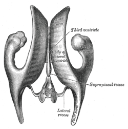
Colpocephaly is a cephalic disorder involving the disproportionate enlargement of the occipital horns of the lateral ventricles and is usually diagnosed early after birth due to seizures. It is a nonspecific finding and is associated with multiple neurological syndromes, including agenesis of the corpus callosum, Chiari malformation, lissencephaly, and microcephaly. Although the exact cause of colpocephaly is not known yet, it is commonly believed to occur as a result of neuronal migration disorders during early brain development, intrauterine disturbances, perinatal injuries, and other central nervous system disorders. Individuals with colpocephaly have various degrees of motor disabilities, visual defects, spasticity, and moderate to severe intellectual disability. No specific treatment for colpocephaly exists, but patients may undergo certain treatments to improve their motor function or intellectual disability.

<span class="mw-page-title-main">Lumbar puncture</span> Procedure to collect cerebrospinal fluid

Lumbar puncture (LP), also known as a spinal tap, is a medical procedure in which a needle is inserted into the spinal canal, most commonly to collect cerebrospinal fluid (CSF) for diagnostic testing. The main reason for a lumbar puncture is to help diagnose diseases of the central nervous system, including the brain and spine. Examples of these conditions include meningitis and subarachnoid hemorrhage. It may also be used therapeutically in some conditions. Increased intracranial pressure is a contraindication, due to risk of brain matter being compressed and pushed toward the spine. Sometimes, lumbar puncture cannot be performed safely. It is regarded as a safe procedure, but post-dural-puncture headache is a common side effect if a small atraumatic needle is not used.

Normal-pressure hydrocephalus (NPH), also called malresorptive hydrocephalus, is a form of communicating hydrocephalus in which excess cerebrospinal fluid (CSF) occurs in the ventricles, and with normal or slightly elevated cerebrospinal fluid pressure. As the fluid builds up, it causes the ventricles to enlarge and the pressure inside the head to increase, compressing surrounding brain tissue and leading to neurological complications. The disease presents in a classic triad of symptoms, which are memory impairment, urinary frequency, and balance problems/gait deviations. The disease was first described by Salomón Hakim and Adams in 1965.

<span class="mw-page-title-main">Craniotomy</span> Surgical operation on skull

A craniotomy is a surgical operation in which a bone flap is temporarily removed from the skull to access the brain. Craniotomies are often critical operations, performed on patients who are suffering from brain lesions, such as tumors, blood clots, removal of foreign bodies such as bullets, or traumatic brain injury (TBI), and can also allow doctors to surgically implant devices, such as deep brain stimulators for the treatment of Parkinson's disease, epilepsy, and cerebellar tremor. The procedure is also used in epilepsy surgery to remove the parts of the brain that are causing epilepsy.

<span class="mw-page-title-main">Cerebral shunt</span> Surgical implant to treat hydrocephalus

A cerebral shunt is a device permanently implanted inside the head and body to drain excess fluid away from the brain. They are commonly used to treat hydrocephalus, the swelling of the brain due to excess buildup of cerebrospinal fluid (CSF). If left unchecked, the excess CSF can lead to an increase in intracranial pressure (ICP), which can cause intracranial hematoma, cerebral edema, crushed brain tissue or herniation. The drainage provided by a shunt can alleviate or prevent these problems in patients with hydrocephalus or related diseases.

<span class="mw-page-title-main">Intraventricular hemorrhage</span> Medical condition

Intraventricular hemorrhage (IVH), also known as intraventricular bleeding, is a bleeding into the brain's ventricular system, where the cerebrospinal fluid is produced and circulates through towards the subarachnoid space. It can result from physical trauma or from hemorrhagic stroke.

<span class="mw-page-title-main">External ventricular drain</span> Medical device

An external ventricular drain (EVD), also known as a ventriculostomy or extraventricular drain, is a device used in neurosurgery to treat hydrocephalus and relieve elevated intracranial pressure when the normal flow of cerebrospinal fluid (CSF) inside the brain is obstructed. An EVD is a flexible plastic catheter placed by a neurosurgeon or neurointensivist and managed by intensive care unit (ICU) physicians and nurses. The purpose of external ventricular drainage is to divert fluid from the ventricles of the brain and allow for monitoring of intracranial pressure. An EVD must be placed in a center with full neurosurgical capabilities, because immediate neurosurgical intervention can be needed if a complication of EVD placement, such as bleeding, is encountered.

<span class="mw-page-title-main">Mollaret's meningitis</span> Medical condition

Mollaret's meningitis is a recurrent or chronic inflammation of the protective membranes covering the brain and spinal cord, known collectively as the meninges. Since Mollaret's meningitis is a recurrent, benign (non-cancerous), aseptic meningitis, it is also referred to as benign recurrent lymphocytic meningitis. It was named for Pierre Mollaret, the French neurologist who first described it in 1944.

<span class="mw-page-title-main">Meningitis</span> Inflammation of the membranes around the brain and spinal cord

Meningitis is acute or chronic inflammation of the protective membranes covering the brain and spinal cord, collectively called the meninges. The most common symptoms are fever, headache, and neck stiffness. Other symptoms include confusion or altered consciousness, nausea, vomiting, and an inability to tolerate light or loud noises. Young children often exhibit only nonspecific symptoms, such as irritability, drowsiness, or poor feeding. A non-blanching rash may also be present.

<span class="mw-page-title-main">Cerebrospinal fluid leak</span> Medical condition

A cerebrospinal fluid leak is a medical condition where the cerebrospinal fluid (CSF) surrounding the brain or spinal cord leaks out of one or more holes or tears in the dura mater. A cerebrospinal fluid leak can be either cranial or spinal, and these are two different disorders. A spinal CSF leak can be caused by one or more meningeal diverticula or CSF-venous fistulas not associated with an epidural leak.

Bobble-head doll syndrome is a rare neurological movement disorder in which patients, usually children around age 3, begin to bob their head and shoulders forward and back, or sometimes side-to-side, involuntarily, in a manner reminiscent of a bobblehead doll. The syndrome is related to cystic lesions and swelling of the third ventricle in the brain. Symptoms of bobble-head doll syndrome are diverse but can be grouped into two categories: physical and neurological. The most common form of treatment is surgical implanting of a shunt to relieve the swelling of the brain.

Citrobacter koseri, formerly known as Citrobacter diversus, is a Gram-negative, non-spore-forming bacillus. It is a facultative anaerobe capable of aerobic respiration. It is motile via peritrichous flagella. It is a member of the family of Enterobacteriaceae. The members of this family are the part of the normal flora and commonly found in the digestive tracts of humans and animals. C. koseri may act as an opportunistic pathogen in individuals who are immunocompromised.

<span class="mw-page-title-main">Neonatal meningitis</span> Medical condition

Neonatal meningitis is a serious medical condition in infants that is rapidly fatal if untreated. Meningitis is an inflammation of the meninges, the protective membranes of the central nervous system, is more common in the neonatal period than any other time in life, and is an important cause of morbidity and mortality globally. Mortality is roughly half in developing countries and ranges from 8%-12.5% in developed countries.

<span class="mw-page-title-main">Aqueductal stenosis</span> Narrowing of the aqueduct of Sylvius

Aqueductal stenosis is a narrowing of the aqueduct of Sylvius which blocks the flow of cerebrospinal fluid (CSF) in the ventricular system. Blockage of the aqueduct can lead to hydrocephalus, specifically as a common cause of congenital and/or obstructive hydrocephalus.

<span class="mw-page-title-main">Intracerebroventricular injection</span> Injection into the cerebrospinal fluid

Intracerebroventricular injection is a route of administration for drugs via injection into the cerebral ventricles so that it reaches the cerebrospinal fluid (CSF). This route of administration is often used to bypass the blood-brain barrier because it can prevent important medications from reaching the central nervous system. This injection method is widely used in diseased mice models to study the effect of drugs, plasmid DNA, and viral vectors on the central nervous system. In humans, ICV injection can be used for the administration of drugs for various reasons. Examples include the treatment of Spinal Muscular Atrophy (SMA), the administration of chemotherapy in gliomas, and the administration of drugs for long-term pain management. ICV injection is also used in the creation of diseased animal models specifically to model neurological disorders.

References

  1. 1 2 Hoefnagel D, Dammers R, Ter Laak-Poort MP, Avezaat CJ (March 2008). "Risk factors for infections related to external ventricular drainage". Acta Neurochir (Wien). 150 (3): 209–14, discussion 214. doi:10.1007/s00701-007-1458-9. PMID   18278575. S2CID   24671596.
  2. 1 2 3 4 Agrawal A, Cincu R, Timothy J (2008). "Current Concepts and Approach to Ventriculitis". Infectious Diseases in Clinical Practice. 16 (2): 100–104. doi:10.1097/IPC.0b013e318142ce2c. ISSN   1056-9103. S2CID   71993640.
  3. Schroeder S, Stuerenburg HJ, Escherich F, Pfeiffer G (November 2000). "Lysozyme in ventriculitis: a marker for diagnosis and disease progression". J. Neurol. Neurosurg. Psychiatry. 69 (5): 696–7. doi:10.1136/jnnp.69.5.696. PMC   1763430 . PMID   11032636.
  4. Wildemann B, Oschmann P, Reiber H (2010). Laboratory Diagnosis in Neurology. Thieme. ISBN   978-3-13-144101-0.
  5. Moon HJ, Kim SD, Lee JB, Lim DJ, Park JY (2007). "Clinical Analysis of External Ventricular Drainage Related Ventriculitis". Journal of Korean Neurosurgical Society . 41 (4): 236. doi: 10.3340/jkns.2007.41.4.236 . ISSN   1225-8245.
  6. Fujikawa A, Tsuchiya K, Honya K, Nitatori T (October 2006). "Comparison of MRI sequences to detect ventriculitis". AJR Am J Roentgenol. 187 (4): 1048–53. doi:10.2214/AJR.04.1923. PMID   16985156.
  7. Kai A, Cooke F, Antoun N, Siddharthan C, Sule O (May 2008). "A rare presentation of ventriculitis and brain abscess caused by Fusobacterium nucleatum". J. Med. Microbiol. 57 (Pt 5): 668–71. doi: 10.1099/jmm.0.47710-0 . PMID   18436604.
  8. Coen M, O'Sullivan M, Bubb WA, Kuchel PW, Sorrell T (December 2005). "Proton nuclear magnetic resonance-based metabonomics for rapid diagnosis of meningitis and ventriculitis". Clin. Infect. Dis. 41 (11): 1582–90. doi: 10.1086/497836 . PMID   16267730.
  9. Dalgic N, Ceylan Y, Sancar M, Telhan L, Kafadar I, Cavusoglu H, Ceylan O, Hasim O (June 2009). "Successful treatment of multidrug-resistant Acinetobacter baumannii ventriculitis with intravenous and intraventricular colistin". Ann Trop Paediatr. 29 (2): 141–7. doi:10.1179/146532809X440761. PMID   19460268. S2CID   8371191.
  10. Muttaiyah S, Ritchie S, Upton A, Roberts S (February 2008). "Clinical parameters do not predict infection in patients with external ventricular drains: a retrospective observational study of daily cerebrospinal fluid analysis". J. Med. Microbiol. 57 (Pt 2): 207–9. doi: 10.1099/jmm.0.47518-0 . PMID   18201987.
  11. Prabhu VC, Kaufman HH, Voelker JL, Aronoff SC, Niewiadomska-Bugaj M, Mascaro S, Hobbs GR (September 1999). "Prophylactic antibiotics with intracranial pressure monitors and external ventricular drains: a review of the evidence". Surg Neurol. 52 (3): 226–36, discussion 236–7. doi:10.1016/s0090-3019(99)00084-1. PMID   10511079.
  12. Lucey MA, Myburgh JA (September 2003). "Antibiotic prophylaxis for external ventricular drains in neurosurgical patients: an audit of compliance with a clinical management protocol". Crit Care Resusc. 5 (3): 182–5. PMID   16573480.