Neurodegenerative disease

Last updated
Neurodegenerative disease
Alzheimer's disease brain comparison.jpg
Normal brain on left contrasted with structural changes shown in brain on right of person with Alzheimer's disease, the most common neurodegenerative disease [1]
Specialty Neurology, Psychiatry

A neurodegenerative disease is caused by the progressive loss of structure or function of neurons, in the process known as neurodegeneration. Such neuronal damage may ultimately involve cell death. Neurodegenerative diseases include amyotrophic lateral sclerosis, multiple sclerosis, Parkinson's disease, Alzheimer's disease, Huntington's disease, multiple system atrophy, tauopathies, and prion diseases. Neurodegeneration can be found in the brain at many different levels of neuronal circuitry, ranging from molecular to systemic. Because there is no known way to reverse the progressive degeneration of neurons, these diseases are considered to be incurable; however research has shown that the two major contributing factors to neurodegeneration are oxidative stress and inflammation. [2] [3] [4] [5] Biomedical research has revealed many similarities between these diseases at the subcellular level, including atypical protein assemblies (like proteinopathy) and induced cell death. [6] [7] These similarities suggest that therapeutic advances against one neurodegenerative disease might ameliorate other diseases as well.

Contents

Within neurodegenerative diseases, it is estimated that 55 million people worldwide had dementia in 2019, and that by 2050 this figure will increase to 139 million people. [8]

Specific disorders

The consequences of neurodegeneration can vary widely depending on the specific region affected, ranging from issues related to movement to the development of dementia. [9] [10]

Alzheimer's disease

Comparison of brain tissue between healthy individual and Alzheimer's disease patient, demonstrating extent of neuronal death Alzheimers brain.jpg
Comparison of brain tissue between healthy individual and Alzheimer's disease patient, demonstrating extent of neuronal death

Alzheimer's disease (AD) is a chronic neurodegenerative disease that results in the loss of neurons and synapses in the cerebral cortex and certain subcortical structures, resulting in gross atrophy of the temporal lobe, parietal lobe, and parts of the frontal cortex and cingulate gyrus. [11] It is the most common neurodegenerative disease. [1] Even with billions of dollars being used to find a treatment for Alzheimer's disease, no effective treatments have been found. [12] However, clinical trials have developed certain compounds that could potentially change the future of Alzheimer's disease treatments. [13] Within clinical trials stable and effective AD therapeutic strategies have a 99.5% failure rate. [14] Reasons for this failure rate include inappropriate drug doses, invalid target and participant selection, and inadequate knowledge of pathophysiology of AD. Currently, diagnoses of Alzheimer's is subpar, and better methods need to be utilized for various aspects of clinical diagnoses. [15] Alzheimer's has a 20% misdiagnosis rate. [15]

AD pathology is primarily characterized by the presence of amyloid plaques and neurofibrillary tangles. Plaques are made up of small peptides, typically 39–43 amino acids in length, called amyloid beta (also written as A-beta or Aβ). Amyloid beta is a fragment from a larger protein called amyloid precursor protein (APP), a transmembrane protein that penetrates through the neuron's membrane. APP appears to play roles in normal neuron growth, survival and post-injury repair. [16] [17] APP is cleaved into smaller fragments by enzymes such as gamma secretase and beta secretase. [18] One of these fragments gives rise to fibrils of amyloid beta which can self-assemble into the dense extracellular amyloid plaques. [19] [20]

Parkinson's disease

Parkinson's disease (PD) is the second most common neurodegenerative disorder. [21] It typically manifests as bradykinesia, rigidity, resting tremor and posture instability. The crude prevalence rate of PD has been reported to range from 15 per 100,000 to 12,500 per 100,000, and the incidence of PD from 15 per 100,000 to 328 per 100,000, with the disease being less common in Asian countries.

PD is primarily characterized by death of dopaminergic neurons in the substantia nigra, a region of the midbrain. The cause of this selective cell death is unknown. Notably, alpha-synuclein-ubiquitin complexes and aggregates are observed to accumulate in Lewy bodies within affected neurons. It is thought that defects in protein transport machinery and regulation, such as RAB1, may play a role in this disease mechanism. [22] Impaired axonal transport of alpha-synuclein may also lead to its accumulation in Lewy bodies. Experiments have revealed reduced transport rates of both wild-type and two familial Parkinson's disease-associated mutant alpha-synucleins through axons of cultured neurons. [23] Membrane damage by alpha-synuclein could be another Parkinson's disease mechanism. [24]

The main known risk factor is age. Mutations in genes such as α-synuclein (SNCA), leucine-rich repeat kinase 2 (LRRK2), glucocerebrosidase (GBA), and tau protein (MAPT) can also cause hereditary PD or increase PD risk. [25] While PD is the second most common neurodegenerative disorder, problems with diagnoses still persist. [26] Problems with the sense of smell is a widespread symptom of Parkinson's disease (PD), however, some neurologists question its efficacy. [26] This assessment method is a source of controversy among medical professionals. [26] The gut microbiome might play a role in the diagnosis of PD, and research suggests various ways that could revolutionize the future of PD treatment. [27]

Huntington's disease

Huntington's disease (HD) is a rare autosomal dominant neurodegenerative disorder caused by mutations in the huntingtin gene (HTT). HD is characterized by loss of medium spiny neurons and astrogliosis. [28] [29] [30] The first brain region to be substantially affected is the striatum, followed by degeneration of the frontal and temporal cortices. [31] The striatum's subthalamic nuclei send control signals to the globus pallidus, which initiates and modulates motion. The weaker signals from subthalamic nuclei thus cause reduced initiation and modulation of movement, resulting in the characteristic movements of the disorder, notably chorea. [32] Huntington's disease presents itself later in life even though the proteins that cause the disease works towards manifestation from their early stages in the humans affected by the proteins. [33] Along with being a neurodegenerative disorder, HD has links to problems with neurodevelopment. [33]

HD is caused by polyglutamine tract expansion in the huntingtin gene, resulting in the mutant huntingtin. Aggregates of mutant huntingtin form as inclusion bodies in neurons, and may be directly toxic. Additionally, they may damage molecular motors and microtubules to interfere with normal axonal transport, leading to impaired transport of important cargoes such as BDNF. [23] Huntington's disease currently has no effective treatments that would modify the disease. [34]

Multiple sclerosis

Multiple sclerosis (MS) is a chronic debilitating demyelinating disease of the central nervous system, caused by an autoimmune attack resulting in the progressive loss of myelin sheath on neuronal axons. [35] The resultant decrease in the speed of signal transduction leads to a loss of functionality that includes both cognitive and motor impairment depending on the location of the lesion. [35] The progression of MS occurs due to episodes of increasing inflammation, which is proposed to be due to the release of antigens such as myelin oligodendrocyte glycoprotein, myelin basic protein, and proteolipid protein, causing an autoimmune response. [36] This sets off a cascade of signaling molecules that result in T cells, B cells, and macrophages to cross the blood-brain barrier and attack myelin on neuronal axons leading to inflammation. [37] Further release of antigens drives subsequent degeneration causing increased inflammation. [38] Multiple sclerosis presents itself as a spectrum based on the degree of inflammation, a majority of patients experience early relapsing and remitting episodes of neuronal deterioration following a period of recovery. Some of these individuals may transition to a more linear progression of the disease, while about 15% of others begin with a progressive course on the onset of multiple sclerosis. The inflammatory response contributes to the loss of the grey matter, and as a result current literature devotes itself to combatting the auto-inflammatory aspect of the disease. [37] While there are several proposed causal links between EBV and the HLA-DRB1*15:01 allele to the onset of MS – they may contribute to the degree of autoimmune attack and the resultant inflammation – they do not determine the onset of MS. [37]

Amyotrophic lateral sclerosis

Amyotrophic lateral sclerosis (ALS), commonly referred to Lou Gehrig's disease, is a rare neurodegenerative disorder characterized by the gradual loss of both upper motor neurons (UMNs) and lower motor neurons (LMNs). [39] Although initial symptoms may vary, most patients develop skeletal muscle weakness that progresses to involve the entire body. [39] The precise etiology of ALS remains unknown. In 1993, missense mutations in the gene encoding the antioxidant enzyme superoxide dismutase 1 (SOD1) were discovered in a subsets of patients with familial ALS. More recently, TAR DNA-binding protein 43 (TDP-43) and Fused in Sarcoma (FUS) protein aggregates have been implicated in some cases of the disease, and a mutation in chromosome 9 (C9orf72) is thought to be the most common known cause of sporadic ALS. Early diagnosis of ALS is harder than with other neurodegenerative diseases as there are no highly effective means of determining its early onset. [39] Currently, there is research being done regarding the diagnosis of ALS through upper motor neuron tests. [40] The Penn Upper Motor Neuron Score (PUMNS) consists of 28 criteria with a score range of 0–32. [40] A higher score indicates a higher level of burden present on the upper motor neurons. [40] The PUMNS has proven quite effective in determining the burden that exists on upper motor neurons in affected patients. [40]

Independent research provided in vitro evidence that the primary cellular sites where SOD1 mutations act are located on astrocytes. [41] [42] Astrocytes then cause the toxic effects on the motor neurons. The specific mechanism of toxicity still needs to be investigated, but the findings are significant because they implicate cells other than neuron cells in neurodegeneration. [43]

Batten disease

Batten disease is a rare and fatal recessive neurodegenerative disorder that begins in childhood. [44] Batten disease is the common name for a group of lysosomal storage disorders known as neuronal ceroid lipofuscinoses (NCLs) – each caused by a specific gene mutation, [44] of which there are thirteen. [45] Since Batten disease is quite rare, its worldwide prevalence is about 1 in every 100,000 live births. [46] In North America, CLN3 disease (juvenile NCL) typically manifests between the ages of 4 and 7. [47] Batten disease is characterized by motor impairment, epilepsy, dementia, vision loss, and shortened lifespan. [48] A loss of vision is common first sign of Batten disease. [47] Loss of vision is typically preceded by cognitive and behavioral changes, seizures, and loss of the ability to walk. [47] It is common for people to establish cardiac arrhythmias and difficulties eating food as the disease progresses. [47] Batten disease diagnosis depends on a conflation of many criteria: clinical signs and symptoms, evaluations of the eye, electroencephalograms (EEG), and brain magnetic resonance imaging (MRI) results. [46] The diagnosis provided by these results are corroborated by genetic and biochemical testing. [46] No effective treatments were available to prevent the disease from being widespread before the past few years. [46] In recent years, more models have been created to expedite the research process for methods to treat Batten disease. [46]

Creutzfeldt–Jakob disease

Creutzfeldt–Jakob disease (CJD) is a prion disease that is characterized by rapidly progressive dementia. [49] Misfolded proteins called prions aggregate in brain tissue leading to nerve cell death. [50] Variant Creutzfeldt–Jakob disease (vCJD) is the infectious form that comes from the meat of a cow that was infected with bovine spongiform encephalopathy, also called mad cow disease. [51]

Risk factor

The greatest risk factor for neurodegenerative diseases is aging. Mitochondrial DNA mutations as well as oxidative stress both contribute to aging. [52] Many of these diseases are late-onset, meaning there is some factor that changes as a person ages for each disease. [6] One constant factor is that in each disease, neurons gradually lose function as the disease progresses with age. It has been proposed that DNA damage accumulation provides the underlying causative link between aging and neurodegenerative disease. [53] [54] About 20–40% of healthy people between 60 and 78 years old experience discernable decrements in cognitive performance in several domains including working, spatial, and episodic memory, and processing speed. [55]

Risks from viral exposures according to one biobank study Hazard ratio lag for replicated viral infection-neurodegenerative disease pairs.jpg
Risks from viral exposures according to one biobank study

A study using electronic health records indicates that 45 (with 22 of these being replicated with the UK Biobank) viral exposures can significantly elevate risks of neurodegenerative disease, including up to 15 years after infection. [56] [57]

Mechanisms

Genetics

Many neurodegenerative diseases are caused by genetic mutations, most of which are located in completely unrelated genes. In many of the different diseases, the mutated gene has a common feature: a repeat of the CAG nucleotide triplet. CAG codes for the amino acid glutamine. A repeat of CAG results in a polyglutamine (polyQ) tract. Diseases associated with such mutations are known as trinucleotide repeat disorders. [58] [59]

Polyglutamine repeats typically cause dominant pathogenesis. Extra glutamine residues can acquire toxic properties through a variety of ways, including irregular protein folding and degradation pathways, altered subcellular localization, and abnormal interactions with other cellular proteins. [58] PolyQ studies often use a variety of animal models because there is such a clearly defined trigger – repeat expansion. Extensive research has been done using the models of nematode (C. elegans), and fruit fly (Drosophila), mice, and non-human primates. [59] [60]

Nine inherited neurodegenerative diseases are caused by the expansion of the CAG trinucleotide and polyQ tract, including Huntington's disease and the spinocerebellar ataxias. [61]

Epigenetics

The presence of epigenetic modifications for certain genes has been demonstrated in this type of pathology. An example is FKBP5 gene, which progressively increases its expression with age and has been related to Braak staging and increased tau pathology both in vitro and in mouse models of AD. [62]

Protein misfolding

Several neurodegenerative diseases are classified as proteopathies as they are associated with the aggregation of misfolded proteins. Protein toxicity is one of the key mechanisms of many neurodegenrative diseases. [63]

Intracellular mechanisms

Protein degradation pathways

Parkinson's disease and Huntington's disease are both late-onset and associated with the accumulation of intracellular toxic proteins. Diseases caused by the aggregation of proteins are known as proteopathies, and they are primarily caused by aggregates in the following structures: [6]

  • cytosol, e.g. Parkinson's and Huntington's
  • nucleus, e.g. Spinocerebellar ataxia type 1 (SCA1)
  • endoplasmic reticulum (ER), (as seen with neuroserpin mutations that cause familial encephalopathy with neuroserpin inclusion bodies)
  • extracellularly excreted proteins, amyloid-beta in Alzheimer's disease

There are two main avenues eukaryotic cells use to remove troublesome proteins or organelles:

  • ubiquitin–proteasome: protein ubiquitin along with enzymes is key for the degradation of many proteins that cause proteopathies including polyQ expansions and alpha-synucleins. Research indicates proteasome enzymes may not be able to correctly cleave these irregular proteins, which could possibly result in a more toxic species. This is the primary route cells use to degrade proteins. [6]
    • Decreased proteasome activity is consistent with models in which intracellular protein aggregates form. It is still unknown whether or not these aggregates are a cause or a result of neurodegeneration. [6]
  • autophagy–lysosome pathways: a form of programmed cell death (PCD), this becomes the favorable route when a protein is aggregate-prone meaning it is a poor proteasome substrate. This can be split into two forms of autophagy: macroautophagy and chaperone-mediated autophagy (CMA). [6]
    • macroautophagy is involved with nutrient recycling of macromolecules under conditions of starvation, certain apoptotic pathways, and if absent, leads to the formation of ubiquinated inclusions. Experiments in mice with neuronally confined macroautophagy-gene knockouts develop intraneuronal aggregates leading to neurodegeneration. [6]
    • chaperone-mediated autophagy defects may also lead to neurodegeneration. Research has shown that mutant proteins bind to the CMA-pathway receptors on lysosomal membrane and in doing so block their own degradation as well as the degradation of other substrates. [6]

Membrane damage

Damage to the membranes of organelles by monomeric or oligomeric proteins could also contribute to these diseases. Alpha-synuclein can damage membranes by inducing membrane curvature, [24] and cause extensive tubulation and vesiculation when incubated with artificial phospholipid vesicles. [24] The tubes formed from these lipid vesicles consist of both micellar as well as bilayer tubes. Extensive induction of membrane curvature is deleterious to the cell and would eventually lead to cell death. Apart from tubular structures, alpha-synuclein can also form lipoprotein nanoparticles similar to apolipoproteins.

Mitochondrial dysfunction

The most common form of cell death in neurodegeneration is through the intrinsic mitochondrial apoptotic pathway. This pathway controls the activation of caspase-9 by regulating the release of cytochrome c from the mitochondrial intermembrane space. Reactive oxygen species (ROS) are normal byproducts of mitochondrial respiratory chain activity. ROS concentration is mediated by mitochondrial antioxidants such as manganese superoxide dismutase (SOD2) and glutathione peroxidase. Over production of ROS (oxidative stress) is a central feature of all neurodegenerative disorders. In addition to the generation of ROS, mitochondria are also involved with life-sustaining functions including calcium homeostasis, PCD, mitochondrial fission and fusion, lipid concentration of the mitochondrial membranes, and the mitochondrial permeability transition. Mitochondrial disease leading to neurodegeneration is likely, at least on some level, to involve all of these functions. [64]

There is strong evidence that mitochondrial dysfunction and oxidative stress play a causal role in neurodegenerative disease pathogenesis, including in four of the more well known diseases Alzheimer's, Parkinson's, Huntington's, and amyotrophic lateral sclerosis. [52]

Neurons are particularly vulnerable to oxidative damage due to their strong metabolic activity associated with high transcription levels, high oxygen consumption, and weak antioxidant defense. [65] [66]

DNA damage

The brain metabolizes as much as a fifth of consumed oxygen, and reactive oxygen species produced by oxidative metabolism are a major source of DNA damage in the brain. Damage to a cell's DNA is particularly harmful because DNA is the blueprint for protein production and unlike other molecules it cannot simply be replaced by re-synthesis. The vulnerability of post-mitotic neurons to DNA damage (such as oxidative lesions or certain types of DNA strand breaks), coupled with a gradual decline in the activities of repair mechanisms, could lead to accumulation of DNA damage with age and contribute to brain aging and neurodegeneration. [67] DNA single-strand breaks are common and are associated with the neurodegenerative disease ataxia-oculomotor apraxia. [68] [66] Increased oxidative DNA damage in the brain is associated with Alzheimer's disease and Parkinson's disease. [68] Defective DNA repair has been linked to neurodegenerative disorders such as Alzheimer's disease, amyotrophic lateral sclerosis, ataxia telangiectasia, Cockayne syndrome, Parkinson's disease and xeroderma pigmentosum. [68] [67]

Axonal transport

Axonal swelling, and axonal spheroids have been observed in many different neurodegenerative diseases. This suggests that defective axons are not only present in diseased neurons, but also that they may cause certain pathological insult due to accumulation of organelles. Axonal transport can be disrupted by a variety of mechanisms including damage to: kinesin and cytoplasmic dynein, microtubules, cargoes, and mitochondria. [23] When axonal transport is severely disrupted a degenerative pathway known as Wallerian-like degeneration is often triggered. [69]

Programmed cell death

Programmed cell death (PCD) is death of a cell in any form, mediated by an intracellular program. [70] This process can be activated in neurodegenerative diseases including Parkinson's disease, amytrophic lateral sclerosis, Alzheimer's disease and Huntington's disease. [71] PCD observed in neurodegenerative diseases may be directly pathogenic; alternatively, PCD may occur in response to other injury or disease processes. [7]

Apoptosis (type I)

Apoptosis is a form of programmed cell death in multicellular organisms. It is one of the main types of programmed cell death (PCD) and involves a series of biochemical events leading to a characteristic cell morphology and death.

  • Extrinsic apoptotic pathways: Occur when factors outside the cell activate cell surface death receptors (e.g., Fas) that result in the activation of caspases-8 or -10. [7]
  • Intrinsic apoptotic pathways: Result from mitochondrial release of cytochrome c or endoplasmic reticulum malfunctions, each leading to the activation of caspase-9. The nucleus and Golgi apparatus are other organelles that have damage sensors, which can lead the cells down apoptotic pathways. [7] [72]

Caspases (cysteine-aspartic acid proteases) cleave at very specific amino acid residues. There are two types of caspases: initiators and effectors. Initiator caspases cleave inactive forms of effector caspases. This activates the effectors that in turn cleave other proteins resulting in apoptotic initiation. [7]

Autophagic (type II)

Autophagy is a form of intracellular phagocytosis in which a cell actively consumes damaged organelles or misfolded proteins by encapsulating them into an autophagosome, which fuses with a lysosome to destroy the contents of the autophagosome. Because many neurodegenerative diseases show unusual protein aggregates, it is hypothesized that defects in autophagy could be a common mechanism of neurodegeneration. [7]

Cytoplasmic (type III)

PCD can also occur via non-apoptotic processes, also known as Type III or cytoplasmic cell death. For example, type III PCD might be caused by trophotoxicity, or hyperactivation of trophic factor receptors. Cytotoxins that induce PCD can cause necrosis at low concentrations, or aponecrosis (combination of apoptosis and necrosis) at higher concentrations. It is still unclear exactly what combination of apoptosis, non-apoptosis, and necrosis causes different kinds of aponecrosis. [7]

Transglutaminase

Transglutaminases are human enzymes ubiquitously present in the human body and in the brain in particular. [73]

The main function of transglutaminases is bind proteins and peptides intra- and intermolecularly, by a type of covalent bonds termed isopeptide bonds, in a reaction termed transamidation or crosslinking. [73]

Transglutaminase binding of these proteins and peptides make them clump together. The resulting structures are turned extremely resistant to chemical and mechanical disruption. [73]

Most relevant human neurodegenerative diseases share the property of having abnormal structures made up of proteins and peptides. [73]

Each of these neurodegenerative diseases have one (or several) specific main protein or peptide. In Alzheimer's disease, these are amyloid-beta and tau. In Parkinson's disease, it is alpha-synuclein. In Huntington's disease, it is huntingtin. [73]

Transglutaminase substrates: Amyloid-beta, tau, alpha-synuclein and huntingtin have been proved to be substrates of transglutaminases in vitro or in vivo, that is, they can be bonded by trasglutaminases by covalent bonds to each other and potentially to any other transglutaminase substrate in the brain. [73]

Transglutaminase augmented expression: It has been proved that in these neurodegenerative diseases (Alzheimer's disease, Parkinson's disease, and Huntington's disease) the expression of the transglutaminase enzyme is increased. [73]

Presence of isopeptide bonds in these structures: The presence of isopeptide bonds (the result of the transglutaminase reaction) have been detected in the abnormal structures that are characteristic of these neurodegenerative diseases. [73]

Co-localization: Co-localization of transglutaminase mediated isopeptide bonds with these abnormal structures has been detected in the autopsy of brains of patients with these diseases. [73]

Management

The process of neurodegeneration is not well understood, so the diseases that stem from it have, as yet, no cures.

Animal models in research

In the search for effective treatments (as opposed to palliative care), investigators employ animal models of disease to test potential therapeutic agents. Model organisms provide an inexpensive and relatively quick means to perform two main functions: target identification and target validation. Together, these help show the value of any specific therapeutic strategies and drugs when attempting to ameliorate disease severity. An example is the drug Dimebon by Medivation, Inc. In 2009 this drug was in phase III clinical trials for use in Alzheimer's disease, and also phase II clinical trials for use in Huntington's disease. [59] In March 2010, the results of a clinical trial phase III were released; the investigational Alzheimer's disease drug Dimebon failed in the pivotal CONNECTION trial of patients with mild-to-moderate disease. [74] With CONCERT, the remaining Pfizer and Medivation Phase III trial for Dimebon (latrepirdine) in Alzheimer's disease failed in 2012, effectively ending the development in this indication. [75]

In another experiment using a rat model of Alzheimer's disease, it was demonstrated that systemic administration of hypothalamic proline-rich peptide (PRP)-1 offers neuroprotective effects and can prevent neurodegeneration in hippocampus amyloid-beta 25–35. This suggests that there could be therapeutic value to PRP-1. [76]

Other avenues of investigation

Protein degradation offers therapeutic options both in preventing the synthesis and degradation of irregular proteins. There is also interest in upregulating autophagy to help clear protein aggregates implicated in neurodegeneration. Both of these options involve very complex pathways that we are only beginning to understand. [6]

The goal of immunotherapy is to enhance aspects of the immune system. Both active and passive vaccinations have been proposed for Alzheimer's disease and other conditions; however, more research must be done to prove safety and efficacy in humans. [77]

A current therapeutic target for the treatment of Alzheimer's disease is the protease β-secretase [78] [ non-primary source needed ], which is involved in the amyloidogenic processing pathway that leads to the pathological accumulation of proteins in the brain. When the gene that encodes for amyloid precursor protein (APP) is spliced by α-secretase [79] [ non-primary source needed ] rather than β-secretase, the toxic protein β amyloid is not produced. Targeted inhibition [80] of β-secretase can potentially prevent the neuronal death that is responsible for the symptoms of Alzheimer's disease.

Dr. Antonio Barbera, a former obstetrics and gynaecology doctor, is prescribing table tennis for patients who are suffering from a serious neurological disorder. [81]

See also

Related Research Articles

<span class="mw-page-title-main">Alpha-synuclein</span> Protein found in humans

Alpha-synuclein(aSyn) is a protein that, in humans, is encoded by the SNCA gene. Alpha-synuclein is a neuronal protein that regulates synaptic vesicle trafficking and subsequent neurotransmitter release.

Degenerative disease is the result of a continuous process based on degenerative cell changes, affecting tissues or organs, which will increasingly deteriorate over time.

<span class="mw-page-title-main">Excitotoxicity</span> Process that kills nerve cells

In excitotoxicity, nerve cells suffer damage or death when the levels of otherwise necessary and safe neurotransmitters such as glutamate become pathologically high, resulting in excessive stimulation of receptors. For example, when glutamate receptors such as the NMDA receptor or AMPA receptor encounter excessive levels of the excitatory neurotransmitter, glutamate, significant neuronal damage might ensue. Excess glutamate allows high levels of calcium ions (Ca2+) to enter the cell. Ca2+ influx into cells activates a number of enzymes, including phospholipases, endonucleases, and proteases such as calpain. These enzymes go on to damage cell structures such as components of the cytoskeleton, membrane, and DNA. In evolved, complex adaptive systems such as biological life it must be understood that mechanisms are rarely, if ever, simplistically direct. For example, NMDA in subtoxic amounts induces neuronal survival of otherwise toxic levels of glutamate.

Molecular neuroscience is a branch of neuroscience that observes concepts in molecular biology applied to the nervous systems of animals. The scope of this subject covers topics such as molecular neuroanatomy, mechanisms of molecular signaling in the nervous system, the effects of genetics and epigenetics on neuronal development, and the molecular basis for neuroplasticity and neurodegenerative diseases. As with molecular biology, molecular neuroscience is a relatively new field that is considerably dynamic.

<span class="mw-page-title-main">Neuroprotection</span> Relative preservation of neuronal structure and/or function

Neuroprotection refers to the relative preservation of neuronal structure and/or function. In the case of an ongoing insult the relative preservation of neuronal integrity implies a reduction in the rate of neuronal loss over time, which can be expressed as a differential equation. It is a widely explored treatment option for many central nervous system (CNS) disorders including neurodegenerative diseases, stroke, traumatic brain injury, spinal cord injury, and acute management of neurotoxin consumption. Neuroprotection aims to prevent or slow disease progression and secondary injuries by halting or at least slowing the loss of neurons. Despite differences in symptoms or injuries associated with CNS disorders, many of the mechanisms behind neurodegeneration are the same. Common mechanisms of neuronal injury include decreased delivery of oxygen and glucose to the brain, energy failure, increased levels in oxidative stress, mitochondrial dysfunction, excitotoxicity, inflammatory changes, iron accumulation, and protein aggregation. Of these mechanisms, neuroprotective treatments often target oxidative stress and excitotoxicity—both of which are highly associated with CNS disorders. Not only can oxidative stress and excitotoxicity trigger neuron cell death but when combined they have synergistic effects that cause even more degradation than on their own. Thus limiting excitotoxicity and oxidative stress is a very important aspect of neuroprotection. Common neuroprotective treatments are glutamate antagonists and antioxidants, which aim to limit excitotoxicity and oxidative stress respectively.

β-Methylamino-<small>L</small>-alanine Chemical compound

β-Methylamino-L-alanine, or BMAA, is a non-proteinogenic amino acid produced by cyanobacteria. BMAA is a neurotoxin. Its potential role in various neurodegenerative disorders is the subject of scientific research.

<span class="mw-page-title-main">Proteinopathy</span> Medical condition

In medicine, proteinopathy, or proteopathy, protein conformational disorder, or protein misfolding disease, is a class of diseases in which certain proteins become structurally abnormal, and thereby disrupt the function of cells, tissues and organs of the body. Often the proteins fail to fold into their normal configuration; in this misfolded state, the proteins can become toxic in some way or they can lose their normal function. The proteinopathies include such diseases as Creutzfeldt–Jakob disease and other prion diseases, Alzheimer's disease, Parkinson's disease, amyloidosis, multiple system atrophy, and a wide range of other disorders. The term proteopathy was first proposed in 2000 by Lary Walker and Harry LeVine.

<span class="mw-page-title-main">VPS35</span> Protein-coding gene in the species Homo sapiens

Vacuolar protein sorting ortholog 35 (VPS35) is a protein involved in autophagy and is implicated in neurodegenerative diseases, such as Parkinson's disease (PD) and Alzheimer's disease (AD). VPS35 is part of a complex called the retromer, which is responsible for transporting select cargo proteins between vesicular structures and the Golgi apparatus. Mutations in the VPS35 gene (VPS35) cause aberrant autophagy, where cargo proteins fail to be transported and dysfunctional or unnecessary proteins fail to be degraded. There are numerous pathways affected by altered VPS35 levels and activity, which have clinical significance in neurodegeneration. There is therapeutic relevance for VPS35, as interventions aimed at correcting VPS35 function are in speculation.

<span class="mw-page-title-main">Brain mitochondrial carrier protein 1</span> Protein-coding gene in the species Homo sapiens

Brain mitochondrial carrier protein 1 is a protein that in humans is encoded by the SLC25A14 gene.

<span class="mw-page-title-main">TMEM106B</span> Protein-coding gene in the species Homo sapiens

Transmembrane protein 106B is a protein that is encoded by the TMEM106B gene. It is found primarily within neurons and oligodendrocytes in the central nervous system with its subcellular location being in lysosomal membranes. TMEM106B helps facilitate important functions for maintaining a healthy lysosome, and therefore certain mutations and polymorphisms can lead to issues with proper lysosomal function. Lysosomes are in charge of clearing out mis-folded proteins and other debris, and thus, play an important role in neurodegenerative diseases that are driven by the accumulation of various mis-folded proteins and aggregates. Due to its impact on lysosomal function, TMEM106B has been investigated and found to be associated to multiple neurodegenerative diseases.

Nervous system diseases, also known as nervous system or neurological disorders, refers to a small class of medical conditions affecting the nervous system. This category encompasses over 600 different conditions, including genetic disorders, infections, cancer, seizure disorders, conditions with a cardiovascular origin, congenital and developmental disorders, and degenerative disorders.

Neuroinflammation is inflammation of the nervous tissue. It may be initiated in response to a variety of cues, including infection, traumatic brain injury, toxic metabolites, or autoimmunity. In the central nervous system (CNS), including the brain and spinal cord, microglia are the resident innate immune cells that are activated in response to these cues. The CNS is typically an immunologically privileged site because peripheral immune cells are generally blocked by the blood–brain barrier (BBB), a specialized structure composed of astrocytes and endothelial cells. However, circulating peripheral immune cells may surpass a compromised BBB and encounter neurons and glial cells expressing major histocompatibility complex molecules, perpetuating the immune response. Although the response is initiated to protect the central nervous system from the infectious agent, the effect may be toxic and widespread inflammation as well as further migration of leukocytes through the blood–brain barrier may occur.

<span class="mw-page-title-main">Estrogen and neurodegenerative diseases</span>

Neurodegenerative diseases can disrupt the normal human homeostasis and result in abnormal estrogen levels. For example, neurodegenerative diseases can cause different physiological effects in males and females. In particular, estrogen studies have revealed complex interactions with neurodegenerative diseases. Estrogen was initially proposed to be a possible treatment for certain types of neurodegenerative diseases but a plethora of harmful side effects such as increased susceptibility to breast cancer and coronary heart disease overshadowed any beneficial outcomes. On the other hand, Estrogen Replacement Therapy has shown some positive effects with postmenopausal women. Estrogen and estrogen-like molecules form a large family of potentially beneficial alternatives that can have dramatic effects on human homeostasis and disease. Subsequently, large-scale efforts were initiated to screen for useful estrogen family molecules. Furthermore, scientists discovered new ways to synthesize estrogen-like compounds that can avoid many side effects.

<span class="mw-page-title-main">Synucleinopathy</span> Medical condition

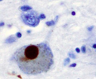
Synucleinopathies are neurodegenerative diseases characterised by the abnormal accumulation of aggregates of alpha-synuclein protein in neurons, nerve fibres or glial cells. There are three main types of synucleinopathy: Parkinson's disease (PD), dementia with Lewy bodies (DLB), and multiple system atrophy (MSA). Other rare disorders, such as various neuroaxonal dystrophies, also have α-synuclein pathologies. Additionally, autopsy studies have shown that around 6% of sporadic Alzheimer's Disease exhibit α-synuclein positive Lewy pathology, and are sub-classed as Alzheimer's Disease with Amygdalar Restricted Lewy Bodies (AD/ALB).

<span class="mw-page-title-main">Epigenetics of neurodegenerative diseases</span> Field of study

Neurodegenerative diseases are a heterogeneous group of complex disorders linked by the degeneration of neurons in either the peripheral nervous system or the central nervous system. Their underlying causes are extremely variable and complicated by various genetic and/or environmental factors. These diseases cause progressive deterioration of the neuron resulting in decreased signal transduction and in some cases even neuronal death. Peripheral nervous system diseases may be further categorized by the type of nerve cell affected by the disorder. Effective treatment of these diseases is often prevented by lack of understanding of the underlying molecular and genetic pathology. Epigenetic therapy is being investigated as a method of correcting the expression levels of misregulated genes in neurodegenerative diseases.

<span class="mw-page-title-main">Pathophysiology of Parkinson's disease</span> Medical condition

The pathophysiology of Parkinson's disease is death of dopaminergic neurons as a result of changes in biological activity in the brain with respect to Parkinson's disease (PD). There are several proposed mechanisms for neuronal death in PD; however, not all of them are well understood. Five proposed major mechanisms for neuronal death in Parkinson's Disease include protein aggregation in Lewy bodies, disruption of autophagy, changes in cell metabolism or mitochondrial function, neuroinflammation, and blood–brain barrier (BBB) breakdown resulting in vascular leakiness.

<span class="mw-page-title-main">Virginia Man-Yee Lee</span> American neuroscientist and biochemist

Virginia Man-Yee Lee is a Chinese-born American biochemist and neuroscientist who specializes in the research of Alzheimer's disease. She is the current John H. Ware 3rd Endowed Professor in Alzheimer's Research at the Department of Pathology and Laboratory Medicine, and the director of the Center for Neurodegenerative Disease Research and co-director of the Marian S. Ware Alzheimer Drug Discovery Program at the Perelman School of Medicine, University of Pennsylvania. She received the 2020 Breakthrough Prize in Life Sciences.

The neuroscience of aging is the study of the changes in the nervous system that occur with ageing. Aging is associated with many changes in the central nervous system, such as mild atrophy of the cortex that is considered non-pathological. Aging is also associated with many neurological and neurodegenerative disease such as amyotrophic lateral sclerosis, dementia, mild cognitive impairment, Parkinson's disease, and Creutzfeldt–Jakob disease.

Ted M. Dawson is an American neurologist and neuroscientist. He is the Leonard and Madlyn Abramson Professor in Neurodegenerative Diseases and Director of the Institute for Cell Engineering at Johns Hopkins University School of Medicine. He has joint appointments in the Department of Neurology, Neuroscience and Department of Pharmacology and Molecular Sciences.

<span class="mw-page-title-main">Animal models of Parkinson's disease</span> Models used in Parkinsons disease research

Animal models of Parkinson's disease are essential in the research field and widely used to study Parkinson's disease. Parkinson's disease is a neurodegenerative disorder, characterized by the loss of dopaminergic neurons in the substantia nigra pars compacta (SNpc). The loss of the dopamine neurons in the brain, results in motor dysfunction, ultimately causing the four cardinal symptoms of PD: tremor, rigidity, postural instability, and bradykinesia. It is the second most prevalent neurodegenerative disease, following Alzheimer's disease. It is estimated that nearly one million people could be living with PD in the United States.

References

  1. 1 2 Erkkinen, Michael G.; Kim, Mee-Ohk; Geschwind, Michael D (April 2018). "Clinical Neurology and Epidemiology of the Major Neurodegenerative Diseases". Cold Spring Harbor Perspectives in Biology . 10 (4): 20. doi:10.1101/cshperspect.a033118. PMC   5880171 . PMID   28716886.
  2. Pereira, TMC; Côco, LZ; Ton, AMM; Meyrelles, SS; Campos-Toimil, M; Campagnaro, BP; Vasquez, EC (20 November 2021). "The Emerging Scenario of the Gut-Brain Axis: The Therapeutic Actions of the New Actor Kefir against Neurodegenerative Diseases". Antioxidants. 10 (11): 1845. doi: 10.3390/antiox10111845 . PMC   8614795 . PMID   34829716.
  3. Stephenson, J; Nutma, E; van der Valk, P; Amor, S (June 2018). "Inflammation in CNS neurodegenerative diseases". Immunology. 154 (2): 204–219. doi:10.1111/imm.12922. PMC   5980185 . PMID   29513402.
  4. Singh, A; Kukreti, R; Saso, L; Kukreti, S (22 April 2019). "Oxidative Stress: A Key Modulator in Neurodegenerative Diseases". Molecules. 24 (8): 1583. doi: 10.3390/molecules24081583 . PMC   6514564 . PMID   31013638.
  5. "What is Neurodegenerative Disease?". JPND Research. 17 July 2014. Retrieved February 7, 2015.
  6. 1 2 3 4 5 6 7 8 9 Rubinsztein DC (October 2006). "The roles of intracellular protein-degradation pathways in neurodegeneration". Nature. 443 (7113): 780–6. Bibcode:2006Natur.443..780R. doi:10.1038/nature05291. PMID   17051204. S2CID   4411895.
  7. 1 2 3 4 5 6 7 Bredesen DE, Rao RV, Mehlen P (October 2006). "Cell death in the nervous system". Nature. 443 (7113): 796–802. Bibcode:2006Natur.443..796B. doi:10.1038/nature05293. PMC   3970704 . PMID   17051206.
  8. Global status report on the public health response to dementia (PDF). Geneva: World Health Organization. 2021. ISBN   978-92-4-003324-5 . Retrieved 14 October 2022.
  9. Ovádi, Judit; Orosz, Ferenc (2009). Protein folding and misfolding: neurodegenerative diseases. Focus on structural biology. Dordrecht, Netherlands, New York, London: Springer. ISBN   978-1-4020-9434-7.
  10. Cell Biochemistry and Biophysics. Springer Science and Business Media LLC. doi:10.1007/12013.1559-0283.
  11. Wenk GL (2003). "Neuropathologic changes in Alzheimer's disease". The Journal of Clinical Psychiatry. 64 (Suppl 9): 7–10. PMID   12934968.
  12. Lock MM (27 October 2013). The Alzheimer conundrum : entanglements of dementia and aging. Princeton. ISBN   978-1-4008-4846-1. OCLC   859536969.{{cite book}}: CS1 maint: location missing publisher (link)
  13. Delrieu J, Ousset PJ, Caillaud C, Vellas B (January 2012). "'Clinical trials in Alzheimer's disease': immunotherapy approaches". Journal of Neurochemistry. 120 (Suppl 1): 186–93. doi: 10.1111/j.1471-4159.2011.07458.x . PMID   21883222.
  14. Svob Strac, Dubravka; Konjevod, Marcela; Sagud, Marina; Nikolac Perkovic, Matea; Nedic Erjavec, Gordana; Vuic, Barbara; Simic, Goran; Vukic, Vana; Mimica, Ninoslav; Pivac, Nela (2021). "Personalizing the Care and Treatment of Alzheimer's Disease: An Overview". Pharmacogenomics and Personalized Medicine. 14: 631–653. doi: 10.2147/PGPM.S284615 . ISSN   1178-7066. PMC   8169052 . PMID   34093032.
  15. 1 2 Archer MC, Hall PH, Morgan JC (2017). "[P2–430]: Accuracy of Clinical Diagnosis of Alzheimer's Disease in Alzheimer's Disease Centers (ADCS)". Alzheimer's & Dementia. 13 (7S_Part_16): P800–P801. doi: 10.1016/j.jalz.2017.06.1086 . S2CID   54359937.
  16. Priller C, Bauer T, Mitteregger G, Krebs B, Kretzschmar HA, Herms J (July 2006). "Synapse formation and function is modulated by the amyloid precursor protein". The Journal of Neuroscience. 26 (27): 7212–21. doi:10.1523/JNEUROSCI.1450-06.2006. PMC   6673945 . PMID   16822978.
  17. Turner PR, O'Connor K, Tate WP, Abraham WC (May 2003). "Roles of amyloid precursor protein and its fragments in regulating neural activity, plasticity and memory". Progress in Neurobiology. 70 (1): 1–32. doi:10.1016/S0301-0082(03)00089-3. PMID   12927332. S2CID   25376584.
  18. Hooper NM (April 2005). "Roles of proteolysis and lipid rafts in the processing of the amyloid precursor protein and prion protein". Biochemical Society Transactions. 33 (Pt 2): 335–8. doi:10.1042/BST0330335. PMID   15787600.
  19. Tiraboschi P, Hansen LA, Thal LJ, Corey-Bloom J (June 2004). "The importance of neuritic plaques and tangles to the development and evolution of AD". Neurology. 62 (11): 1984–9. doi:10.1212/01.WNL.0000129697.01779.0A. PMID   15184601. S2CID   25017332.
  20. Ohnishi S, Takano K (March 2004). "Amyloid fibrils from the viewpoint of protein folding". Cellular and Molecular Life Sciences. 61 (5): 511–524. doi:10.1007/s00018-003-3264-8. PMID   15004691. S2CID   25739126.
  21. Elbaz A, Carcaillon L, Kab S, Moisan F (January 2016). "Epidemiology of Parkinson's disease". Revue Neurologique. 172 (1): 14–26. doi:10.1016/j.neurol.2015.09.012. PMID   26718594.
  22. "Parkinson's Disease Mechanism Discovered," HHMI Research News June 22, 2006.
  23. 1 2 3 De Vos KJ, Grierson AJ, Ackerley S, Miller CC (2008). "Role of axonal transport in neurodegenerative diseases". Annual Review of Neuroscience. 31: 151–73. doi:10.1146/annurev.neuro.31.061307.090711. PMID   18558852.
  24. 1 2 3 Varkey J, Isas JM, Mizuno N, Jensen MB, Bhatia VK, Jao CC, et al. (October 2010). "Membrane curvature induction and tubulation are common features of synucleins and apolipoproteins". The Journal of Biological Chemistry. 285 (42): 32486–93. doi: 10.1074/jbc.M110.139576 . PMC   2952250 . PMID   20693280.
  25. Davis AA, Andruska KM, Benitez BA, Racette BA, Perlmutter JS, Cruchaga C (January 2016). "Variants in GBA, SNCA, and MAPT influence Parkinson disease risk, age at onset, and progression". Neurobiology of Aging. 37: 209.e1–209.e7. doi:10.1016/j.neurobiolaging.2015.09.014. PMC   4688052 . PMID   26601739.
  26. 1 2 3 Schmidt, Nele; Paschen, Laura; Witt, Karsten (2020-11-16). Mirabella, Giovanni (ed.). "Invalid Self-Assessment of Olfactory Functioning in Parkinson's Disease Patients May Mislead the Neurologist". Parkinson's Disease. 2020: 1–5. doi: 10.1155/2020/7548394 . ISSN   2042-0080. PMC   7683170 . PMID   33274040.
  27. Hill-Burns, Erin M.; Debelius, Justine W.; Morton, James T.; Wissemann, William T.; Lewis, Matthew R.; Wallen, Zachary D.; Peddada, Shyamal D.; Factor, Stewart A.; Molho, Eric; Zabetian, Cyrus P.; Knight, Rob (May 2017). "Parkinson's disease and Parkinson's disease medications have distinct signatures of the gut microbiome: PD, Medications, and Gut Microbiome". Movement Disorders. 32 (5): 739–749. doi:10.1002/mds.26942. PMC   5469442 . PMID   28195358.
  28. Purves D, Augustine GA, Fitzpatrick D, Hall W, LaMantia AS, McNamara JO, Williams SM (2001). "Modulation of Movement by the Basal Ganglia - Circuits within the Basal Ganglia System". In Dale Purves (ed.). Neuroscience (2nd ed.). Sunderland, MA: Sinauer Associates. ISBN   978-0-87893-742-4.
  29. Estrada Sánchez AM, Mejía-Toiber J, Massieu L (April 2008). "Excitotoxic neuronal death and the pathogenesis of Huntington's disease". Archives of Medical Research. 39 (3): 265–76. doi:10.1016/j.arcmed.2007.11.011. PMID   18279698.
  30. Lobsiger CS, Cleveland DW (November 2007). "Glial cells as intrinsic components of non-cell-autonomous neurodegenerative disease". Nature Neuroscience. 10 (11): 1355–60. doi:10.1038/nn1988. PMC   3110080 . PMID   17965655.
  31. Purves D, Augustine GA, Fitzpatrick D, Hall W, LaMantia AS, McNamara JO, Williams SM (2001). "Modulation of Movement by the Basal Ganglia - Box A. Huntington's Disease". In Dale Purves (ed.). Neuroscience (2nd ed.). Sunderland, MA: Sinauer Associates. ISBN   978-0-87893-742-4.
  32. Crossman AR (May 2000). "Functional anatomy of movement disorders". Journal of Anatomy. 196 ( Pt 4) (4): 519–25. doi:10.1046/j.1469-7580.2000.19640519.x. PMC   1468094 . PMID   10923984.
  33. 1 2 Barnat, Monia; Capizzi, Mariacristina; Aparicio, Esther; Boluda, Susana; Wennagel, Doris; Kacher, Radhia; Kassem, Rayane; Lenoir, Sophie; Agasse, Fabienne; Braz, Barbara Y.; Liu, Jeh-Ping (2020-08-14). "Huntington's disease alters human neurodevelopment". Science. 369 (6505): 787–793. Bibcode:2020Sci...369..787B. doi:10.1126/science.aax3338. ISSN   0036-8075. PMC   7859879 . PMID   32675289.
  34. Labbadia J, Morimoto RI (August 2013). "Huntington's disease: underlying molecular mechanisms and emerging concepts". Trends in Biochemical Sciences. 38 (8): 378–85. doi:10.1016/j.tibs.2013.05.003. PMC   3955166 . PMID   23768628.
  35. 1 2 "Multiple Sclerosis: Hope Through Research | National Institute of Neurological Disorders and Stroke". www.ninds.nih.gov. Retrieved 2020-11-30.
  36. Kaufman, David Myland; Milstein, Mark J. (2013-01-01), Kaufman, David Myland; Milstein, Mark J. (eds.), "Chapter 15 - Multiple Sclerosis", Kaufman's Clinical Neurology for Psychiatrists (Seventh Edition), Philadelphia: W.B. Saunders, pp. 329–349, ISBN   978-0-7234-3748-2 , retrieved 2020-12-07
  37. 1 2 3 Stys, Peter K.; Tsutsui, Shigeki (2019-12-13). "Recent advances in understanding multiple sclerosis". F1000Research. 8: 2100. doi: 10.12688/f1000research.20906.1 . ISSN   2046-1402. PMC   6915812 . PMID   31885862.
  38. Irvine, K. A.; Blakemore, W. F. (2008). "Remyelination protects axons from demyelination-associated axon degeneration". Brain. 131 (6): 1464–1477. CiteSeerX   10.1.1.328.2931 . doi: 10.1093/brain/awn080 . PMID   18490361.
  39. 1 2 3 Min YG, Choi SJ, Hong YH, Kim SM, Shin JY, Sung JJ (September 2020). "Dissociated leg muscle atrophy in amyotrophic lateral sclerosis/motor neuron disease: the 'split-leg' sign". Scientific Reports. 10 (1): 15661. Bibcode:2020NatSR..1015661M. doi:10.1038/s41598-020-72887-7. PMC   7518279 . PMID   32973334.
  40. 1 2 3 4 Quinn C, Edmundson C, Dahodwala N, Elman L (April 2020). "Reliable and efficient scale to assess upper motor neuron disease burden in amyotrophic lateral sclerosis". Muscle & Nerve. 61 (4): 508–511. doi:10.1002/mus.26764. PMID   31743477. S2CID   208186566.
  41. Nagai M, Re DB, Nagata T, Chalazonitis A, Jessell TM, Wichterle H, Przedborski S (May 2007). "Astrocytes expressing ALS-linked mutated SOD1 release factors selectively toxic to motor neurons". Nature Neuroscience. 10 (5): 615–22. doi:10.1038/nn1876. PMC   3799799 . PMID   17435755.
  42. Di Giorgio FP, Carrasco MA, Siao MC, Maniatis T, Eggan K (May 2007). "Non-cell autonomous effect of glia on motor neurons in an embryonic stem cell-based ALS model". Nature Neuroscience. 10 (5): 608–14. doi:10.1038/nn1885. PMC   3139463 . PMID   17435754.
  43. Julien JP (May 2007). "ALS: astrocytes move in as deadly neighbors". Nature Neuroscience. 10 (5): 535–7. doi: 10.1038/nn0507-535 . PMID   17453052. S2CID   2987257.
  44. 1 2 "Batten Disease Fact Sheet | National Institute of Neurological Disorders and Stroke". www.ninds.nih.gov. Retrieved 30 November 2020.
  45. Johnson TB, Cain JT, White KA, Ramirez-Montealegre D, Pearce DA, Weimer JM (March 2019). "Therapeutic landscape for Batten disease: current treatments and future prospects". Nature Reviews. Neurology. 15 (3): 161–178. doi:10.1038/s41582-019-0138-8. PMC   6681450 . PMID   30783219.
  46. 1 2 3 4 5 Johnson, Tyler B.; Cain, Jacob T.; White, Katherine A.; Ramirez-Montealegre, Denia; Pearce, David A.; Weimer, Jill M. (March 2019). "Therapeutic landscape for Batten disease: current treatments and future prospects". Nature Reviews Neurology. 15 (3): 161–178. doi:10.1038/s41582-019-0138-8. ISSN   1759-4758. PMC   6681450 . PMID   30783219.
  47. 1 2 3 4 Masten MC, Williams JD, Vermilion J, Adams HR, Vierhile A, Collins A, et al. (June 2020). "The CLN3 Disease Staging System: A new tool for clinical research in Batten disease". Neurology. 94 (23): e2436–e2440. doi:10.1212/WNL.0000000000009454. PMC   7455368 . PMID   32300063.
  48. Hartnett L (2019-09-30). "Batten disease". Learning Disability Practice. 22 (5): 22. doi:10.7748/ldp.22.5.22.s16. S2CID   241832253.
  49. "Creutzfeldt-Jakob Disease Fact Sheet | National Institute of Neurological Disorders and Stroke". www.ninds.nih.gov. National Institute of Health. Retrieved 31 March 2022.
  50. "Creutzfeldt-Jakob disease - Symptoms and causes". Mayo Clinic. Retrieved 31 March 2022.
  51. Research, Center for Biologics Evaluation and (12 April 2019). "Variant Creutzfeldt-Jakob Disease (vCJD) and Factor VIII (pdFVIII) Questions and Answers". FDA. Retrieved 31 March 2022.
  52. 1 2 Lin MT, Beal MF (October 2006). "Mitochondrial dysfunction and oxidative stress in neurodegenerative diseases". Nature. 443 (7113): 787–95. Bibcode:2006Natur.443..787L. doi:10.1038/nature05292. PMID   17051205. S2CID   4421515.
  53. Bernstein C, Bernstein H (1991). Aging, Sex, and DNA Repair. San Diego: Academic Press. pp. 121–139. ISBN   978-0120928606.
  54. Maynard S, Fang EF, Scheibye-Knudsen M, Croteau DL, Bohr VA (September 2015). "DNA Damage, DNA Repair, Aging, and Neurodegeneration". Cold Spring Harbor Perspectives in Medicine. 5 (10): a025130. doi:10.1101/cshperspect.a025130. PMC   4588127 . PMID   26385091.
  55. Camandola S, Mattson MP (June 2017). "Brain metabolism in health, aging, and neurodegeneration". The EMBO Journal. 36 (11): 1474–1492. doi:10.15252/embj.201695810. PMC   5452017 . PMID   28438892.
  56. 1 2 Levine, Kristin S.; Leonard, Hampton L.; Blauwendraat, Cornelis; Iwaki, Hirotaka; Johnson, Nicholas; Bandres-Ciga, Sara; Ferrucci, Luigi; Faghri, Faraz; Singleton, Andrew B.; Nalls, Mike A. (19 January 2023). "Virus exposure and neurodegenerative disease risk across national biobanks". Neuron. 111 (7): 1086–1093.e2. doi: 10.1016/j.neuron.2022.12.029 . ISSN   0896-6273. PMC   10079561 . PMID   36669485.
  57. Leblanc, Pascal; Vorberg, Ina Maja (2022-08-04). "Viruses in neurodegenerative diseases: More than just suspects in crimes". PLOS Pathogens. 18 (8): e1010670. doi: 10.1371/journal.ppat.1010670 . ISSN   1553-7374. PMC   9352104 . PMID   35925897.
  58. 1 2 Thompson LM (April 2008). "Neurodegeneration: a question of balance". Nature. 452 (7188): 707–8. Bibcode:2008Natur.452..707T. doi: 10.1038/452707a . PMID   18401401. S2CID   205037169.
  59. 1 2 3 Marsh JL, Lukacsovich T, Thompson LM (March 2009). "Animal models of polyglutamine diseases and therapeutic approaches". The Journal of Biological Chemistry. 284 (12): 7431–5. doi: 10.1074/jbc.R800065200 . PMC   2658038 . PMID   18957429.
  60. Orr HT (March 2009). "Unstable nucleotide repeat minireview series: a molecular biography of unstable repeat disorders". The Journal of Biological Chemistry. 284 (12): 7405. doi: 10.1074/jbc.R800067200 . PMC   2658033 . PMID   18957428.
  61. Zoghbi HY, Orr HT (March 2009). "Pathogenic mechanisms of a polyglutamine-mediated neurodegenerative disease, spinocerebellar ataxia type 1". The Journal of Biological Chemistry. 284 (12): 7425–9. doi: 10.1074/jbc.R800041200 . PMC   2658037 . PMID   18957430.
  62. Nabais, Marta F.; Laws, Simon M.; Lin, Tian; Vallerga, Costanza L.; Armstrong, Nicola J.; Blair, Ian P.; Kwok, John B.; Mather, Karen A.; Mellick, George D.; Sachdev, Perminder S.; Wallace, Leanne (2021-03-26). "Meta-analysis of genome-wide DNA methylation identifies shared associations across neurodegenerative disorders". Genome Biology. 22 (1): 90. doi: 10.1186/s13059-021-02275-5 . ISSN   1474-760X. PMC   8004462 . PMID   33771206.
  63. Chung, CG; Lee, H; Lee, SB (September 2018). "Mechanisms of protein toxicity in neurodegenerative diseases". Cellular and Molecular Life Sciences. 75 (17): 3159–3180. doi: 10.1007/s00018-018-2854-4 . PMC   6063327 . PMID   29947927.
  64. DiMauro S, Schon EA (2008). "Mitochondrial disorders in the nervous system". Annual Review of Neuroscience. 31: 91–123. doi:10.1146/annurev.neuro.30.051606.094302. PMID   18333761.
  65. Liu Z, Zhou T, Ziegler AC, Dimitrion P, Zuo L (2017). "Oxidative Stress in Neurodegenerative Diseases: From Molecular Mechanisms to Clinical Applications". Oxidative Medicine and Cellular Longevity. 2017: 2525967. doi: 10.1155/2017/2525967 . PMC   5529664 . PMID   28785371.
  66. 1 2 Wang H, Dharmalingam P, Vasquez V, Mitra J, Boldogh I, Rao KS, et al. (January 2017). "Chronic oxidative damage together with genome repair deficiency in the neurons is a double whammy for neurodegeneration: Is damage response signaling a potential therapeutic target?". Mechanisms of Ageing and Development. 161 (Pt A): 163–176. doi:10.1016/j.mad.2016.09.005. PMC   5316312 . PMID   27663141.
  67. 1 2 Madabhushi R, Pan L, Tsai LH (July 2014). "DNA damage and its links to neurodegeneration". Neuron. 83 (2): 266–282. doi:10.1016/j.neuron.2014.06.034. PMC   5564444 . PMID   25033177.
  68. 1 2 3 Jeppesen DK, Bohr VA, Stevnsner T (July 2011). "DNA repair deficiency in neurodegeneration". Progress in Neurobiology. 94 (2): 166–200. doi:10.1016/j.pneurobio.2011.04.013. PMC   3123739 . PMID   21550379.
  69. Coleman MP & Freeman MF 'Wallerian degeneration, WldS and Nmnat' Annual Review of Neuroscience 2010, 33: 245-67
  70. Engelberg-Kulka H, Amitai S, Kolodkin-Gal I, Hazan R (October 2006). "Bacterial programmed cell death and multicellular behavior in bacteria". PLOS Genetics. 2 (10): e135. doi: 10.1371/journal.pgen.0020135 . PMC   1626106 . PMID   17069462.
  71. Vila M, Przedborski S (May 2003). "Targeting programmed cell death in neurodegenerative diseases". Nature Reviews. Neuroscience. 4 (5): 365–75. doi:10.1038/nrn1100. PMID   12728264. S2CID   33018251.
  72. Green DR, Kroemer G (October 2005). "Pharmacological manipulation of cell death: clinical applications in sight?". The Journal of Clinical Investigation. 115 (10): 2610–7. doi:10.1172/JCI26321. PMC   1236695 . PMID   16200193.
  73. 1 2 3 4 5 6 7 8 9 Caccamo D, Currò M, Condello S, Ferlazzo N, Ientile R (February 2010). "Critical role of transglutaminase and other stress proteins during neurodegenerative processes". Amino Acids. 38 (2): 653–8. doi:10.1007/s00726-009-0428-3. PMID   19960212. S2CID   19739739.
  74. Dimebon Disappoints in Phase 3 Trial
  75. Sweetlove M: Phase III CONCERT Trial of Latrepirdine. Negative results. Pharm Med 2012;26(2):113-115
  76. Galoyan AA, Sarkissian JS, Chavushyan VA, Meliksetyan IB, Avagyan ZE, Poghosyan MV, et al. (September 2008). "Neuroprotection by hypothalamic peptide proline-rich peptide-1 in Abeta25-35 model of Alzheimer's disease". Alzheimer's & Dementia. 4 (5): 332–44. doi:10.1016/j.jalz.2007.10.019. PMID   18790460. S2CID   39817779.
  77. Brody DL, Holtzman DM (2008). "Active and passive immunotherapy for neurodegenerative disorders". Annual Review of Neuroscience. 31: 175–93. doi:10.1146/annurev.neuro.31.060407.125529. PMC   2561172 . PMID   18352830.
  78. Pastorino L, Ikin AF, Lamprianou S, Vacaresse N, Revelli JP, Platt K, et al. (April 2004). "BACE (beta-secretase) modulates the processing of APLP2 in vivo". Molecular and Cellular Neurosciences. 25 (4): 642–9. doi:10.1016/j.mcn.2003.12.013. PMID   15080893. S2CID   54334969.
  79. Esch FS, Keim PS, Beattie EC, Blacher RW, Culwell AR, Oltersdorf T, et al. (June 1990). "Cleavage of amyloid beta peptide during constitutive processing of its precursor". Science. 248 (4959): 1122–4. Bibcode:1990Sci...248.1122E. doi:10.1126/science.2111583. PMID   2111583.
  80. Schenk D, Basi GS, Pangalos MN (September 2012). "Treatment strategies targeting amyloid β-protein". Cold Spring Harbor Perspectives in Medicine. 2 (9): a006387. doi: 10.1101/cshperspect.a006387 . PMC   3426815 . PMID   22951439.
  81. "Colorado doctor prescribes ping pong treatment for neurodegenerative disorders: 'Doing something good'". Fox News. January 12, 2024.