Cockayne syndrome

Last updated
Cockayne syndrome
Other namesNeill-Dingwall syndrome
Cockayne syndrome picture.png
Specialty Medical genetics, neurology, dermatology   OOjs UI icon edit-ltr-progressive.svg

Cockayne syndrome (CS), also called Neill-Dingwall syndrome, is a rare and fatal autosomal recessive neurodegenerative disorder characterized by growth failure, impaired development of the nervous system, abnormal sensitivity to sunlight (photosensitivity), eye disorders and premature aging. [1] [2] [3] Failure to thrive and neurological disorders are criteria for diagnosis, while photosensitivity, hearing loss, eye abnormalities, and cavities are other very common features. [3] Problems with any or all of the internal organs are possible. It is associated with a group of disorders called leukodystrophies, which are conditions characterized by degradation of neurological white matter. There are two primary types of Cockayne syndrome: Cockayne syndrome type A (CSA), arising from mutations in the ERCC8 gene, and Cockayne syndrome type B (CSB), resulting from mutations in the ERCC6 gene. [4]

Contents

The underlying disorder is a defect in a DNA repair mechanism. [5] Unlike other defects of DNA repair, patients with CS are not predisposed to cancer or infection. [6] Cockayne syndrome is a rare but destructive disease usually resulting in death within the first or second decade of life. The mutation of specific genes in Cockayne syndrome is known, but the widespread effects and its relationship with DNA repair is yet to be well understood. [6]

It is named after English physician Edward Alfred Cockayne (1880–1956) who first described it in 1936 and re-described in 1946. [7] Neill-Dingwall syndrome was named after Mary M. Dingwall and Catherine A. Neill. [7] These two scientists described the case of two brothers with Cockayne syndrome and asserted it was the same disease described by Cockayne. In their article, the two contributed to the signs of the disease through their discovery of calcifications in the brain. They also compared Cockayne syndrome to what is now known as Hutchinson–Gilford progeria syndrome (HGPS), then called progeria, due to the advanced aging that characterizes both disorders. [7]

Types

Causes

If hyperoxia or excess oxygen occurs in the body, the cellular metabolism produces several highly reactive forms of oxygen called free radicals. This can cause oxidative damage to cellular components including the DNA. In normal cells, our body repairs the damaged sections. In the case of this disease, due to subtle defects in transcription, children's genetic machinery for synthesizing proteins needed by the body does not operate at normal capacity. Over time, went this theory, results in developmental failure and death. Every minute, the body pumps 10 to 20 liters of oxygen through the blood, carrying it to billions of cells in our bodies. In its normal molecular form, oxygen is harmless. However, cellular metabolism involving oxygen can generate several highly reactive free radicals. These free radicals can cause oxidative damage to cellular components including the DNA. In an average human cell, several thousand lesions occur in the DNA every day. Many of these lesions result from oxidative damage. Each lesion—a damaged section of DNA—must be snipped out and the DNA repaired to preserve its normal function. Unrepaired DNA can lose its ability to code for proteins. Mutations also can result. These mutations can activate oncogenes or silence tumor suppressor genes. According to research, oxidative damage to active genes is not preferentially repaired, and in the most severe cases, the repair is slowed throughout the whole genome. The resulting accumulation of oxidative damage could impair the normal functions of the DNA and may even result in triggering a program of cell death (apoptosis). The children with this disease do not repair the active genes where oxidative damage occurs. Normally, oxidative damage repair is faster in the active genes (which make up less than five percent of the genome) than in inactive regions of the DNA. The resulting accumulation of oxidative damage could impair the normal functions of the DNA and may even result in triggering a program of cell death (apoptosis). [12]

Genetics

Cockayne syndrome has an autosomal recessive pattern of inheritance. Autorecessive.svg
Cockayne syndrome has an autosomal recessive pattern of inheritance.

Cockayne syndrome is classified genetically as follows: [13]

Type OMIM Gene
A 216400 ERCC8 (also called CSA)
B 133540 ERCC6 (also called CSB)
C 216411 none known

Mechanism

In contrast to cells with normal repair capability, CSA and CSB deficient cells are unable to preferentially repair cyclobutane pyrimidine dimers induced by the action of ultraviolet (UV) light on the template strand of actively transcribed genes. [14] This deficiency reflects the loss of ability to perform the DNA repair process known as transcription coupled nucleotide excision repair (TC-NER). [15]

Within the damaged cell, the CSA protein normally localizes to sites of DNA damage, particularly inter-strand cross-links, double-strand breaks and some monoadducts. [16] CSB protein is also normally recruited to DNA damaged sites, and its recruitment is most rapid and robust as follows: interstrand crosslinks > double-strand breaks > monoadducts > oxidative damage. [16] CSB protein forms a complex with another DNA repair protein, SNM1A (DCLRE1A), a 5' – 3' exonuclease, that localizes to inter-strand cross-links in a transcription dependent manner. [17] The accumulation of CSB protein at sites of DNA double-strand breaks occurs in a transcription dependent manner and facilitates homologous recombinational repair of the breaks. [18] During the G0/G1 phase of the cell cycle, DNA damage can trigger a CSB-dependent recombinational repair process that uses an RNA (rather than DNA) template. [19]

The premature aging features of CS are likely due, at least in part, to the deficiencies in DNA repair (see DNA damage theory of aging). [15]

Diagnosis

People with this syndrome have smaller than normal head sizes (microcephaly), are of short stature (dwarfism), their eyes appear sunken, and they have an "aged" look. They often have long limbs with joint contractures (inability to relax the muscle at a joint), a hunched back (kyphosis), and they may be very thin (cachetic), due to a loss of subcutaneous fat. Their small chin, large ears, and pointy, thin nose often give an aged appearance. [9] The skin of those with Cockayne syndrome is also frequently affected: hyperpigmentation, varicose or spider veins (telangiectasia), [9] and serious sensitivity to sunlight are common, even in individuals without XP-CS. Often patients with Cockayne Syndrome will severely burn or blister with very little heat exposure. The eyes of patients can be affected in various ways and eye abnormalities are common in CS. Cataracts and cloudiness of the cornea (corneal opacity) are common. The loss of and damage to the nerves of the optic nerve, causing optic atrophy, can occur. [3] Nystagmus, or involuntary eye movement, and pupils that fail to dilate demonstrate a loss of control of voluntary and involuntary muscle movement. [9] A salt and pepper retinal pigmentation is also a typical sign. Diagnosis is determined by a specific test for DNA repair, which measures the recovery of RNA after exposure to UV radiation. Despite being associated with genes involved in nucleotide excision repair (NER), unlike xeroderma pigmentosum, CS is not associated with an increased risk of cancer. [6]

Laboratory Studies

In Cockayne syndrome patients, UV-irradiated cells show decreased DNA and RNA synthesis. https://emedicine.medscape.com/article/1115866-workup#c5 Laboratory studies are mainly useful to eliminate other disorders. For example, skeletal radiography, endocrinologic tests, and chromosomal breakage studies can help in excluding disorders included in the differential diagnosis.[ citation needed ]

Imaging Studies

Brain CT scanning in Cockayne syndrome patients may reveal calcifications and cortical atrophy. [15]

Other Tests

Prenatal evaluation is possible. Amniotic fluid cell culturing is used to demonstrate that fetal cells are deficient in RNA synthesis after UV irradiation.[ citation needed ]

Neurology

Imaging studies reveal a widespread absence of the myelin sheaths of the neurons in the white matter of the brain and general atrophy of the cortex. [6] Calcifications have also been found in the putamen, an area of the forebrain that regulates movements and aids in some forms of learning, [9] along with the cortex. [7] Additionally, atrophy of the central area of the cerebellum found in patients with Cockayne syndrome could also result in the lack of muscle control, particularly involuntary, and poor posture typically seen.[ citation needed ]

Treatment

There is no permanent cure for this syndrome, although patients can be symptomatically treated. Treatment usually involves physical therapy and minor surgeries to the affected organs, such as cataract removal. [3] Also wearing high-factor sunscreen and protective clothing is recommended because Cockayne Syndrome patients are very sensitive to UV radiation. [20] Optimal nutrition can also help. Genetic counseling for the parents is recommended, as the disorder has a 25% chance of being passed to any future children, and prenatal testing is also a possibility. [3] Another important aspect is the prevention of recurrence of CS in other siblings. Identification of gene defects involved makes it possible to offer genetic counseling and antenatal diagnostic testing to the parents who already have one affected child. [21]

Currently, there are two ongoing projects focused on the development of gene therapy for Cockayne syndrome. The first project, led by the Viljem Julijan Association for Children with Rare Diseases, aims to develop gene therapy specifically for Cockayne syndrome type B. [22] The second project, led by the Riaan Research Initiative, is dedicated to the development of gene therapy for Cockayne syndrome type A. [23]

Prognosis

The prognosis for those with Cockayne syndrome is poor, as death typically occurs by the age of 12. [24] The prognosis for Cockayne syndrome varies by disease type. There are three types of Cockayne syndrome according to the severity and onset of the symptoms. However, the differences between the types are not always clear-cut, and some researchers believe the signs and symptoms reflect a spectrum instead of distinct types: Cockayne syndrome Type A (CSA) is marked by normal development until a child is 1 or 2 years old, at which point growth slows and developmental delays are noticed. Symptoms are not apparent until they are 1 year. Life expectancy for type A is approximately 10 to 20 years. These symptoms are seen in CS type 1 children. Cockayne syndrome type B (CSB), also known as "cerebro-oculo-facio-skeletal (COFS) syndrome" (or "Pena-Shokeir syndrome type B"), is the most severe subtype. Symptoms are present at birth and normal brain development stops after birth. The average lifespan for children with type B is up to 7 years of age. These symptoms are seen in CS type 2 children. Cockayne syndrome type C (CSC) appears later in childhood with milder symptoms than the other types and a slower progression of the disorder. People with this type of Cockayne syndrome live into adulthood, with an average lifespan of 40 to 50 years. These symptoms are seen in CS type 3. [15]

Epidemiology

Cockayne syndrome is rare worldwide. No racial predilection is reported for Cockayne syndrome. No sexual predilection is described for Cockayne syndrome; the male-to-female ratio is equal. Cockayne syndrome I (CS-A) manifests in childhood. Cockayne syndrome II (CS-B) manifests at birth or in infancy, and it has a worse prognosis. [15]

Recent research

The recent research on Jan 2018 mentions different CS features that are seen globally with similarities and differences: CS has an incidence of 1 in 250,000 live births, and a prevalence of approximately 1 per 2.5 million, which is remarkably consistent across various regions globally: [25] [26]

Affected partsClinical featurespathology
Face Wizened faceies. Sunken eyes, large ears, thin pointy nose. Small chin. Dental caries, enamel hypoplasia
Skin, hair, nails Photosensitivity. Wrinkled and aged appearing skin. Thin dry hair, prematurely gray hair. Poor venous access.
Central nervous system Microcephaly usually beginning at age 2. Mental retardation with low IQ. Delayed milestones.Tremors, ataxia, seizures, strokes, and subdural hemorrhages. Demyelination – is patchy and segmental– "Metachromatic leukodystrophy". Both oligodendroglia and Schwann cells are affected. Affects cerebral white matter, corpus callosum, brainstem, spinal cord, and peripheral nerves. Neuronal loss at multiple sites, especially the cerebellum. Loss of anterior horn cells due to anterograde and/or retrograde degeneration.

Calcification [55–95%] of the cerebral cortex (especially depths of sulci, basal ganglia, cerebellum, thalamus; also of the arteries, arterioles, and capillaries).

Vascular changes - String vessels, especially in areas of Metachromatic leukodystrophy, calcification in leptomeningeal vessels, accelerated atherosclerosis and arteriolosclerosis. Gliosis is present.

Astrocytes and microglia may show irregular cytoplasm, multiple nuclei. May be seen as a high-intensity white matter on FLAIR MRI sequences signals. No major brain malformations. Relative sparing of the cerebral cortex, slight thinning of cortical ribbon may be seen. Normal gyral pattern with widening of sulci. Lamination, neuronal size, and configuration of the neocortex are preserved. May show parietal occipital dominance. Severe cerebellar atrophy. Loss of Purkinje, granular neurons, and in some cases neurons in the dentate nucleus. Dendrites of Purkinje cells may be grossly deformed ("cactus flowers"), ferruginated dendrites. Dendrites have fewer higher order branches. Purkinje "axonal torpedoes" may be present. Ventricular enlargement, enlarged cisterna magna are seen. Amyloid plaques, neurofibrillary tangles, Hirano bodies not commonly seen, although ubiquitin reactivity of axons present

Hearing and vestibular systems Sensorineural, high tone hearing loss [60–90%]. Mixed conductive and sensorineural hearing loss (44%) Most commonly bilateral, rarely unilateralLoss of hair cells in the cochlea, particularly in the basal turn. Loss of neurons in spiral ganglion. Atrophy of auditory pathways. Scala communis, thickened stapes curare, widened prototympanum. Loss of hair cells in pars superior. Loss of neurons in vestibular ganglion. Collapse of the endolymphatic duct of pars inferior
Vision Corneal opacification.

Cataracts [36–86%]. Usually bilateral, most develop by 4 years of age. Pigmentary retinopathy ("salt and pepper")[43–89%]. Miotic pupils, Optic disk pallor, Enophthalmos, Narrow palpebral fissures.

Patchy loss of melanin pigment granules. Lipofuscin deposition, large pigment laden cells in a perivascular distribution. Retinal pigment epithelial atrophy and hyperplasia. Loss of cells in ganglion and outer nuclear cell layers. Both the outer and inner segments of photoreceptors are affected. Optic nerve atrophy, with partial demyelination, axonal loss, and gliosis
Musculoskeletal system Cachectic dwarfism. Contractures. Kyphosis, scoliosis. Stooped posture. Muscle wasting. Denervation myopathy, disuse atrophy
Cardiovascular system Accelerated hypertension. Aortic root dilatation. Cardiomyopathy. Increased intima medial thickening. Atherosclerosis, arteriosclerosis.
Gastrointestinal system Severe reflux. Abnormal gastrointestinal motility. Many have percutaneous gastrostomy tubes. Hepatomegaly, splenomegaly, elevated liver enzymes. Altered metabolism of drugs-
Renal system Renal failure Renal arteries show changes in advanced atherosclerosis and arteriolosclerosis. Unilateral or hypoplastic kidneys.
Reproductive system --
Males Micropenis, smaller testicular size-
Females Ovarian atrophy. A successful pregnancy has been reported.-
Endocrine systems Normal secondary sexual characteristics. Normal growth hormone, thyroid-stimulating hormone, calcium levels Normal pituitary gland and thyroid gland
Eccrine systems Decreased production of sweat, tears, saliva -

See also

Related Research Articles

<span class="mw-page-title-main">Werner syndrome</span> Medical condition

Werner syndrome (WS) or Werner's syndrome, also known as "adult progeria", is a rare, autosomal recessive disorder which is characterized by the appearance of premature aging.

<span class="mw-page-title-main">Helicase</span> Class of enzymes to unpack an organisms genes

Helicases are a class of enzymes thought to be vital to all organisms. Their main function is to unpack an organism's genetic material. Helicases are motor proteins that move directionally along a nucleic acid phosphodiester backbone, separating two hybridized nucleic acid strands, using energy from ATP hydrolysis. There are many helicases, representing the great variety of processes in which strand separation must be catalyzed. Approximately 1% of eukaryotic genes code for helicases.

<span class="mw-page-title-main">Xeroderma pigmentosum</span> Medical condition

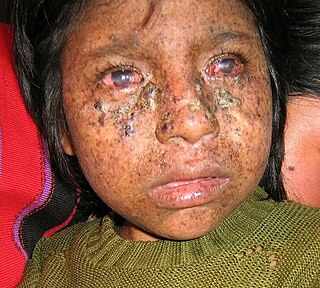
Xeroderma pigmentosum (XP) is a genetic disorder in which there is a decreased ability to repair DNA damage such as that caused by ultraviolet (UV) light. Symptoms may include a severe sunburn after only a few minutes in the sun, freckling in sun-exposed areas, dry skin and changes in skin pigmentation. Nervous system problems, such as hearing loss, poor coordination, loss of intellectual function and seizures, may also occur. Complications include a high risk of skin cancer, with about half having skin cancer by age 10 without preventative efforts, and cataracts. There may be a higher risk of other cancers such as brain cancers.

<span class="mw-page-title-main">Nucleotide excision repair</span> DNA repair mechanism

Nucleotide excision repair is a DNA repair mechanism. DNA damage occurs constantly because of chemicals, radiation and other mutagens. Three excision repair pathways exist to repair single stranded DNA damage: Nucleotide excision repair (NER), base excision repair (BER), and DNA mismatch repair (MMR). While the BER pathway can recognize specific non-bulky lesions in DNA, it can correct only damaged bases that are removed by specific glycosylases. Similarly, the MMR pathway only targets mismatched Watson-Crick base pairs.

Chromosome instability syndromes are a group of inherited conditions associated with chromosomal instability and breakage. They often lead to an increased tendency to develop certain types of malignancies.

<span class="mw-page-title-main">XPB</span> Mammalian protein found in Homo sapiens

XPB is an ATP-dependent DNA helicase in humans that is a part of the TFIIH transcription factor complex.

A DNA repair-deficiency disorder is a medical condition due to reduced functionality of DNA repair.

<span class="mw-page-title-main">ERCC2</span> Mammalian protein found in humans

ERCC2, or XPD is a protein involved in transcription-coupled nucleotide excision repair.

<span class="mw-page-title-main">DeSanctis–Cacchione syndrome</span> Medical condition

DeSanctis–Cacchione syndrome is a genetic disorder characterized by the skin and eye symptoms of xeroderma pigmentosum (XP) occurring in association with microcephaly, progressive intellectual disability, slowed growth and sexual development, deafness, choreoathetosis, ataxia and quadriparesis.

<span class="mw-page-title-main">ERCC1</span> Protein-coding gene in the species Homo sapiens

DNA excision repair protein ERCC-1 is a protein that in humans is encoded by the ERCC1 gene. Together with ERCC4, ERCC1 forms the ERCC1-XPF enzyme complex that participates in DNA repair and DNA recombination.

The enzyme DNA-(apurinic or apyrimidinic site) lyase, also referred to as DNA-(apurinic or apyrimidinic site) 5'-phosphomonoester-lyase or DNA AP lyase catalyzes the cleavage of the C-O-P bond 3' from the apurinic or apyrimidinic site in DNA via β-elimination reaction, leaving a 3'-terminal unsaturated sugar and a product with a terminal 5'-phosphate. In the 1970s, this class of enzyme was found to repair at apurinic or apyrimidinic DNA sites in E. coli and in mammalian cells. The major active enzyme of this class in bacteria, and specifically, E. coli is endonuclease type III. This enzyme is part of a family of lyases that cleave carbon-oxygen bonds.

<span class="mw-page-title-main">XPC (gene)</span> Protein-coding gene in the species Homo sapiens

Xeroderma pigmentosum, complementation group C, also known as XPC, is a protein which in humans is encoded by the XPC gene. XPC is involved in the recognition of bulky DNA adducts in nucleotide excision repair. It is located on chromosome 3.

<span class="mw-page-title-main">XPA</span> Protein-coding gene in the species Homo sapiens

DNA repair protein complementing XP-A cells is a protein that in humans is encoded by the XPA gene.

<span class="mw-page-title-main">ERCC6</span> Gene of the species Homo sapiens

DNA excision repair protein ERCC-6 is a protein that in humans is encoded by the ERCC6 gene. The ERCC6 gene is located on the long arm of chromosome 10 at position 11.23.

<span class="mw-page-title-main">ERCC5</span> Protein-coding gene in the species Homo sapiens

DNA repair protein complementing XP-G cells is a protein that in humans is encoded by the ERCC5 gene.

<span class="mw-page-title-main">ERCC4</span> Protein-coding gene in the species Homo sapiens

ERCC4 is a protein designated as DNA repair endonuclease XPF that in humans is encoded by the ERCC4 gene. Together with ERCC1, ERCC4 forms the ERCC1-XPF enzyme complex that participates in DNA repair and DNA recombination.

<span class="mw-page-title-main">ERCC8 (gene)</span> Protein-coding gene in humans

DNA excision repair protein ERCC-8 is a protein that in humans is encoded by the ERCC8 gene.

<span class="mw-page-title-main">Trichothiodystrophy</span> Medical condition

Trichothiodystrophy (TTD) is an autosomal recessive inherited disorder characterised by brittle hair and intellectual impairment. The word breaks down into tricho – "hair", thio – "sulphur", and dystrophy – "wasting away" or literally "bad nourishment". TTD is associated with a range of symptoms connected with organs of the ectoderm and neuroectoderm. TTD may be subclassified into four syndromes: Approximately half of all patients with trichothiodystrophy have photosensitivity, which divides the classification into syndromes with or without photosensitivity; BIDS and PBIDS, and IBIDS and PIBIDS. Modern covering usage is TTD-P (photosensitive), and TTD.

Progeroid syndromes (PS) are a group of rare genetic disorders that mimic physiological aging, making affected individuals appear to be older than they are. The term progeroid syndrome does not necessarily imply progeria, which is a specific type of progeroid syndrome.

<span class="mw-page-title-main">Hereditary cancer syndrome</span> Inherited genetic condition that predisposes a person to cancer

A hereditary cancer syndrome is a genetic disorder in which inherited genetic mutations in one or more genes predispose the affected individuals to the development of cancer and may also cause early onset of these cancers. Hereditary cancer syndromes often show not only a high lifetime risk of developing cancer, but also the development of multiple independent primary tumors.

References

  1. Bertola; Cao, H; Albano, Lm; Oliveira, Dp; Kok, F; Marques-Dias, Mj; Kim, Ca; Hegele, Ra (2006). "Cockayne syndrome type A: novel mutations in eight typical patients". Journal of Human Genetics. 51 (8): 701–5. doi: 10.1007/s10038-006-0011-7 . PMID   16865293.
  2. James, William; Berger, Timothy; Elston, Dirk (2005). Andrews' Diseases of the Skin: Clinical Dermatology (10th ed.). Saunders. p.  575. ISBN   978-0-7216-2921-6.
  3. 1 2 3 4 5 Bender M, Potocki L, Metry D. What syndrome is this? Cockayne syndrome. Pediatric Dermatology [serial online]. November 2003;20(6):538-540. Available from: MEDLINE with Full Text, Ipswich, MA. Accessed April 30, 2015.
  4. Laugel, Vincent (2013). "Cockayne syndrome: the expanding clinical and mutational spectrum". Mechanisms of Ageing and Development. 134 (5–6): 161–170. doi:10.1016/j.mad.2013.02.006. ISSN   1872-6216. PMID   23428416. S2CID   19137836.
  5. Hoeijmakers JH (October 2009). "DNA damage, aging, and cancer". N. Engl. J. Med. 361 (15): 1475–85. doi:10.1056/NEJMra0804615. PMID   19812404.
  6. 1 2 3 4 Nance M, Berry S (1 January 1992). "Cockayne syndrome: review of 140 cases". American Journal of Medical Genetics. 42 (1): 68–84. doi:10.1002/ajmg.1320420115. PMID   1308368.
  7. 1 2 3 4 Neill CA, Dingwall MM. A Syndrome Resembling Progeria: A Review of Two Cases. Archives of Disease in Childhood. 1950;25(123):213-223.
  8. 1 2 3 4 5 6 Cockayne Syndrome. Genetics Home Reference http://ghr.nlm.nih.gov/condition/cockayne-syndrome Published April 28, 2015. Reviewed May 2010. Accessed April 30, 2015.
  9. 1 2 3 4 5 6 Javadzadeh M. Cockayne Syndrome. Iran J Child Neurol. Autumn 2014;8;4(Suppl.1):18-19.
  10. Cerebrooculofacioskeletal Syndrome 2. Online Mendelian Inheritance in Man. https://omim.org/entry/610756. Published 2/12/2007.
  11. Laugel V. Cockayne Syndrome. 2000 Dec 28 [Updated 2012 Jun 14]. In: Pagon RA, Adam MP, Ardinger HH, et al., editors. GeneReviews® [Internet]. Seattle (WA): University of Washington, Seattle; 1993-2015. Available from:
  12. "Cell Injury and Death". The Lecturio Medical Concept Library. Retrieved 7 July 2021.
  13. Vessoni, Alexandre Teixeira; Guerra, Camila Chaves Coelho; Kajitani, Gustavo Satoru; Nascimento, Livia Luz Souza; Garcia, Camila Carrião Machado (2020). "Cockayne Syndrome: The many challenges and approaches to understand a multifaceted disease". Genetics and Molecular Biology. 43 (1 suppl. 1): e20190085. doi:10.1590/1678-4685-GMB-2019-0085. ISSN   1415-4757. PMC   7250278 . PMID   32453336.
  14. van Hoffen A, Natarajan AT, Mayne LV, van Zeeland AA, Mullenders LH, Venema J (1993). "Deficient repair of the transcribed strand of active genes in Cockayne's syndrome cells". Nucleic Acids Res. 21 (25): 5890–5. doi:10.1093/nar/21.25.5890. PMC   310470 . PMID   8290349.
  15. 1 2 3 4 5 Karikkineth A, Scheibye-Knudsen M, Fivenson E, Croteau D, Bohr B (January 2017). "Cockayne syndrome: Clinical features, model systems and pathways". Ageing Research Reviews. 33: 3–17. doi:10.1016/j.arr.2016.08.002. PMC   5195851 . PMID   27507608.
  16. 1 2 Iyama T, Wilson DM (2016). "Elements That Regulate the DNA Damage Response of Proteins Defective in Cockayne Syndrome". J. Mol. Biol. 428 (1): 62–78. doi:10.1016/j.jmb.2015.11.020. PMC   4738086 . PMID   26616585.
  17. Iyama T, Lee SY, Berquist BR, Gileadi O, Bohr VA, Seidman MM, McHugh PJ, Wilson DM (2015). "CSB interacts with SNM1A and promotes DNA interstrand crosslink processing". Nucleic Acids Res. 43 (1): 247–58. doi:10.1093/nar/gku1279. PMC   4288174 . PMID   25505141.
  18. Batenburg NL, Thompson EL, Hendrickson EA, Zhu XD (2015). "Cockayne syndrome group B protein regulates DNA double-strand break repair and checkpoint activation". EMBO J. 34 (10): 1399–416. doi:10.15252/embj.201490041. PMC   4491999 . PMID   25820262.
  19. Wei L, Nakajima S, Böhm S, Bernstein KA, Shen Z, Tsang M, Levine AS, Lan L (2015). "DNA damage during the G0/G1 phase triggers RNA-templated, Cockayne syndrome B-dependent homologous recombination". Proc. Natl. Acad. Sci. U.S.A. 112 (27): E3495–504. Bibcode:2015PNAS..112E3495W. doi: 10.1073/pnas.1507105112 . PMC   4500203 . PMID   26100862.
  20. Kyllermen, Marten. Cockayne Syndrome. Swedish Information Centre for Rare Diseases. 2012: 4.0. http://www.socialstyrelsen.se/rarediseases/cockaynesyndrome#anchor_17 Archived 2015-09-24 at the Wayback Machine
  21. Title: Cockayne Syndrome Authors: Dr Nita R Sutay, Dr Md Ashfaque Tinmaswala, Dr Manjiri Karlekar, Dr Swati Jhahttp://jmscr.igmpublication.org/v3-i7/35%20jmscr.pdf
  22. admin (2020-10-23). "Cockayne syndrome type b | Viljem Julijan Association". Cure for Cockayne Syndrome – type B. Retrieved 2023-07-06.
  23. "Riaan Research Initiative — Cockayne Syndrome" . Retrieved 2023-07-06.
  24. "Cockayne syndrome | Genetic and Rare Diseases Information Center (GARD) – an NCATS Program".
  25. Karikkineth, A. C.; Scheibye-Knudsen, M.; Fivenson, E.; Croteau, D. L.; Bohr, V. A. (2016). "Cockayne syndrome: Clinical features, model systems and pathways". Ageing Research Reviews. 33: 3–17. doi:10.1016/j.arr.2016.08.002. PMC   5195851 . PMID   27507608.
  26. Kubota, Masaya; Ohta, Sayaka; Ando, Aki; Koyama, Akiko; Terashima, Hiroshi; Kashii, Hirofumi; Hoshino, Hideki; Sugita, Katsuo; Hayashi, Masaharu (June 2015). "Nationwide survey of Cockayne syndrome in Japan: Incidence, clinical course and prognosis: Cockayne syndrome in Japan". Pediatrics International. 57 (3): 339–347. doi:10.1111/ped.12635. PMID   25851792. S2CID   5311897.
  27. "Orphanet: CAMFAK syndrome".