Integrative neuroscience

Last updated

Integrative neuroscience is the study of neuroscience that works to unify functional organization data to better understand complex structures and behaviors. [1] The relationship between structure and function, and how the regions and functions connect to each other. Different parts of the brain carrying out different tasks, interconnecting to come together allowing complex behavior. [2] Integrative neuroscience works to fill gaps in knowledge that can largely be accomplished with data sharing, to create understanding of systems, currently being applied to simulation neuroscience: Computer Modeling of the brain that integrates functional groups together. [3] [1]

Contents

Overview

The roots of integrative neuroscience originated from the Rashevsky-Rosen school of relational biology [4] that characterizes functional organization mathematically by abstracting away the structure (i.e., physics and chemistry). It was further expanded by Chauvet [5] who introduced hierarchical and functional integration.

Hierarchical integration is structural involving spatiotemporal dynamic continuity in Euclidean space to bring about functional organization, viz.

Hierarchical organization + Hierarchical integration = Functional organization

However, functional integration is relational and as such this requires a topology not restricted to Euclidean space, but rather occupying vector spaces [6] This means that for any given functional organization the methods of functional analysis enable a relational organization to be mapped by the functional integration, viz.

Functional organization + Functional integration = Relational Organization

Thus hierarchical and functional integration entails a "neurobiology of cognitive semantics" where hierarchical organization is associated with the neurobiology and relational organization is associated with the cognitive semantics. Relational organization throws away the matter; "function dictates structure", hence material aspects are entailed, while in reductionism the causal nexus between structure and dynamics entails function that obviates functional integration because the causal entailment in the brain of hierarchical integration is absent from the structure.

If integrative neuroscience is studied from the viewpoint of functional organization of hierarchical levels then it is defined as causal entailment in the brain of hierarchical integration. If it is studied from the viewpoint of relational organization then it is defined as semantic entailment in the brain of functional integration.

It aims to present studies of functional organization of particular brain systems across scale through hierarchical integration leading to species-typical behaviors under normal and pathological states. As such, integrative neuroscience aims for a unified understanding of brain function across scale.

Spivey's continuity of mind thesis [7] extends integrative neuroscience to the domain of continuity psychology.

Motivation

With data building up, it ends up in its respective specializations with very little overlap. [1] With the creation of a standardized integrated database of neuroscience data, lead to statical models that would otherwise not be possible, for example, understanding and treating psychiatric disorders. [8]

It provides a framework for linking the great diversity of specializations within contemporary neuroscience, including

This diversity is inevitable, yet has arguably created a void: neglect of the primary role of the nervous system in enabling the animal to survive and prosper. Integrative neuroscience aims to fill this perceived void.

Experimental methods

Identifying different brain regions through correlation and causal methods, combine to contribute an overall brain function and location map. Using different data collected from different methods combine to create a better interconnected and integrative understanding of the brain.

Correlation

The relationship between brain states and behavioral states. [9] Observed through spatial and temporal differences. That pin point places in the brain affected by an action or stimuli, and the timing of the response. [10] Tools used for this include fMRI and EEG, more information below.

Functional magnetic resonance imaging

Functional magnetic resonance imaging (fMRI) measures blood oxygen dependent response (BOLD), using magnetic resonance to observe blood oxygenated areas. Active areas are associated with increased blood flow, presenting a correlation relationship. [11] [12] The spatial localization of fMRI allows accurate information down to the nuclei and Brodmann areas. [13] Certain activities such as the visual system are so rapid lasting only fractions of seconds, while other brain functions can take days or months such as memory. fMRI measures in the frame of seconds, making it difficult to measure extremely fast processes. [14]

Electroencephalography

Electroencephalography (EEG) allows you to see the electrical activity of the brain over time, can only measure presented stimuli responses, stimuli the experimenter presents. it uses electrode sensors places on the surface on the skull to measure synchronous neuron firing. It can not be certain activity is caused by stimuli only a correlation between a given function and brain area. EEG measures overall changes in wide regions, lacking specificity. [15]

Causal

Brain activity is directly caused by stimulation of a specific region, as proven through experimentation.

TMS

TMS (Transcranial magnetic stimulation) uses a magnetic coil releasing a burst of magnetic field that activated activity in a specific brain area. It is useful in exciting a specific area in the cortex and recording the MEPs (Motor Evoked Potentials) that occurs as a result. [16] [17] It gives certain causal relationships, but is limited to the cortex making it impossible to reach any deeper than the surface of the brain. [17]

Lesions studies

When patients have natural lesions, it is an opportunity to watch how a lesion in a given region affects functionality. Or in non-human experimentation, lesions can be created by removing sections of the brain. These methods are not reversible, unlike brain studying techniques, and does not accurately show what that section of the brain are disabled due to the disruption of homeostasis in the brain. With a permeate lesion, the brain chemically adjusted and restores homeostasis [18] Relying on natural occurrences has little control over variables such as location and size. And in cases with damage in multiple areas, differentiation is not certain with lack of mass data.

Electrode stimulation

Cortical Stimulation Mapping, invasive brain surgery that probes at area of the cortex to relate different regions to function. [19] Typically occurs during open brain surgery where electrodes are inserted in areas and observations are made. This method is limited by number of patients having open brain surgery that consent to such experimentation, and to what area of the brain is being operated on. Also performed in mice with full range over the brain.

Applications

Human Brain Project

Since the 'decade of the brain' there has been an explosion of insights into the brain and their application in most areas of medicine. With this explosion, the need for integration of data across studies, modalities and levels of understanding is increasingly recognized. A concrete exemplar of the value of large-scale data sharing has been provided by the Human Brain Project.

Medical

The importance of large-scale integration of brain information for new approaches to medicine has been recognized. [20] Rather than relying mainly on symptom information, a combination of brain and gene information may ultimately be required for understanding what treatment is best suited to which individual person.

Behavioral

There is also work studying empathy and social behavior trends to better understand how empathy plays a role in behavioral science, and how the brain responds to empathy, produces empathy, and develops empathy over time. Combining these functional units and the social behavior and impact work to create a better understanding of the complex behaviors that create the human experience. [21]

Related Research Articles

Brocas area Speech production region in the dominant hemisphere of the hominid brain

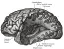
Broca's area, or the Broca area, is a region in the frontal lobe of the dominant hemisphere, usually the left, of the brain with functions linked to speech production.

Cognitive neuroscience is the scientific field that is concerned with the study of the biological processes and aspects that underlie cognition, with a specific focus on the neural connections in the brain which are involved in mental processes. It addresses the questions of how cognitive activities are affected or controlled by neural circuits in the brain. Cognitive neuroscience is a branch of both neuroscience and psychology, overlapping with disciplines such as behavioral neuroscience, cognitive psychology, physiological psychology and affective neuroscience. Cognitive neuroscience relies upon theories in cognitive science coupled with evidence from neurobiology, and computational modeling.

Neuropsychology is a branch of psychology that is concerned with how a person's cognition and behavior are related to the brain and the rest of the nervous system. Professionals in this branch of psychology often focus on how injuries or illnesses of the brain affect cognitive and behavioral functions.

Behavioral neuroscience, also known as biological psychology, biopsychology, or psychobiology, is the application of the principles of biology to the study of physiological, genetic, and developmental mechanisms of behavior in humans and other animals.

A mirror neuron is a neuron that fires both when an animal acts and when the animal observes the same action performed by another. Thus, the neuron "mirrors" the behavior of the other, as though the observer were itself acting. Such neurons have been directly observed in human and primate species, and birds.

Language processing in the brain

Language processing refers to the way humans use words to communicate ideas and feelings, and how such communications are processed and understood. Language processing is considered to be a uniquely human ability that is not produced with the same grammatical understanding or systematicity in even human's closest primate relatives.

Functional integration is the study of how brain regions work together to process information and effect responses. Though functional integration frequently relies on anatomic knowledge of the connections between brain areas, the emphasis is on how large clusters of neurons – numbering in the thousands or millions – fire together under various stimuli. The large datasets required for such a whole-scale picture of brain function have motivated the development of several novel and general methods for the statistical analysis of interdependence, such as dynamic causal modelling and statistical linear parametric mapping. These datasets are typically gathered in human subjects by non-invasive methods such as EEG/MEG, fMRI, or PET. The results can be of clinical value by helping to identify the regions responsible for psychiatric disorders, as well as to assess how different activities or lifestyles affect the functioning of the brain.

Relational frame theory (RFT) is a psychological theory of human language. It was developed originally by Steven C. Hayes of University of Nevada, Reno and has been extended in research, notably by Dermot Barnes-Holmes and colleagues of Ghent University.

Social neuroscience is an interdisciplinary field devoted to understanding the relationship between social experiences and biological systems. Humans are fundamentally a social species, rather than individualists. As such, Homo sapiens create emergent organizations beyond the individual—structures that range from dyads, families, and groups to cities, civilizations, and cultures. In this regard, studies indicate that various social influences including life events, poverty, unemployment and loneliness can influence health related biomarkers. The term "social neuroscience" can be traced to a publication entitled "Social Neuroscience Bulletin" that was published quarterly between 1988 and 1994. The term was subsequently popularized in an article by John Cacioppo and Gary Berntson, published in the American Psychologist in 1992. Cacioppo and Berntson are considered as the legitimate fathers of social neuroscience. Still a young field, social neuroscience is closely related to affective neuroscience and cognitive neuroscience, focusing on how the brain mediates social interactions. The biological underpinnings of social cognition are investigated in social cognitive neuroscience.

Inferior temporal gyrus One of three gyri of the temporal lobe of the brain

The inferior temporal gyrus is one of three gyri of the temporal lobe and is located below the middle temporal gyrus, connected behind with the inferior occipital gyrus; it also extends around the infero-lateral border on to the inferior surface of the temporal lobe, where it is limited by the inferior sulcus. This region is one of the higher levels of the ventral stream of visual processing, associated with the representation of objects, places, faces, and colors. It may also be involved in face perception, and in the recognition of numbers.

In cognitive neuroscience, visual modularity is an organizational concept concerning how vision works. The way in which the primate visual system operates is currently under intense scientific scrutiny. One dominant thesis is that different properties of the visual world require different computational solutions which are implemented in anatomically/functionally distinct regions that operate independently – that is, in a modular fashion.

The study of memory incorporates research methodologies from neuropsychology, human development and animal testing using a wide range of species. The complex phenomenon of memory is explored by combining evidence from many areas of research. New technologies, experimental methods and animal experimentation have led to an increased understanding of the workings of memory.

Resting state fMRI

Resting state fMRI is a method of functional magnetic resonance imaging (fMRI) that is used in brain mapping to evaluate regional interactions that occur in a resting or task-negative state, when an explicit task is not being performed. A number of resting-state conditions are identified in the brain, one of which is the default mode network. These resting brain state conditions are observed through changes in blood flow in the brain which creates what is referred to as a blood-oxygen-level dependent (BOLD) signal that can be measured using fMRI.

Pain empathy is a specific subgroup of empathy that involves recognizing and understanding another person's pain. Empathy is the mental ability that allows one person to understand another person's mental and emotional state and how to effectively respond to that person. When a person receives cues that another person is in pain, neural pain circuits within the brain are activated. There are several cues that can communicate pain to another person: visualization of the injury causing event, the injury itself, behavioral efforts of the injured to avoid further harm, and displays of pain and distress such as facial expressions, crying, and screaming. From an evolutionary perspective, pain empathy is beneficial for human group survival since it provides motivation for non-injured people to offer aid to the injured and to avoid injury themselves.

Biological basis of personality

The biological basis of personality is the collection of brain systems and mechanisms that underlie human personality. Human neurobiology, especially as it relates to complex traits and behaviors, is not well understood, but research into the neuroanatomical and functional underpinnings of personality are an active field of research. Animal models of behavior, molecular biology, and brain imaging techniques have provided some insight into human personality, especially trait theories.

Russell Poldrack

Russell "Russ" Alan Poldrack is an American psychologist and neuroscientist. He is a professor of Psychology at Stanford University, member of the Stanford Neuroscience Institute and director of the Stanford Center for Reproducible Neuroscience.

Dorsomedial prefrontal cortex

The dorsomedial prefrontal cortex (dmPFC or DMPFC is a section of the prefrontal cortex in some species' brain anatomy. It includes portions of Brodmann areas BA8, BA9, BA10, BA24 and BA32, although some authors identify it specifically with BA8 and BA9 Some notable sub-components include the dorsal anterior cingulate cortex, the prelimbic cortex, and the infralimbic cortex.

Neuromorality is an emerging field of neuroscience that studies the connection between morality and neuronal function. Scientists use fMRI and psychological assessment together to investigate the neural basis of moral cognition and behavior. Evidence shows that the central hub of morality is the prefrontal cortex guiding activity to other nodes of the neuromoral network. A spectrum of functional characteristics within this network to give rise to both altruistic and psychopathological behavior. Evidence from the investigation of neuromorality has applications in both clinical neuropsychiatry and forensic neuropsychiatry.

Salience network Large-scale brain network involved in detecting and attending to relevant stimuli

The salience network (SN), also known anatomically as the midcingulo-insular network (M-CIN), is a large scale brain network of the human brain that is primarily composed of the anterior insula (AI) and dorsal anterior cingulate cortex (dACC). It is involved in detecting and filtering salient stimuli, as well as in recruiting relevant functional networks. Together with its interconnected brain networks, the SN contributes to a variety of complex functions, including communication, social behavior, and self-awareness through the integration of sensory, emotional, and cognitive information.

Social cognitive neuroscience is the scientific study of the biological processes underpinning social cognition. Specifically, it uses the tools of neuroscience to study "the mental mechanisms that create, frame, regulate, and respond to our experience of the social world". Social cognitive neuroscience uses the epistemological foundations of cognitive neuroscience, and is closely related to social neuroscience. Social cognitive neuroscience employs human neuroimaging, typically using functional magnetic resonance imaging (fMRI). Human brain stimulation techniques such as transcranial magnetic stimulation and transcranial direct-current stimulation are also used. In nonhuman animals, direct electrophysiological recordings and electrical stimulation of single cells and neuronal populations are utilized for investigating lower-level social cognitive processes.

References

  1. 1 2 3 Integrative neuroscience : bringing together biological, psychological and clinical models of the human brain. Gordon, Evian. Amsterdam: Harwood Academic Publishers. 2000. ISBN   9780203304761. OCLC   567985508.CS1 maint: others (link)
  2. Kotchoubey, Boris; Tretter, Felix; Braun, Hans A.; Buchheim, Thomas; Draguhn, Andreas; Fuchs, Thomas; Hasler, Felix; Hastedt, Heiner; Hinterberger, Thilo; Northoff, Georg; Rentschler, Ingo (2016). "Methodological Problems on the Way to Integrative Human Neuroscience". Frontiers in Integrative Neuroscience. 10: 41. doi:10.3389/fnint.2016.00041. ISSN   1662-5145. PMC   5126073 . PMID   27965548.
  3. Fan, Xue; Markram, Henry (2019-05-07). "A Brief History of Simulation Neuroscience". Frontiers in Neuroinformatics. 13: 32. doi:10.3389/fninf.2019.00032. ISSN   1662-5196. PMC   6513977 . PMID   31133838.
  4. Louie, AH (2009). More Than Life Itself: A Synthetic Continuation in Relational Biology. Frankfurt [Germany]: Ontos Verlag.
  5. Chauvet, Gilbert (1996). Theoretical Systems in Biology: Hierarchal and Functional Integration. Oxford [United Kingdom]: Pergamon Press.
  6. Brzychczy, S.; Poznanski, RR (2013). Mathematical Neuroscience. Amsterdam [The Netherlands]: Elsevier BV.
  7. Spivey, M.J. (2007). The Continuity of the Mind. New York [New York]: Oxford University Press.
  8. Gordon, Evian (June 2003). "Integrative Neuroscience in Psychiatry: The Role of a Standardized Database". Australasian Psychiatry. 11 (2): 156–163. doi:10.1046/j.1039-8562.2003.00533.x. ISSN   1039-8562. S2CID   145382687.
  9. Dijkstra, Nadine; de Bruin, Leon (2016-07-19). "Cognitive Neuroscience and Causal Inference: Implications for Psychiatry". Frontiers in Psychiatry. 7: 129. doi:10.3389/fpsyt.2016.00129. ISSN   1664-0640. PMC   4949233 . PMID   27486408.
  10. Woods, Adam J.; Hamilton, Roy H.; Kranjec, Alexander; Minhaus, Preet; Bikson, Marom; Yu, Jonathan; Chatterjee, Anjan (2014-05-15). "Space, time, and causality in the human brain". NeuroImage. 92: 285–297. doi:10.1016/j.neuroimage.2014.02.015. ISSN   1095-9572. PMC   4008651 . PMID   24561228.
  11. "Introduction to FMRI — Nuffield Department of Clinical Neurosciences". www.ndcn.ox.ac.uk. Retrieved 2019-11-26.
  12. Logothetis, Nikos K.; Pauls, Jon; Augath, Mark; Trinath, Torsten; Oeltermann, Axel (2001). "Neurophysiological investigation of the basis of the fMRI signal". Nature. 412 (6843): 150–157. Bibcode:2001Natur.412..150L. doi:10.1038/35084005. PMID   11449264. S2CID   969175. Our results show unequivocally that a spatially localized increase in the BOLD contrast directly and monotonically reflects an increase in neural activity.
  13. Carr, Valerie A.; Rissman, Jesse; Wagner, Anthony D. (2010). "Imaging the Human Medial Temporal Lobe with High-Resolution fMRI". Neuron. 65 (3): 298–308. doi:10.1016/j.neuron.2009.12.022. PMC   2844113 . PMID   20159444.
  14. Huettel, S. A.; Song, A. W.; McCarthy, G. (2009), Functional Magnetic Resonance Imaging (2 ed.), Massachusetts: Sinauer, ISBN   978-0-87893-286-3
  15. Niedermeyer E.; da Silva F.L. (2004). Electroencephalography: Basic Principles, Clinical Applications, and Related Fields. Lippincott Williams & Wilkins. ISBN   978-0-7817-5126-1
  16. van Dun, Kim; Bodranghien, Florian; Manto, Mario; Mariën, Peter (2017-06-01). "Targeting the Cerebellum by Noninvasive Neurostimulation: a Review". The Cerebellum. 16 (3): 695–741. doi:10.1007/s12311-016-0840-7. ISSN   1473-4230. PMID   28032321. S2CID   3999098.
  17. 1 2 Groppa, S.; Oliviero, A.; Eisen, A.; Quartarone, A.; Cohen, L.G.; Mall, V.; Kaelin-Lang, A.; Mima, T.; Rossi, S.; Thickbroom, G.W.; Rossini, P.M. (May 2012). "A practical guide to diagnostic transcranial magnetic stimulation: Report of an IFCN committee". Clinical Neurophysiology. 123 (5): 858–882. doi:10.1016/j.clinph.2012.01.010. ISSN   1388-2457. PMC   4890546 . PMID   22349304.
  18. Vaidya, Avinash R.; Pujara, Maia S.; Petrides, Michael; Murray, Elisabeth A.; Fellows, Lesley K. (2019). "Lesion Studies in Contemporary Neuroscience". Trends in Cognitive Sciences. 23 (8): 653–671. doi:10.1016/j.tics.2019.05.009. PMC   6712987 . PMID   31279672.
  19. Lesser, Ronald P.; Arroyo, Santiago; Crone, Nathan; Gordon, Barry (1998). "Motor and Sensory Mapping of the Frontal and Occipital Lobes". Epilepsia. 39: S69–S80. doi: 10.1111/j.1528-1157.1998.tb05127.x . PMID   9637595.
  20. Insel, Thomas R; Volkow, Nora D; Landis, Story C; Li, Ting-Kai; Battey, James F; Sieving, Paul (2003). "Limits to growth: why neuroscience needs large-scale science". Nature Neuroscience. 7 (5): 426–427. doi:10.1038/nn0504-426. PMID   15114352. S2CID   30158264.
  21. Hein, Grit; Singer, Tania (2010), "Neuroscience meets social psychology: An integrative approach to human empathy and prosocial behavior.", Prosocial motives, emotions, and behavior: The better angels of our nature, American Psychological Association, pp. 109–125, doi:10.1037/12061-006, ISBN   978-1433805462, S2CID   142743763