Related Research Articles

The lungs are the primary organs of the respiratory system in humans and most other animals including a few fish, and some snails. In mammals and most other vertebrates, two lungs are located near the backbone on either side of the heart. Their function in the respiratory system is to extract oxygen from the air and transfer it into the bloodstream, and to release carbon dioxide from the bloodstream into the atmosphere, in a process of gas exchange. Respiration is driven by different muscular systems in different species. Mammals, reptiles and birds use their different muscles to support and foster breathing. In earlier tetrapods, air was driven into the lungs by the pharyngeal muscles via buccal pumping, a mechanism still seen in amphibians. In humans, the main muscle of respiration that drives breathing is the diaphragm. The lungs also provide airflow that makes vocal sounds including human speech possible.

A pulmonary alveolus also known as an air sac or air space is one of millions of hollow cup-shaped cavities in the lungs where oxygen is exchanged for carbon dioxide. Alveoli make up the functional tissue of the lungs known as the lung parenchyma, which takes up 90 percent of the total lung volume.

The parasympathetic nervous system (PSNS) is one of the three divisions of the autonomic nervous system, the others being the sympathetic nervous system and the enteric nervous system.

The sympathetic nervous system (SNS) is one of two divisions of the autonomic nervous system, along with the parasympathetic nervous system. The enteric nervous system is sometimes considered part of the autonomic nervous system, and sometimes considered an independent system.

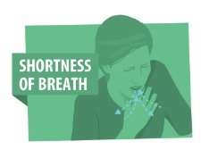
Shortness of breath (SOB), also known as dyspnea is a feeling of not being able to breathe well enough. The American Thoracic Society defines it as "a subjective experience of breathing discomfort that consists of qualitatively distinct sensations that vary in intensity", and recommends evaluating dyspnea by assessing the intensity of the distinct sensations, the degree of distress involved, and its burden or impact on activities of daily living. Distinct sensations include effort/work, chest tightness, and air hunger. The tripod position is a often assumed.

The phrenic nerve is a mixed motor/sensory nerve which originates from the C3-C5 spinal nerves in the neck. The nerve is important for breathing because it provides exclusive motor control of the diaphragm, the primary muscle of respiration. In humans, the right and left phrenic nerves are primarily supplied by the C4 spinal nerve, but there is also contribution from the C3 and C5 spinal nerves. From its origin in the neck, the nerve travels downward into the chest to pass between the heart and lungs towards the diaphragm.
Hyperventilation occurs when the rate or tidal volume of breathing eliminates more carbon dioxide than the body can produce. This leads to hypocapnia, a reduced concentration of carbon dioxide dissolved in the blood. The body normally attempts to compensate for this homeostatically, but if this fails or is overridden, the blood pH will rise, leading to respiratory alkalosis. The symptoms of respiratory alkalosis include: dizziness, tingling in the lips, hands or feet, headache, weakness, fainting, and seizures. In extreme cases it may cause carpopedal spasms, a flapping and contraction of the hands and feet.
A chemoreceptor, also known as chemosensor, is a specialized sensory receptor cell which transduces a chemical substance to generate a biological signal. This signal may be in the form of an action potential, if the chemoreceptor is a neuron, or in the form of a neurotransmitter that can activate a nerve fiber if the chemoreceptor is a specialized cell, such as taste receptors, or an internal peripheral chemoreceptor, such as the carotid bodies. In physiology, a chemoreceptor detects changes in the normal environment, such as an increase in blood levels of carbon dioxide (hypercapnia) or a decrease in blood levels of oxygen (hypoxia), and transmits that information to the central nervous system which engages body responses to restore homeostasis.
In physiology, respiration is the movement of oxygen from the outside environment to the cells within tissues, and the removal of carbon dioxide in the opposite direction.
The control of ventilation refers to the physiological mechanisms involved in the control of breathing, which is the movement of air into and out of the lungs. Ventilation facilitates respiration. Respiration refers to the utilization of oxygen and balancing of carbon dioxide by the body as a whole, or by individual cells in cellular respiration.

Afferent nerve fibers are the axons carried by a sensory nerve that relay sensory information from sensory receptors to regions of the brain. Afferent projections arrive at a particular brain region. Efferent nerve fibers are carried by efferent nerves and exit a region to act on muscles and glands.

A nociceptor is a sensory neuron that responds to damaging or potentially damaging stimuli by sending “possible threat” signals to the spinal cord and the brain. If the brain perceives the threat as credible, it creates the sensation of pain to direct attention to the body part, so the threat can hopefully be mitigated; this process is called nociception.

Hypoxemia is an abnormally low level of oxygen in the blood. More specifically, it is oxygen deficiency in arterial blood. Hypoxemia has many causes, and often causes hypoxia as the blood is not supplying enough oxygen to the tissues of the body.
The cough reflex has both sensory (afferent) mainly via the vagus nerve and motor (efferent) components. Pulmonary irritant receptors in the epithelium of the respiratory tract are sensitive to both mechanical and chemical stimuli. The bronchi and trachea are so sensitive to light touch that slight amounts of foreign matter or other causes of irritation initiate the cough reflex. The larynx and carina are especially sensitive. Terminal bronchioles and even the alveoli are sensitive to chemical stimuli such as sulfur dioxide gas or chlorine gas. Rapidly moving air usually carries with it any foreign matter that is present in the bronchi or trachea. Stimulation of the cough receptors by dust or other foreign particles produces a cough, which is necessary to remove the foreign material from the respiratory tract before it reaches the lungs
The Hering–Breuer inflation reflex, named for Josef Breuer and Ewald Hering, is a reflex triggered to prevent the over-inflation of the lung. Pulmonary stretch receptors present on the wall of bronchi and bronchioles of the airways respond to excessive stretching of the lung during large inspirations.

The respiratory center is located in the medulla oblongata and pons, in the brainstem. The respiratory center is made up of three major respiratory groups of neurons, two in the medulla and one in the pons. In the medulla they are the dorsal respiratory group, and the ventral respiratory group. In the pons, the pontine respiratory group includes two areas known as the pneumotaxic centre and the apneustic centre.
The Bezold–Jarisch reflex involves a variety of cardiovascular and neurological processes which cause hypopnea, hypotension and bradycardia in response to noxious stimuli detected in the cardiac ventricles. The reflex is named after Albert von Bezold and Adolf Jarisch Junior. The significance of the discovery is that it was the first recognition of a chemical (non-mechanical) reflex.
The Golgi tendon reflex (also called inverse stretch reflex, autogenic inhibition, tendon reflex) is an inhibitory effect on the muscle resulting from the muscle tension stimulating Golgi tendon organs (GTO) of the muscle, and hence it is self-induced. The reflex arc is a negative feedback mechanism preventing too much tension on the muscle and tendon. When the tension is extreme, the inhibition can be so great it overcomes the excitatory effects on the muscle's alpha motoneurons causing the muscle to suddenly relax. This reflex is also called the inverse myotatic reflex, because it is the inverse of the stretch reflex.

Breathing is the process of moving air out and in the lungs to facilitate gas exchange with the internal environment, mostly to flush out carbon dioxide and bring in oxygen.
Group A nerve fibers are one of the three classes of nerve fiber as generally classified by Erlanger and Gasser. The other two classes are the group B nerve fibers, and the group C nerve fibers. Group A are heavily myelinated, group B are moderately myelinated, and group C are unmyelinated.
References
- ↑ Sircar, Sabyasachi (2008). Principles of Medical Physiology. pp. 350–51. ISBN 978-1-58890-572-7.
- ↑ Guyton (2011). "Regulation of Respiration". Textbook of Medical Physiology. Saunders Elsevier. ISBN 978-1-4160-4574-8.
- 1 2 Ganong (2016). "Regulation of Respiration". Review of Medical Physiology, 25th ed. McGraw-Hill Education. p. 662. ISBN 978-0-07-184897-8.
- ↑ A.A. Majid, A.N.Kingsworth, Fundamentals of Surgical Practice. Cambridge, MA: Cambridge University Press, 2006. ISBN 0-521-67706-8.
- ↑ J.C. Bennett, F.Plum ed. Cecil Textbook of Medicine, 20th ed., W.B. Saunders, Philadelphia PA, 1996. ISBN 0-7216-3574-1
- ↑ "A.S. Paintal — a celebrated physiologist". The Hindu . Chennai, India. 19 January 2006. Archived from the original on 12 July 2006.
- ↑ Widdicombe, John (2006). "Reflexes from the lungs and airways: Historical perspective". Journal of Applied Physiology. 101 (2): 628–634. doi:10.1152/japplphysiol.00155.2006. PMID 16601307.