Related Research Articles

The respiratory system is a biological system consisting of specific organs and structures used for gas exchange in animals and plants. The anatomy and physiology that make this happen varies greatly, depending on the size of the organism, the environment in which it lives and its evolutionary history. In land animals the respiratory surface is internalized as linings of the lungs. Gas exchange in the lungs occurs in millions of small air sacs; in mammals and reptiles these are called alveoli, and in birds they are known as atria. These microscopic air sacs have a very rich blood supply, thus bringing the air into close contact with the blood. These air sacs communicate with the external environment via a system of airways, or hollow tubes, of which the largest is the trachea, which branches in the middle of the chest into the two main bronchi. These enter the lungs where they branch into progressively narrower secondary and tertiary bronchi that branch into numerous smaller tubes, the bronchioles. In birds the bronchioles are termed parabronchi. It is the bronchioles, or parabronchi that generally open into the microscopic alveoli in mammals and atria in birds. Air has to be pumped from the environment into the alveoli or atria by the process of breathing which involves the muscles of respiration.

Shortness of breath (SOB), also medically known as dyspnea or dyspnoea, is an uncomfortable feeling of not being able to breathe well enough. The American Thoracic Society defines it as "a subjective experience of breathing discomfort that consists of qualitatively distinct sensations that vary in intensity", and recommends evaluating dyspnea by assessing the intensity of its distinct sensations, the degree of distress and discomfort involved, and its burden or impact on the patient's activities of daily living. Distinct sensations include effort/work to breathe, chest tightness or pain, and "air hunger". The tripod position is often assumed to be a sign.
Dead space is the volume of air that is inhaled that does not take part in the gas exchange, because it either remains in the conducting airways or reaches alveoli that are not perfused or poorly perfused. It means that not all the air in each breath is available for the exchange of oxygen and carbon dioxide. Mammals breathe in and out of their lungs, wasting that part of the inhalation which remains in the conducting airways where no gas exchange can occur.

The diving reflex, also known as the diving response and mammalian diving reflex, is a set of physiological responses to immersion that overrides the basic homeostatic reflexes, and is found in all air-breathing vertebrates studied to date. It optimizes respiration by preferentially distributing oxygen stores to the heart and brain, enabling submersion for an extended time.
Exhalation is the flow of the breath out of an organism. In animals, it is the movement of air from the lungs out of the airways, to the external environment during breathing. This happens due to elastic properties of the lungs, as well as the internal intercostal muscles which lower the rib cage and decrease thoracic volume. As the thoracic diaphragm relaxes during exhalation it causes the tissue it has depressed to rise superiorly and put pressure on the lungs to expel the air. During forced exhalation, as when blowing out a candle, expiratory muscles including the abdominal muscles and internal intercostal muscles generate abdominal and thoracic pressure, which forces air out of the lungs.
In physiology, respiration is the movement of oxygen from the outside environment to the cells within tissues, and the removal of carbon dioxide in the opposite direction that's to the environment.

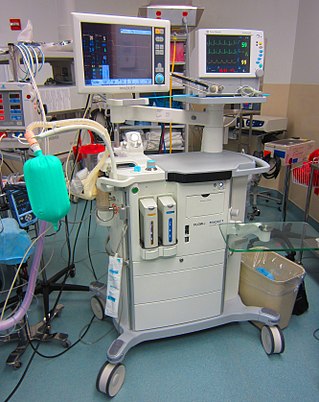
Oxygen therapy, also known as supplemental oxygen, is the use of oxygen as medical treatment. Acute indications for therapy include hypoxemia, carbon monoxide toxicity and cluster headache. It may also be prophylactically given to maintain blood oxygen levels during the induction of anesthesia. Oxygen therapy is often useful in chronic hypoxemia caused by conditions such as severe COPD or cystic fibrosis. Oxygen can be delivered via nasal cannula or face mask, or via high pressure conditions such as in endotracheal intubation or hyperbaric chamber. It can also be given through bypassing the airway, such as in ECMO therapy.
The control of ventilation is the physiological mechanisms involved in the control of breathing, which is the movement of air into and out of the lungs. Ventilation facilitates respiration. Respiration refers to the utilization of oxygen and balancing of carbon dioxide by the body as a whole, or by individual cells in cellular respiration.
A photoplethysmogram (PPG) is an optically obtained plethysmogram that can be used to detect blood volume changes in the microvascular bed of tissue. A PPG is often obtained by using a pulse oximeter which illuminates the skin and measures changes in light absorption. A conventional pulse oximeter monitors the perfusion of blood to the dermis and subcutaneous tissue of the skin.
Vital signs are a group of the four to six most crucial medical signs that indicate the status of the body's vital (life-sustaining) functions. These measurements are taken to help assess the general physical health of a person, give clues to possible diseases, and show progress toward recovery. The normal ranges for a person's vital signs vary with age, weight, gender, and overall health.

Hypoxemia is an abnormally low level of oxygen in the blood. More specifically, it is oxygen deficiency in arterial blood. Hypoxemia has many causes, and often causes hypoxia as the blood is not supplying enough oxygen to the tissues of the body.

A respiratory examination, or lung examination, is performed as part of a physical examination, in response to respiratory symptoms such as shortness of breath, cough, or chest pain, and is often carried out with a cardiac examination.

Diaphragmatic breathing, abdominal breathing, belly breathing, or deep breathing, is breathing that is done by contracting the diaphragm, a muscle located horizontally between the thoracic cavity and abdominal cavity. Air enters the lungs as the diaphragm strongly contracts, but unlike during traditional relaxed breathing (eupnea) the intercostal muscles of the chest do minimal work in this process. The belly also expands during this type of breathing to make room for the contraction of the diaphragm.

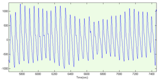
A pneumograph, also known as a pneumatograph or spirograph, is a device for recording velocity and force of chest movements during respiration.

The respiratory center is located in the medulla oblongata and pons, in the brainstem. The respiratory center is made up of three major respiratory groups of neurons, two in the medulla and one in the pons. In the medulla they are the dorsal respiratory group, and the ventral respiratory group. In the pons, the pontine respiratory group includes two areas known as the pneumotaxic center and the apneustic center.

Breathing is the process of moving air into and from the lungs to facilitate gas exchange with the internal environment, mostly to flush out carbon dioxide and bring in oxygen.
Central sleep apnea (CSA) or central sleep apnea syndrome (CSAS) is a sleep-related disorder in which the effort to breathe is diminished or absent, typically for 10 to 30 seconds either intermittently or in cycles, and is usually associated with a reduction in blood oxygen saturation. CSA is usually due to an instability in the body's feedback mechanisms that control respiration. Central sleep apnea can also be an indicator of Arnold–Chiari malformation.
Respiratory inductance plethysmography (RIP) is a method of evaluating pulmonary ventilation by measuring the movement of the chest and abdominal wall.
The rapid shallow breathing index (RSBI) or Yang Tobin index is a tool that is used in the weaning of mechanical ventilation on intensive care units. The RSBI is defined as the ratio of respiratory frequency to tidal volume (f/VT). People on a ventilator who cannot tolerate independent breathing tend to breathe rapidly and shallowly, and will therefore have a high RSBI. The index was introduced in 1991 by Karl Yang and Martin J. Tobin.
Intermittent Mandatory Ventilation (IMV) refers to any mode of mechanical ventilation where a regular series of breaths are scheduled but the ventilator senses patient effort and reschedules mandatory breaths based on the calculated need of the patient. Similar to continuous mandatory ventilation in parameters set for the patients pressures and volumes but distinct in its ability to support a patient by either supporting their own effort or providing support when patient effort is not sensed. IMV is frequently paired with additional strategies to improve weaning from ventilator support or to improve cardiovascular stability in patients who may need full life support.
References
- ↑ "OSA -" . Retrieved 30 September 2016.
- ↑ "Vital Signs 101". www.hopkinsmedicine.org. 14 June 2022.
- ↑ Simoes EA, Roark R, Berman S, Esler LL, Murphy J (October 1991). "Respiratory rate: measurement of variability over time and accuracy at different counting periods". Archives of Disease in Childhood. 66 (10): 1199–1203. doi:10.1136/adc.66.10.1199. PMC 1793530 . PMID 1953002.
- ↑ Grenvik A, Ballou S, McGinley E, Millen JE, Cooley WL, Safar P (October 1972). "Impedance pneumography. Comparison between chest impedance changes and respiratory volumines in 11 healthy volunteers". Chest. 62 (4): 439–443. doi:10.1378/chest.62.4.439. PMID 5077999.
- ↑ Charlton PH, Birrenkott DA, Bonnici T, Pimentel MA, Johnson AE, Alastruey J, et al. (2018). "Breathing Rate Estimation From the Electrocardiogram and Photoplethysmogram: A Review". IEEE Reviews in Biomedical Engineering. 11: 2–20. doi:10.1109/rbme.2017.2763681. PMC 7612521 . PMID 29990026. S2CID 11186066.
- ↑ Bailón R, Sõrnmo L, Laguna P. "Advanced Methods and Tools for ECG Data Analysis - ECG Derived Respiratory Frequency Estimation - Chapter 8". www.robots.ox.ac.uk. Retrieved 2016-02-23.
- ↑ Karlen W, Raman S, Ansermino JM, Dumont GA (July 2013). "Multiparameter respiratory rate estimation from the photoplethysmogram". IEEE Transactions on Bio-Medical Engineering. 60 (7): 1946–1953. doi:10.1109/TBME.2013.2246160. PMID 23399950. S2CID 8033758.
- ↑ Lapi S, Lavorini F, Borgioli G, Calzolai M, Masotti L, Pistolesi M, Fontana GA (January 2014). "Respiratory rate assessments using a dual-accelerometer device". Respiratory Physiology & Neurobiology. 191: 60–66. doi:10.1016/j.resp.2013.11.003. PMID 24263211. S2CID 22222399.
- ↑ Napoli NJ, Rodrigues VR, Davenport PW (2022). "Characterizing and Modeling Breathing Dynamics: Flow Rate, Rhythm, Period, and Frequency". Frontiers in Physiology. 12: 772295. doi: 10.3389/fphys.2021.772295 . PMC 8899297 . PMID 35264974.
- 1 2 Barrett KE, Barman SM, Boitano S, Brooks H (2012-04-05). Ganong's Review of Medical Physiology (24 ed.). p. 619. ISBN 978-0071780032.
- ↑ DeBoer SL (4 November 2004). Emergency Newborn Care. Trafford Publishing. p. 30. ISBN 978-1-4120-3089-2.
- ↑ Lindh WQ, Pooler M, Tamparo C, Dahl BM (9 March 2009). Delmar's Comprehensive Medical Assisting: Administrative and Clinical Competencies. Cengage Learning. p. 573. ISBN 978-1-4354-1914-8.
- 1 2 Rodríguez-Molinero A, Narvaiza L, Ruiz J, Gálvez-Barrón C (December 2013). "Normal respiratory rate and peripheral blood oxygen saturation in the elderly population". Journal of the American Geriatrics Society. 61 (12): 2238–2240. doi:10.1111/jgs.12580. PMID 24329828. S2CID 44700347.
- ↑ Mower WR, Sachs C, Nicklin EL, Safa P, Baraff LJ (November 1996). "A comparison of pulse oximetry and respiratory rate in patient screening". Respiratory Medicine. 90 (10): 593–599. doi: 10.1016/S0954-6111(96)90017-7 . PMID 8959116.
- ↑ Smith GB, Prytherch DR, Meredith P, Schmidt PE, Featherstone PI (April 2013). "The ability of the National Early Warning Score (NEWS) to discriminate patients at risk of early cardiac arrest, unanticipated intensive care unit admission, and death" (PDF). Resuscitation. 84 (4): 465–470. doi:10.1016/j.resuscitation.2012.12.016. PMID 23295778.
| Authority control: National |
|---|