Vital signs

Last updated
Vital signs
Maquet Flow-I anesthesia machine.jpg
An anesthetic machine with integrated systems for monitoring of several vital parameters, including blood pressure and heart rate
Purposeassess the general physical health of a person

Vital signs (also known as vitals) are a group of the four to six most crucial medical signs that indicate the status of the body's vital (life-sustaining) functions. These measurements are taken to help assess the general physical health of a person, give clues to possible diseases, and show progress toward recovery. [1] [2] The normal ranges for a person's vital signs vary with age, weight, sex, and overall health. [3]

Contents

There are four primary vital signs: body temperature, blood pressure, pulse (heart rate), and breathing rate (respiratory rate), often notated as BT, BP, HR, and RR. However, depending on the clinical setting, the vital signs may include other measurements called the "fifth vital sign" or "sixth vital sign."

Early warning scores have been proposed that combine the individual values of vital signs into a single score. This was done in recognition that deteriorating vital signs often precede cardiac arrest and/or admission to the intensive care unit. Used appropriately, a rapid response team can assess and treat a deteriorating patient and prevent adverse outcomes. [4] [5] [6]

Primary vital signs

There are four primary vital signs which are standard in most medical settings: [7]

  1. Body temperature
  2. Heart rate or Pulse
  3. Respiratory rate
  4. Blood pressure

The equipment needed is a thermometer, a sphygmomanometer, and a watch. [8] Although a pulse can be taken by hand, a stethoscope may be required for a clinician to take a patient's apical pulse. [9]

Temperature

Temperature recording gives an indication of core body temperature, which is normally tightly controlled (thermoregulation), as it affects the rate of chemical reactions. Body temperature is maintained through a balance of the heat produced by the body and the heat lost from the body. [10]

Oral glass thermometer showing a body temperature in degF Thermometer (Oral Glass).png
Oral glass thermometer showing a body temperature in °F

Temperature can be recorded in order to establish a baseline for the individual's normal body temperature for the site and measuring conditions.

Temperature can be measured from the mouth, rectum, axilla (armpit), ear, or skin. Oral, rectal, and axillary temperature can be measured with either a glass or electronic thermometer. [11] Note that rectal temperature measures approximately 0.5 °C higher than oral temperature, and axillary temperature approximately 0.5 °C less than oral temperature. [12] Aural and skin temperature measurements require special devices designed to measure temperature from these locations. [11]

While 37 °C (98.6 °F) is considered "normal" body temperature, there is some variance between individuals. Most have a normal body temperature set point that falls within the range of 36.0 °C to 37.5 °C (96.5–99.5 °F). [13]

The main reason for checking body temperature is to solicit any signs of systemic infection or inflammation in the presence of a fever. Fever is considered temperature of 37.8 °C (100.0 °F) or above. [13] Other causes of elevated temperature include hyperthermia, which results from unregulated heat generation or abnormalities in the body's heat exchange mechanisms. [13]

Temperature depression (hypothermia) also needs to be evaluated. Hypothermia is classified as temperature below 35 °C (95 °F). [12]

It is also recommended to review the trend of the patient's temperature over time. A fever of 38 °C does not necessarily indicate an ominous sign if the patient's previous temperature has been higher.

Pulse

An individual taking their own radial pulse. Pulse (Wrist).png
An individual taking their own radial pulse.

The pulse is the rate at which the heart beats while pumping blood through the arteries, recorded as beats per minute (bpm). [11] It may also be called "heart rate". In addition to providing the heart rate, the pulse should also be evaluated for strength and obvious rhythm abnormalities. [11] The pulse is commonly taken at the wrist (radial artery). Alternative sites include the elbow (brachial artery), the neck (carotid artery), behind the knee (popliteal artery), or in the foot (dorsalis pedis or posterior tibial arteries). [11] The pulse is taken with the index finger and middle finger by pushing with firm yet gentle pressure at the locations described above, and counting the beats felt per 60 seconds (or per 30 seconds and multiplying by two). [11] The pulse rate can also be measured by listening directly to the heartbeat using a stethoscope. The pulse may vary due to exercise, fitness level, disease, emotions, and medications. [11] The pulse also varies with age. A newborn can have a heart rate of 100–⁠160 bpm, an infant (0–⁠5 months old) a heart rate of 90–⁠150 bpm, and a toddler (6–⁠12 months old) a heart rate of 80–140 bpm. [12] A child aged 1–⁠3 years old can have a heart rate of 80–⁠130 bpm, a child aged 3–⁠5 years old a heart rate of 80–⁠120 bpm, an older child (age of 6–10) a heart rate of 70–⁠110 bpm, and an adolescent (age 11–⁠14) a heart rate of 60–105 bpm. [12] An adult (age 15+) can have a heart rate of 60–100 bpm. [12]

Respiratory rate

Average respiratory rates vary between ages, but the normal reference range for people age 18 to 65 is 16–20 breaths per minute. [4] The value of respiratory rate as an indicator of potential respiratory dysfunction has been investigated but findings suggest it is of limited value. Respiratory rate is a clear indicator of acidotic states, as the main function of respiration is removal of CO2 leaving bicarbonate base in circulation.

Blood pressure

Manual sphygmomanometer and stethoscope used to take blood pressure in clinic. Sphygmomanometer.jpg
Manual sphygmomanometer and stethoscope used to take blood pressure in clinic.

Blood pressure is recorded as two readings: a higher systolic pressure, which occurs during the maximal contraction of the heart, and the lower diastolic or resting pressure. [11] In adults, a normal blood pressure is 120/80, with 120 being the systolic and 80 being the diastolic reading. [12] Usually, the blood pressure is read from the left arm unless there is some damage to the arm. The difference between the systolic and diastolic pressure is called the pulse pressure. The measurement of these pressures is now usually done with an aneroid or electronic sphygmomanometer. The classic measurement device is a mercury sphygmomanometer, using a column of mercury measured off in millimeters. In the United States and UK, the common form is millimeters of mercury, while elsewhere SI units of pressure are used. There is no natural 'normal' value for blood pressure, but rather a range of values that on increasing are associated with increased risks. The guideline acceptable reading also takes into account other co-factors for disease. Therefore, elevated blood pressure (hypertension) is variously defined when the systolic number is persistently over 140–160 mmHg. Low blood pressure is hypotension. Blood pressures are also taken at other portions of the extremities. These pressures are called segmental blood pressures and are used to evaluate blockage or arterial occlusion in a limb (see Ankle brachial pressure index).

Other signs

In the U.S., in addition to the above four, many providers are required or encouraged by government technology-in-medicine laws to record the patient's height, weight, and body mass index. [14] In contrast to the traditional vital signs, these measurements are not useful for assessing acute changes in state because of the rate at which they change; however, they are useful for assessing the impact of prolonged illness or chronic health problems.

The definition of vital signs may also vary with the setting of the assessment. Emergency medical technicians (EMTs), in particular, are taught to measure the vital signs of respiration, pulse, skin, pupils, and blood pressure as "the 5 vital signs" in a non-hospital setting. [15]

Fifth vital signs

The "fifth vital sign" may refer to a few different parameters.

Sixth vital signs

There is no standard "sixth vital sign"; its use is more informal and discipline-dependent.

Variations by age

Reference ranges for blood pressure
StageApproximate ageSystolicDiastolic
RangeTypical exampleRangeTypical example
Infants1 to 12 months75-100 [33] 8550–70 [33] 60
Toddlers1 to 4 years80-110 [33] 9550–80 [33] 65
Preschoolers3 to 5 years80-110 [33] 9550–80 [33] 65
School age6 to 13 years85-120 [33] 10055–80 [33] 65
Adolescents13 to 18 years95-140 [33] 11560–90 [33] 75


Children and infants have respiratory and heart rates that are faster than those of adults as shown in the following table :

AgeNormal heart rate
(beats per minute)
Normal respiratory rate
(breaths per minute)
Range [34] Typical exampleRange [35] Typical example
Newborn100–160 [36] 13030–5040
0–5 months90–15012025–4030
6–12 months80–14011020–3025
1–3 years80–13010520–3025
3–5 years80–12010020–3025
6–10 years70–1109015–3020
11–14 years60–1058012–2016
15–20 years60–1008012–30 [37] 20

Monitoring

Monitoring of vital parameters most commonly includes at least blood pressure and heart rate, and preferably also pulse oximetry and respiratory rate. Multimodal monitors that simultaneously measure and display the relevant vital parameters are commonly integrated into the bedside monitors in intensive care units, and the anesthetic machines in operating rooms. These allow for continuous monitoring of a patient, with medical staff being continuously informed of the changes in the general condition of a patient.

While monitoring has traditionally been done by nurses and doctors, a number of companies are developing devices that can be used by consumers themselves. These include Cherish Health, Scanadu and Azoi.

See also

Related Research Articles

In medicine, a pulse represents the tactile arterial palpation of the cardiac cycle (heartbeat) by trained fingertips. The pulse may be palpated in any place that allows an artery to be compressed near the surface of the body, such as at the neck, wrist, at the groin, behind the knee, near the ankle joint, and on foot. Pulse is equivalent to measuring the heart rate. The heart rate can also be measured by listening to the heart beat by auscultation, traditionally using a stethoscope and counting it for a minute. The radial pulse is commonly measured using three fingers. This has a reason: the finger closest to the heart is used to occlude the pulse pressure, the middle finger is used get a crude estimate of the blood pressure, and the finger most distal to the heart is used to nullify the effect of the ulnar pulse as the two arteries are connected via the palmar arches. The study of the pulse is known as sphygmology.

<span class="mw-page-title-main">Blood pressure</span> Pressure exerted by circulating blood upon the walls of arteries

Blood pressure (BP) is the pressure of circulating blood against the walls of blood vessels. Most of this pressure results from the heart pumping blood through the circulatory system. When used without qualification, the term "blood pressure" refers to the pressure in a brachial artery, where it is most commonly measured. Blood pressure is usually expressed in terms of the systolic pressure over diastolic pressure in the cardiac cycle. It is measured in millimeters of mercury (mmHg) above the surrounding atmospheric pressure, or in kilopascals (kPa). The difference between the systolic and diastolic pressures is known as pulse pressure, while the average pressure during a cardiac cycle is known as mean arterial pressure.

<span class="mw-page-title-main">Shock (circulatory)</span> Medical condition of insufficient blood flow

Shock is the state of insufficient blood flow to the tissues of the body as a result of problems with the circulatory system. Initial symptoms of shock may include weakness, fast heart rate, fast breathing, sweating, anxiety, and increased thirst. This may be followed by confusion, unconsciousness, or cardiac arrest, as complications worsen.

<span class="mw-page-title-main">Cardiac output</span> Measurement of blood pumped by the heart

In cardiac physiology, cardiac output (CO), also known as heart output and often denoted by the symbols , , or , is the volumetric flow rate of the heart's pumping output: that is, the volume of blood being pumped by a single ventricle of the heart, per unit time. Cardiac output (CO) is the product of the heart rate (HR), i.e. the number of heartbeats per minute (bpm), and the stroke volume (SV), which is the volume of blood pumped from the left ventricle per beat; thus giving the formula:

<span class="mw-page-title-main">Arterial blood gas test</span> A test of blood taken from an artery that measures the amounts of certain dissolved gases

An arterial blood gas (ABG) test, or arterial blood gas analysis (ABGA) measures the amounts of arterial gases, such as oxygen and carbon dioxide. An ABG test requires that a small volume of blood be drawn from the radial artery with a syringe and a thin needle, but sometimes the femoral artery in the groin or another site is used. The blood can also be drawn from an arterial catheter.

<span class="mw-page-title-main">Heart rate</span> Speed of the heartbeat, measured in beats per minute

Heart rate is the frequency of the heartbeat measured by the number of contractions of the heart per minute. The heart rate varies according to the body's physical needs, including the need to absorb oxygen and excrete carbon dioxide. It is also modulated by numerous factors, including genetics, physical fitness, stress or psychological status, diet, drugs, hormonal status, environment, and disease/illness, as well as the interaction between these factors. It is usually equal or close to the pulse measured at any peripheral point.

<span class="mw-page-title-main">Sphygmomanometer</span> Instrument for measuring blood pressure

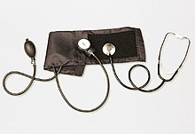
A sphygmomanometer, also known as a blood pressure monitor, or blood pressure gauge, is a device used to measure blood pressure, composed of an inflatable cuff to collapse and then release the artery under the cuff in a controlled manner, and a mercury or aneroid manometer to measure the pressure. Manual sphygmomanometers are used with a stethoscope when using the auscultatory technique.

<span class="mw-page-title-main">Pulse oximetry</span> Measurement of blood oxygen saturation

Pulse oximetry is a noninvasive method for monitoring a person's blood oxygen saturation. Peripheral oxygen saturation (SpO2) readings are typically within 2% accuracy of the more accurate reading of arterial oxygen saturation (SaO2) from arterial blood gas analysis. But the two are correlated well enough that the safe, convenient, noninvasive, inexpensive pulse oximetry method is valuable for measuring oxygen saturation in clinical use.

<span class="mw-page-title-main">Hypovolemic shock</span> Medical condition

Hypovolemic shock is a form of shock caused by severe hypovolemia. It could be the result of severe dehydration through a variety of mechanisms or blood loss. Hypovolemic shock is a medical emergency; if left untreated, the insufficient blood flow can cause damage to organs, leading to multiple organ failure.

At the end of pregnancy, the fetus must take the journey of childbirth to leave the reproductive mother. Upon its entry to the air-breathing world, the newborn must begin to adjust to life outside the uterus. This is true for all viviparous animals; this article discusses humans as the most-researched example.

<span class="mw-page-title-main">Pulmonary artery catheter</span> Catheter for insertion into a pulmonary artery

A pulmonary artery catheter (PAC), also known as a Swan-Ganz catheter or right heart catheter, is a balloon-tipped catheter that is inserted into a pulmonary artery in a procedure known as pulmonary artery catheterization or right heart catheterization. Pulmonary artery catheterization is a useful measure of the overall function of the heart particularly in those with complications from heart failure, heart attack, arrythmias or pulmonary embolism. It is also a good measure for those needing intravenous fluid therapy, for instance post heart surgery, shock, and severe burns. The procedure can also be used to measure pressures in the heart chambers.

<span class="mw-page-title-main">Capnography</span> Monitoring of the concentration of carbon dioxide in respiratory gases

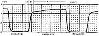
Capnography is the monitoring of the concentration or partial pressure of carbon dioxide (CO
2
) in the respiratory gases. Its main development has been as a monitoring tool for use during anesthesia and intensive care. It is usually presented as a graph of CO
2
(measured in kilopascals, "kPa" or millimeters of mercury, "mmHg") plotted against time, or, less commonly, but more usefully, expired volume (known as volumetric capnography). The plot may also show the inspired CO
2
, which is of interest when rebreathing systems are being used. When the measurement is taken at the end of a breath (exhaling), it is called "end tidal" CO
2
(PETCO2).

<span class="mw-page-title-main">Ankle–brachial pressure index</span> The ratio of the blood pressure at the ankle to the blood pressure in the upper arm

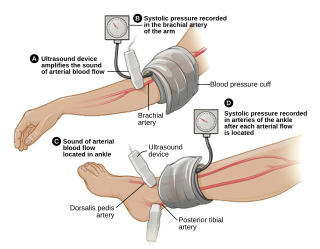
The ankle-brachial pressure index (ABPI) or ankle-brachial index (ABI) is the ratio of the blood pressure at the ankle to the blood pressure in the upper arm (brachium). Compared to the arm, lower blood pressure in the leg suggests blocked arteries due to peripheral artery disease (PAD). The ABPI is calculated by dividing the systolic blood pressure at the ankle by the systolic blood pressure in the arm.

The respiratory rate is the rate at which breathing occurs; it is set and controlled by the respiratory center of the brain. A person's respiratory rate is usually measured in breaths per minute.

Pediatric advanced life support (PALS) is a course offered by the American Heart Association (AHA) for health care providers who take care of children and infants in the emergency room, critical care and intensive care units in the hospital, and out of hospital. The course teaches healthcare providers how to assess injured and sick children and recognize and treat respiratory distress/failure, shock, cardiac arrest, and arrhythmias.

<span class="mw-page-title-main">Oxygen saturation (medicine)</span> Medical measurement

Oxygen saturation is the fraction of oxygen-saturated haemoglobin relative to total haemoglobin in the blood. The human body requires and regulates a very precise and specific balance of oxygen in the blood. Normal arterial blood oxygen saturation levels in humans are 96–100 percent. If the level is below 90 percent, it is considered low and called hypoxemia. Arterial blood oxygen levels below 80 percent may compromise organ function, such as the brain and heart, and should be promptly addressed. Continued low oxygen levels may lead to respiratory or cardiac arrest. Oxygen therapy may be used to assist in raising blood oxygen levels. Oxygenation occurs when oxygen molecules enter the tissues of the body. For example, blood is oxygenated in the lungs, where oxygen molecules travel from the air and into the blood. Oxygenation is commonly used to refer to medical oxygen saturation.

The cardiovascular examination is a portion of the physical examination that involves evaluation of the cardiovascular system. The exact contents of the examination will vary depending on the presenting complaint but a complete examination will involve the heart, lungs, belly and the blood vessels.

<span class="mw-page-title-main">Monitoring (medicine)</span> Observation of a disease, condition or one or several medical parameters over time

In medicine, monitoring is the observation of a disease, condition or one or several medical parameters over time.

The article reviews the evolution of continuous noninvasive arterial pressure measurement (CNAP). The historical gap between ease of use, but intermittent upper arm instruments and bulky, but continuous “pulse writers” (sphygmographs) is discussed starting with the first efforts to measure pulse, published by Jules Harrison in 1835. Such sphygmographs led a shadowy existence in the past, while Riva Rocci's upper arm blood pressure measurement started its triumphant success over 100 years ago. In recent times, CNAP measurement introduced by Jan Penáz in 1973 enabled the first recording of noninvasive beat-to-beat blood pressure resulting in marketed products such as the Finapres™ device and its successors. Recently, a novel method for CNAP monitoring has been designed for patient monitoring in perioperative, critical and emergency care, where blood pressure needs to be measured repeatedly or even continuously to facilitate the best care for patients.

<span class="mw-page-title-main">Blood pressure measurement</span> Techniques for determining blood pressure

Arterial blood pressure is most commonly measured via a sphygmomanometer, which historically used the height of a column of mercury to reflect the circulating pressure. Blood pressure values are generally reported in millimetres of mercury (mmHg), though aneroid and electronic devices do not contain mercury.

References

  1. "Vital Signs".
  2. "Emergency Physicians Home". www.emergencyphysicians.org.
  3. "Vital Signs Table - ProHealthSys". 3 July 2013.
  4. 1 2 National Early Warning Score Development and Implementation Group (NEWSDIG) (2012). National Early Warning Score (NEWS): standardising the assessment of acute-illness severity in the NHS. London: Royal College of Physicians. ISBN   978-1-86016-471-2.
  5. National Institute for Health and Clinical Excellence . Clinical guideline 50: Acutely ill patients in hospital . London, 2007.
  6. "Acute care toolkit 6: the medical patient at risk: recognition and care of the seriously ill or deteriorating medical patient" (PDF). Royal College of Physicians of London. May 2013.
  7. "Vital Signs". Cleveland Clinic. Retrieved 10 Sep 2020.
  8. "Vital Signs (Body Temperature, Pulse Rate, Respiration Rate, Blood Pressure)". www.hopkinsmedicine.org. 2022-06-14. Retrieved 2023-11-06.
  9. "Apical Pulse: What It Is and How to Take It". Cleveland Clinic. Retrieved 2023-11-06.
  10. "Vital Signs: How to Check My Vitals at Home". Cleveland Clinic. Retrieved 2023-11-06.
  11. 1 2 3 4 5 6 7 8 "Vital Signs (Body Temperature, Pulse Rate, Respiration Rate, Blood Pressure)". www.hopkinsmedicine.org. Retrieved 2019-08-30.
  12. 1 2 3 4 5 6 "Normal Vital Signs: Normal Vital Signs, Normal Heart Rate, Normal Respiratory Rate". 2019-07-23.
  13. 1 2 3 LeBlond RF, Brown DD, Suneja M, Szot JF (2014-09-05). DeGowin's diagnostic examination (10th ed.). New York: McGraw-Hill Education. ISBN   9780071814478. OCLC   876336892.
  14. "What should I include when I record vital signs of my patients for MU? - Providers & Professionals - HealthIT.gov". Archived from the original on 2018-03-25. Retrieved 2014-08-24.
  15. Emergency Care, 11th edition, pp. 226–244.
  16. "Pain as the 5Th Vital Sign Toolkit" (PDF).
  17. Lorenz KA, Sherbourne CD, Shugarman LR, Rubenstein LV, Wen L, Cohen A, Goebel JR, Hagenmeier E, Simon B, Lanto A, Asch SM (1 May 2009). "How Reliable is Pain as the Fifth Vital Sign?". J Am Board Fam Med. 22 (3): 291–298. doi: 10.3122/jabfm.2009.03.080162 . PMID   19429735.
  18. Wellbery C (15 October 2006). "Tips From Other Journals - American Family Physician". American Family Physician. 74 (8): 1417–1418.
  19. Mularski RA, White-Chu F, Overbay D, Miller L, Asch SM, Ganzini L (2006). "Measuring pain as the 5th vital sign does not improve quality of pain management". J Gen Intern Med. 21 (6): 607–12. doi:10.1111/j.1525-1497.2006.00415.x. PMC   1924634 . PMID   16808744.
  20. "The Fifth Vital Sign: Implementation of the Neonatal Infant Pain Scale" (PDF). Archived from the original (PDF) on 2012-05-26. Retrieved 2023-12-24.
  21. American College of Obstetricians and Gynecologists. (2015). "Menstruation in girls and adolescents: using the menstrual cycle as a vital sign. Committee Opinion No. 651". Obstet Gynecol. 126: 143–6.
  22. "Menstruation in Girls and Adolescents: Using the Menstrual Cycle as a Vital Sign". Pediatrics. 118 (5). American Academy of Pediatrics, Committee on Adolescence, American College of Obstetricians and Gynecologists, Committee on Adolescent Health Care. 2006.
  23. Mower W, Myers G, Nicklin E, Kearin K, Baraff L, Sachs C (1998). "Pulse oximetry as a fifth vital sign in emergency geriatric assessment". Acad Emerg Med. 5 (9): 858–65. doi: 10.1111/j.1553-2712.1998.tb02813.x . PMID   9754497.
  24. Mower W, Sachs C, Nicklin E, Baraff L (1997). "Pulse oximetry as a fifth pediatric vital sign". Pediatrics. 99 (5): 681–6. CiteSeerX   10.1.1.575.2200 . doi:10.1542/peds.99.5.681. PMID   9113944.
  25. Neff T (1988). "Routine oximetry. A fifth vital sign?". Chest. 94 (2): 227. doi: 10.1378/chest.94.2.227a . PMID   3396392.
  26. "Mining Vital Signs from Wearable Healthcare Device via Nonlinear Machine Learning". University of Hull. Archived from the original on 2016-08-17. Retrieved 2016-05-14.
  27. Vardi A, Levin I, Paret G, Barzilay, Z (2000). "The sixth vital sign: end-tidal CO2 in pediatric trauma patients during transport". Harefuah. 139 (3–4): 85–7, 168. PMID   10979461.
  28. Holcomb JB, Salinas J, McManus JM, Miller CC, Cooke WH, Convertino VA (2005). "Manual vital signs reliably predict need for life-saving interventions in trauma patients". J Trauma. 59 (4): 821–8, discussion 828–9. doi:10.1097/01.ta.0000188125.44129.7c. PMID   16374268.
  29. Bierman A (2001). "Functional Status: The Sixth Vital Sign". J Gen Intern Med. 16 (11): 785–6. doi:10.1111/j.1525-1497.2001.10918.x. PMC   1495293 . PMID   11722694.
  30. "Nursing care of dyspnea: the 6th vital sign in individuals with chronic obstructive pulmonary disease (COPD)". National Guideline Clearinghouse. Archived from the original on 2009-01-17. Retrieved 2009-01-16.
  31. Studenski S, Perera S, Wallace D, et al. (2003). "Physical performance measures in the clinical setting" (PDF). J Am Geriatr Soc. 51 (9): 314–322. doi:10.1046/j.1532-5415.2003.51104.x. PMID   12588574. S2CID   31083716.
  32. Bellelli G, Trabucchi M (May 1, 2008). "Delirium as the Sixth Vital Sign". Journal of the American Medical Directors Association. 9 (4): 279, author reply 279–80. doi:10.1016/j.jamda.2007.08.014. PMID   18457806 via www.jamda.com.
  33. 1 2 3 4 5 6 7 8 9 10 PEDIATRIC AGE SPECIFIC Archived 2017-05-16 at the Wayback Machine , page 6. Revised 6/10. By Theresa Kirkpatrick and Kateri Tobias. UCLA Health System
  34. Emergency Care, Page 214
  35. Emergency Care, Page 215
  36. Vorvick L. "Pulse". MedlinePlus. U.S. National Library of Medicine. Retrieved 23 January 2011.
  37. "Normal Vital Signs: Normal Vital Signs, Normal Heart Rate, Normal Respiratory Rate". 2019-07-23.