Popliteal artery

Last updated
Popliteal artery
Popliteal branches from posterior labeled.png
Popliteal artery and branches
Gray609.png
Lymph glands of popliteal fossa.
Details
Source Femoral artery
Branches Anterior tibial, posterior tibial artery, sural, superior genicular (medial, lateral), middle genicular, inferior genicular (medial, lateral)
Vein Popliteal vein
Identifiers
Latin arteria poplitea
MeSH D011150
TA98 A12.2.16.033
TA2 4699
FMA 77155
Anatomical terminology

The popliteal artery is a deeply placed continuation of the femoral artery opening in the distal portion of the adductor magnus muscle. It courses through the popliteal fossa and ends at the lower border of the popliteus muscle, where it branches into the anterior and posterior tibial arteries.

Contents

The deepest (most anterior) structure in the fossa, the popliteal artery runs close to the joint capsule of the knee as it spans the intercondylar fossa. Five genicular branches of the popliteal artery supply the capsule and ligaments of the knee joint. The genicular arteries are the superior lateral, superior medial, middle, inferior lateral, and inferior medial genicular arteries. They participate in the formation of the periarticular genicular anastomosis, a network of vessels surrounding the knee that provides collateral circulation capable of maintaining blood supply to the leg during full knee flexion, which may kink the popliteal artery. [1]

Structure

Popliteal and other leg arteries Blausen 0607 LegArteries.png
Popliteal and other leg arteries

The popliteal artery is the continuation of the femoral artery. [2] It exits the popliteal fossa under the fibrous arch of the soleus muscle. [2]

Relations

Branches

The branches of the popliteal artery are:

Muscular branches of the popliteal artery supply the hamstring, gastrocnemius, soleus, and plantaris muscles. The superior muscular branches of the popliteal artery have clinically important anastomoses with the terminal part of the deep femoral and gluteal arteries. The cutaneous branches arise either directly by the popliteal artery or indirectly by the muscular branches. Genicular branches are five in number, two superior, two inferior, and one middle. The middle genicular artery supplies the cruciate ligaments and the synovial membrane of knee joint.

Tibial-fibular trunk

The fibular artery typically arises from the posterior tibial artery. [3] Therefore, the posterior tibial artery proximal to the fibular artery origin is sometimes called the tibial-peroneal trunk or tibial-fibular trunk and it could be said that the popliteal artery bifurcates into the tibial-fibular trunk and anterior tibial artery.

Clinical significance

Additional images

See also

Related Research Articles

<span class="mw-page-title-main">Human leg</span> Lower extremity or limb of the human body (foot, lower leg, thigh and hip)

The leg is the entire lower limb of the human body, including the foot, thigh or sometimes even the hip or buttock region. The major bones of the leg are the femur, tibia, and adjacent fibula. The thigh is between the hip and knee, while the calf (rear) and shin (front) are between the knee and foot.

<span class="mw-page-title-main">Knee</span> Leg joint in primates

In humans and other primates, the knee joins the thigh with the leg and consists of two joints: one between the femur and tibia, and one between the femur and patella. It is the largest joint in the human body. The knee is a modified hinge joint, which permits flexion and extension as well as slight internal and external rotation. The knee is vulnerable to injury and to the development of osteoarthritis.

<span class="mw-page-title-main">Popliteal vein</span> Large leg vein

The popliteal vein is a vein of the lower limb. It is formed from the anterior tibial vein and the posterior tibial vein. It travels medial to the popliteal artery, and becomes the femoral vein. It drains blood from the leg. It can be assessed using medical ultrasound. It can be affected by popliteal vein entrapment.

<span class="mw-page-title-main">Gastrocnemius muscle</span> Calf muscle

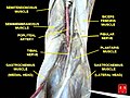
The gastrocnemius muscle is a superficial two-headed muscle that is in the back part of the lower leg of humans. It is located superficial to the soleus in the posterior (back) compartment of the leg. It runs from its two heads just above the knee to the heel, extending across a total of three joints.

The anterior tibial artery is an artery of the leg. It carries blood to the anterior compartment of the leg and dorsal surface of the foot, from the popliteal artery.

<span class="mw-page-title-main">Tibial nerve</span> Branch of the sciatic nerve

The tibial nerve is a branch of the sciatic nerve. The tibial nerve passes through the popliteal fossa to pass below the arch of soleus.

The biceps femoris is a muscle of the thigh located to the posterior, or back. As its name implies, it consists of two heads; the long head is considered part of the hamstring muscle group, while the short head is sometimes excluded from this characterization, as it only causes knee flexion and is activated by a separate nerve.

<span class="mw-page-title-main">Adductor magnus muscle</span> Muscle in the thigh

The adductor magnus is a large triangular muscle, situated on the medial side of the thigh.

The semimembranosus muscle is the most medial of the three hamstring muscles in the thigh. It is so named because it has a flat tendon of origin. It lies posteromedially in the thigh, deep to the semitendinosus muscle. It extends the hip joint and flexes the knee joint.

<span class="mw-page-title-main">Plantaris muscle</span> One of the superficial muscles of the superficial posterior compartment of the leg

The plantaris is one of the superficial muscles of the superficial posterior compartment of the leg, one of the fascial compartments of the leg.

The common fibular nerve is a nerve in the lower leg that provides sensation over the posterolateral part of the leg and the knee joint. It divides at the knee into two terminal branches: the superficial fibular nerve and deep fibular nerve, which innervate the muscles of the lateral and anterior compartments of the leg respectively. When the common fibular nerve is damaged or compressed, foot drop can ensue.

<span class="mw-page-title-main">Popliteal fossa</span> Depression at back of knee joint

The popliteal fossa is a shallow depression located at the back of the knee joint. The bones of the popliteal fossa are the femur and the tibia. Like other flexion surfaces of large joints, it is an area where blood vessels and nerves pass relatively superficially, and with an increased number of lymph nodes.

<span class="mw-page-title-main">Lower extremity of femur</span>

The lower extremity of femur is the lower end of the femur in human and other animals, closer to the knee. It is larger than the upper extremity of femur, is somewhat cuboid in form, but its transverse diameter is greater than its antero-posterior; it consists of two oblong eminences known as the lateral condyle and medial condyle.

In anatomy, arterial tree is used to refer to all arteries and/or the branching pattern of the arteries. This article regards the human arterial tree. Starting from the aorta:

<span class="mw-page-title-main">Descending genicular artery</span>

The descending genicular artery arises from the femoral artery just before its passage through the adductor hiatus.

The sural arteries are two large branches, lateral and medial, which are distributed to the gastrocnemius, soleus, and plantaris muscles. Sural means related to the calf. The term applies to any of four or five arteries arising from the popliteal artery, with distribution to the muscles and integument of the calf, and with anastomoses to the posterior tibial, medial and lateral inferior genicular arteries.

<span class="mw-page-title-main">Posterior compartment of thigh</span> One of the fascial compartments that contains the knee flexors and hip extensors

The posterior compartment of the thigh is one of the fascial compartments that contains the knee flexors and hip extensors known as the hamstring muscles, as well as vascular and nervous elements, particularly the sciatic nerve.

<span class="mw-page-title-main">Patellar network</span> Network of blood vessels around the knee

The patellar network is an intricate network of blood vessels around and above the patella, and on the contiguous ends of the femur and tibia, forming a superficial and a deep plexus.

<span class="mw-page-title-main">Outline of human anatomy</span> Overview of and topical guide to human anatomy

The following outline is provided as an overview of and topical guide to human anatomy:

References

  1. Moore K.L. and Dalley A.F. (2006), Clinically Oriented Anatomy, 5th Edition, Lippincott Williams & Wilkins, Toronto, page 636
  2. 1 2 3 4 5 6 7 8 9 10 Masquelet, Alain-Charles; Sassu, Paolo (2009-01-01), Wei, Fu-Chan; Mardini, Samir (eds.), "CHAPTER 30 - Gastrocnemius flap", Flaps and Reconstructive Surgery, Edinburgh: W.B. Saunders, pp. 411–423, doi:10.1016/b978-0-7216-0519-7.00030-7, ISBN   978-0-7216-0519-7 , retrieved 2021-02-20
  3. Day C, Orme R (2006). "Popliteal artery branching patterns -- an angiographic study". Clin Radiol. 61 (8): 696–9. doi:10.1016/j.crad.2006.03.014. PMID   16843754.
  4. Moore K.L. and Dalley A.F. (2006), Clinically Oriented Anatomy, 5th Edition, Lippincott Williams & Wilkins, Toronto, page 637