Apgar score

Last updated
Apgar score
Virginia Apgar.jpg
Virginia Apgar, creator of the Apgar score

The Apgar score is a quick way for health professionals to evaluate the health of all newborns at 1 and 5 minutes after birth and in response to resuscitation. [1] It was originally developed in 1952 by an anesthesiologist at Columbia University, Virginia Apgar, to address the need for a standardized way to evaluate infants shortly after birth. [2]

Contents

Today, the categories developed by Apgar used to assess the health of a newborn remain largely the same as in 1952, though the way they are implemented and used has evolved over the years. [3] The score is determined through the evaluation of the newborn in five criteria: activity (tone), pulse, grimace, appearance, and respiration. For each criterion, newborns can receive a score from 0 to 2. [1] [3] [4] The list of criteria is a backronym of Apgar's surname.

History and development of the Apgar score

Apgar originally developed the criteria as way to address the lack of a standardized way to assess the need for assistive breathing procedures for newborns. In 1952, after some refinement of her initial system, Apgar presented the Apgar score at a joint meeting between the International Anesthesia Research Society and the International College of Anesthetists, and it was then published in Anesthesia & Analgesia in 1953. [2]

In 1955, efforts to establish a scientific basis to the score increased. Alongside Duncan Holaday and Stanley James, [2] Apgar published a research paper using the scores of 15,348 infants to establish the association between a low Apgar score (0-2) and laboratory findings characteristics of asphyxia. [5]

The Apgar score is no longer used as a way to determine the need for newborn resuscitation because supportive measures must be implemented prior to 1 minute after birth, the first time-point at which the Apgar score is determined. [4] However, the Apgar score is a method of assessment endorsed by the American College of Obstetricians and Gynecologist and the American Academy of Pediatrics. [3]

Criteria

The five criteria of the Apgar score: [4]
Score of 0Score of 1Score of 2Component of backronym
Skin colorblue or pale all overblue at extremities,
body pink
(acrocyanosis)
no cyanosis
body and extremities pink
Appearance
Pulse rate absent< 100 beats per minute 100 beats per minutePulse
Reflex  irritability grimaceno response to stimulationgrimace or suction on aggressive stimulationcry on stimulationGrimace
Muscle Tonenonesome flexion flexed arms and legs that resist extensionActivity
Respiratory effort absentweak, irregular, gaspingstrong, robust cryRespiration

Interpretation of scores

Mind map showing summary for the Apgar score Apgar score.png
Mind map showing summary for the Apgar score

Various members of the healthcare team, including midwives, nurses, or physicians, may be involved in the Apgar scoring of a neonate. [3] The test is generally done at one and five minutes after birth and may be repeated later if the score is and remains low. Scores of seven and above are generally normal; four to six, fairly low; and three and below are generally regarded as critically low and cause for immediate resuscitative efforts. [6]

A low score on the one-minute mark may show that the neonate requires medical attention, [7] but does not necessarily indicate a long-term problem, particularly if the score improves at the five-minute mark. A constellation of factors may contribute to a low Apgar score value. [8] An Apgar score that remains below three at five minutes and later times, such as 10, 15, or 30 minutes, does not provide supporting evidence for a specific illness but can sometimes be among the first indicators of neonatal encephalopathy. [8] [6] [9] However, the Apgar test's purpose is to determine quickly whether or not a newborn needs immediate medical care. It is not designed to predict long-term health issues. [10]

A score of 10 is uncommon, due to the prevalence of transient cyanosis, and does not substantially differ from a score of nine. Transient cyanosis is common, particularly in babies born at high altitude. [11]

Implementation of scores

In cases where a newborn needs resuscitation, it should be initiated before the Apgar score is assigned at the one-minute mark. Therefore, the Apgar score is not used to determine if initial resuscitation is needed, rather it is used to determine if resuscitation efforts should be continued. Variation between the one-minute and five-minute Apgar scores can be used to assess an infant's response to resuscitation. If the score is below seven at the five-minute mark, the Neonatal Resuscitation Program guidelines specify that the infant's Apgar score should be reassessed at five-minute intervals for up to 20 minutes. [4]

Exceedingly few infants who have an Apgar score of 0 at 10 minutes of age survive with intact neurological function. As a result, the 2011 Neonatal Resuscitation Program suggests that if no pulse is appreciable at 10 minutes of life, "discontinuation of resuscitative efforts may be appropriate." [6] However, in a recent retrospective study including 17 infants with an Apgar score of 0 at 10 minutes who received therapeutic hypothermia, 4 of the 8 surviving babies had no neurological abnormalities and only 1 infant had severe abnormalities, as assessed through brain MRI. [12]

During neonatal resuscitation, Apgar scores may not accurately represent the condition of the neonate as resuscitation measures (i.e. positive pressure ventilation and chest compressions) may artificially elevate scores. As a result, the American Academy of Pediatrics and the American College of Obstetricians and Gynecologists encourage the use of an expanded Apgar score report, which records resuscitation efforts utilized at each time point. [6]

A systematic review that analyzed the relationship between umbilical cord pH and neonatal outcomes found that low cord pH is strongly correlated with mortality, morbidity and cerebral palsy in childhood. [13] To reduce the risk of negative outcomes, it is recommended to obtain a sample of the umbilical artery blood gas when a newborn has an Apgar score of five or less at the five-minute mark. [4]

Newborn crying right after birth HumanNewborn.JPG
Newborn crying right after birth

Recognizing the importance of skin to skin contact (SSC), the World Health Organization (WHO), as part of the Baby Friendly Hospital Initiative, recommends that the Apgar score should be done while SSC is underway whenever possible. [14] This recommendation was adopted by the American Academy of Pediatrics in 2009. [14]

Limitations

There are numerous factors that contribute to the Apgar score, several of which are subjective. Examples of subjective factors include but are not limited to color, tone, and reflex irritability. [15] Preterm infants may receive a lower score in these categories due to lack of maturity rather than asphyxia. Other factors that may contribute to variability among infants are birth defects, sedation of the mother during labor, gestational age or trauma. Inappropriately using the Apgar score has led to errors in diagnosing asphyxia. [4]

Various studies have shown that the Apgar score has variability between individual medical providers. One study was done in which several health care providers were assigned to give Apgar scores to the same infants. Results showed an Apgar score consistency of 55% to 82% between health care providers. [16] [17] Ideally, to limit variability and bolster consistency, it is preferred that the same individual determine the 1-minute and 5-minute Apgar scores. [3]

Acronym

Some ten years after initial publication, [18] [ citation needed ] a backronym for APGAR was coined in the United States as a mnemonic learning aid: Appearance (skin color), Pulse (heart rate), Grimace (reflex irritability), Activity (muscle tone), and Respiration.

Another eponymous backronym from Virginia Apgar's name is American Pediatric Gross Assessment Record.

Another mnemonic for the test is "How Ready Is This Child?", which summarizes the test criteria as Heart rate, Respiratory effort, Irritability, Tone, and Color. [20]

See also

Related Research Articles

<span class="mw-page-title-main">Umbilical cord</span> Conduit between embryo/fetus and the placenta

In placental mammals, the umbilical cord is a conduit between the developing embryo or fetus and the placenta. During prenatal development, the umbilical cord is physiologically and genetically part of the fetus and normally contains two arteries and one vein, buried within Wharton's jelly. The umbilical vein supplies the fetus with oxygenated, nutrient-rich blood from the placenta. Conversely, the fetal heart pumps low-oxygen, nutrient-depleted blood through the umbilical arteries back to the placenta.

<span class="mw-page-title-main">Virginia Apgar</span> American physician and obstetrical anesthesiologist (1909–1974)

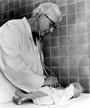
Virginia Apgar was an American physician, obstetrical anesthesiologist and medical researcher, best known as the inventor of the Apgar score, a way to quickly assess the health of a newborn child immediately after birth in order to combat infant mortality. In 1952, she developed the 10-point Apgar score to assist physicians and nurses in assessing the status of newborns. Given at one minute and five minutes after birth, the Apgar test measures a child's breathing, skin color, reflexes, motion, and heart rate. A friend said, "She probably did more than any other physician to bring the problem of birth defects out of back rooms." She was a leader in the fields of anesthesiology and teratology, and introduced obstetrical considerations to the established field of neonatology.

Fetal distress, also known as non-reassuring fetal status, is a condition during pregnancy or labor in which the fetus shows signs of inadequate oxygenation. Due to its imprecision, the term "fetal distress" has fallen out of use in American obstetrics. The term "non-reassuring fetal status" has largely replaced it. It is characterized by changes in fetal movement, growth, heart rate, and presence of meconium stained fluid.

<span class="mw-page-title-main">Cardiotocography</span> Technical means of recording the fetal heartbeat and the uterine contractions during pregnancy

Cardiotocography (CTG) is a technique used to monitor the fetal heartbeat and uterine contractions during pregnancy and labour. The machine used to perform the monitoring is called a cardiotocograph.

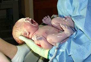
At the end of pregnancy, the fetus must take the journey of childbirth to leave the reproductive mother. Upon its entry to the air-breathing world, the newborn must begin to adjust to life outside the uterus. This is true for all viviparous animals; this article discusses humans as the most-researched example.

Transient tachypnea of the newborn is a respiratory problem that can be seen in the newborn shortly after delivery. It is caused by retained fetal lung fluid due to impaired clearance mechanisms. It is the most common cause of respiratory distress in term neonates. It consists of a period of tachypnea (rapid breathing. Usually, this condition resolves over 24–72 hours. Treatment is supportive and may include supplemental oxygen and antibiotics. The chest x-ray shows hyperinflation of the lungs including prominent pulmonary vascular markings, flattening of the diaphragm, and fluid in the horizontal fissure of the right lung.

<span class="mw-page-title-main">Water birth</span> Childbirth that occurs in water

Water birth is labor and sometimes delivery that occurs in water, usually a birthing pool. The American College of Obstetricians and Gynecologists does not recommend birthing in water as the safety has not been determined. Proponents believe childbirth in water results in a more relaxed, less painful experience that promotes a midwife-led model of care. Critics argue that the safety of waterbirth has not been scientifically proven and that a wide range of adverse neonatal outcomes have been documented, including increased mother or child infections and the possibility of infant drowning. A 2018 Cochrane Review of water immersion in the first stages of labor found evidence of fewer epidurals and few adverse effects but insufficient information regarding giving birth in water.

<span class="mw-page-title-main">Neonatology</span> Medical care of newborns, especially the ill or premature

Neonatology is a subspecialty of pediatrics that consists of the medical care of newborn infants, especially the ill or premature newborn. It is a hospital-based specialty and is usually practised in neonatal intensive care units (NICUs). The principal patients of neonatologists are newborn infants who are ill or require special medical care due to prematurity, low birth weight, intrauterine growth restriction, congenital malformations, sepsis, pulmonary hypoplasia, or birth asphyxia.

<span class="mw-page-title-main">Neonatal intensive care unit</span> Intensive care unit specializing in the care of ill or premature newborn infants

A neonatal intensive care unit (NICU), also known as an intensive care nursery (ICN), is an intensive care unit (ICU) specializing in the care of ill or premature newborn infants. The NICU is divided into several areas, including a critical care area for babies who require close monitoring and intervention, an intermediate care area for infants who are stable but still require specialized care, and a step down unit where babies who are ready to leave the hospital can receive additional care before being discharged.

Perinatal asphyxia is the medical condition resulting from deprivation of oxygen to a newborn infant that lasts long enough during the birth process to cause physical harm, usually to the brain. It remains a serious condition which causes significant mortality and morbidity. It is also the inability to establish and sustain adequate or spontaneous respiration upon delivery of the newborn, an emergency condition that requires adequate and quick resuscitation measures. Perinatal asphyxia is also an oxygen deficit from the 28th week of gestation to the first seven days following delivery. It is also an insult to the fetus or newborn due to lack of oxygen or lack of perfusion to various organs and may be associated with a lack of ventilation. In accordance with WHO, perinatal asphyxia is characterised by: profound metabolic acidosis, with a pH less than 7.20 on umbilical cord arterial blood sample, persistence of an Apgar score of 3 at the 5th minute, clinical neurologic sequelae in the immediate neonatal period, or evidence of multiorgan system dysfunction in the immediate neonatal period. Hypoxic damage can occur to most of the infant's organs, but brain damage is of most concern and perhaps the least likely to quickly or completely heal. In more pronounced cases, an infant will survive, but with damage to the brain manifested as either mental, such as developmental delay or intellectual disability, or physical, such as spasticity.

Fetal viability is the ability of a human fetus to survive outside the uterus. Medical viability is generally considered to be between 23 and 24 weeks gestational age. Viability depends upon factors such as birth weight, gestational age, and the availability of advanced medical care. In low-income countries, half of newborns born at or below 32 weeks gestational age died due to a lack of medical access; in high-income countries, the vast majority of newborns born above 24 weeks gestational age survive.

In obstetrics, gestational age is a measure of the age of a pregnancy taken from the beginning of the woman's last menstrual period (LMP), or the corresponding age of the gestation as estimated by a more accurate method, if available. Such methods include adding 14 days to a known duration since fertilization, or by obstetric ultrasonography. The popularity of using this measure of pregnancy is largely due to convenience: menstruation is usually noticed, while there is generally no convenient way to discern when fertilization or implantation occurred.

<span class="mw-page-title-main">Cerebral hypoxia</span> Oxygen shortage of the brain

Cerebral hypoxia is a form of hypoxia, specifically involving the brain; when the brain is completely deprived of oxygen, it is called cerebral anoxia. There are four categories of cerebral hypoxia; they are, in order of increasing severity: diffuse cerebral hypoxia (DCH), focal cerebral ischemia, cerebral infarction, and global cerebral ischemia. Prolonged hypoxia induces neuronal cell death via apoptosis, resulting in a hypoxic brain injury.

<span class="mw-page-title-main">Intrauterine hypoxia</span> Medical condition when the fetus is deprived of sufficient oxygen

Intrauterine hypoxia occurs when the fetus is deprived of an adequate supply of oxygen. It may be due to a variety of reasons such as prolapse or occlusion of the umbilical cord, placental infarction, maternal diabetes and maternal smoking. Intrauterine growth restriction may cause or be the result of hypoxia. Intrauterine hypoxia can cause cellular damage that occurs within the central nervous system. This results in an increased mortality rate, including an increased risk of sudden infant death syndrome (SIDS). Oxygen deprivation in the fetus and neonate have been implicated as either a primary or as a contributing risk factor in numerous neurological and neuropsychiatric disorders such as epilepsy, attention deficit hyperactivity disorder, eating disorders and cerebral palsy.

<span class="mw-page-title-main">Neonatal nursing</span>

Neonatal nursing is a sub-specialty of nursing care for newborn infants up to 28 days after birth. The term neonatal comes from neo, "new", and natal, "pertaining to birth or origin". Neonatal nursing requires a high degree of skill, dedication and emotional strength as they care for newborn infants with a range of problems. These problems vary between prematurity, birth defects, infection, cardiac malformations and surgical issues. Neonatal nurses are a vital part of the neonatal care team and are required to know basic newborn resuscitation, be able to control the newborn's temperature and know how to initiate cardiopulmonary and pulse oximetry monitoring. Most neonatal nurses care for infants from the time of birth until they are discharged from the hospital.

Neonatal encephalopathy (NE), previously known as neonatal hypoxic-ischemic encephalopathy, is defined as a encephalopathy syndrome with signs and symptoms of abnormal neurological function, in the first few days of life in an infant born after 35 weeks of gestation. In this condition there is difficulty initiating and maintaining respirations, a subnormal level of consciousness, and associated depression of tone, reflexes, and possibly seizures. Encephalopathy is a nonspecific response of the brain to injury which may occur via multiple methods, but is commonly caused by birth asphyxia, leading to cerebral hypoxia.

Mild total body hypothermia, induced by cooling a baby to 33-34°C for three days after birth, is nowadays a standardized treatment after moderate to severe hypoxic ischemic encephalopathy in full-term and near to fullterm neonates. It has recently been proven to be the only medical intervention which reduces brain damage, and improves an infant's chance of survival and reduced disability.

Neonatal hypoglycemia occurs when the neonate's blood glucose level is less than the newborn's body requirements for factors such as cellular energy and metabolism. There is inconsistency internationally for diagnostic thresholds. In the US, hypoglycemia is when the blood glucose level is below 30 mg/dL within the first 24 hours of life and below 45 mg/dL thereafter. In the UK, however, lower and more variable thresholds are used. The neonate's gestational age, birth weight, metabolic needs, and wellness state of the newborn has a substantial impact on the neonates blood glucose level. There are known risk factors that can be both maternal and neonatal. This is a treatable condition. Its treatment depends on the cause of the hypoglycemia. Though it is treatable, it can be fatal if gone undetected. Hypoglycemia is the most common metabolic problem in newborns.

The Bloxsom air lock was an incubator used in the treatment of respiratory distress among newly born infants in the 1950s. The device attempted to mimic the rhythm of uterine contractions, which were thought to have a role in stimulating fetal breathing. The device was developed by Dr. Allan Bloxsom, a pediatrician at St. Joseph Hospital and Baylor College of Medicine in Houston, Texas. At its peak, the device was utilized in more than 700 hospitals.

<span class="mw-page-title-main">Neonatal resuscitation</span>

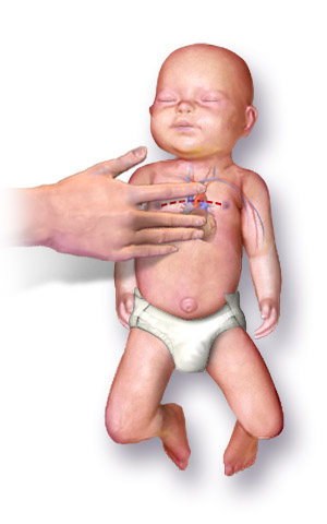
Neonatal resuscitation, also known as newborn resuscitation, is an emergency procedure focused on supporting approximately 10% of newborn children who do not readily begin breathing, putting them at risk of irreversible organ injury and death. Many of the infants who require this support to start breathing breath well on their own after assistance. Through positive airway pressure, and in severe cases chest compressions, medical personnel certified in neonatal resuscitation can often stimulate neonates to begin breathing on their own, with attendant normalization of heart rate.

References

  1. 1 2 "The Apgar Score". www.acog.org. Retrieved 2021-09-10.
  2. 1 2 3 Calmes, Selma H. (May 2015). "Dr. Virginia Apgar and the Apgar Score: How the Apgar Score Came to Be". Anesthesia & Analgesia . 120 (5): 1060–1064. doi: 10.1213/ANE.0000000000000659 . ISSN   0003-2999. PMID   25899273. S2CID   41296774.
  3. 1 2 3 4 5 Simon, Leslie V.; Hashmi, Muhammad F.; Bragg, Bradley N. (2021), "APGAR Score", StatPearls, Treasure Island, Florida: StatPearls Publishing, PMID   29262097 , retrieved 2021-09-10
  4. 1 2 3 4 5 6 "The Apgar Score". www.acog.org. Retrieved 2019-08-01.
  5. Apgar, Virginia (1958-12-13). "Evaluation of the Newborn Infant-Second Report". Journal of the American Medical Association. 168 (15): 1985–1988. doi:10.1001/jama.1958.03000150027007. ISSN   0002-9955. PMID   13598635.
  6. 1 2 3 4 Newborn, American Academy of Pediatrics Committee on Fetus And; Practice, American College of Obstetricians and Gynecologists Committee on Obstetric (2015-10-01). "The Apgar Score". Pediatrics. 136 (4): 819–822. doi: 10.1542/peds.2015-2651 . ISSN   0031-4005. PMID   26416932. S2CID   31689176.
  7. Casey BM, McIntire DD, Leveno KJ (February 2001). "The continuing value of the Apgar score for the assessment of newborn infants". The New England Journal of Medicine. 344 (7): 467–71. doi: 10.1056/NEJM200102153440701 . PMID   11172187.
  8. 1 2 Pediatrics, American Academy of (2014-05-01). "Neonatal Encephalopathy and Neurologic Outcome, Second EditionReport of the American College of Obstetricians and Gynecologists' Task Force on Neonatal Encephalopathy". Pediatrics. 133 (5): e1482–e1488. doi: 10.1542/peds.2014-0724 . ISSN   0031-4005.
  9. Care of the baby and woman immediately after birth. National Institute for Health and Care Excellence (UK). December 2014.
  10. Apgar V (1953). "A proposal for a new method of evaluation of the newborn infant" (PDF). Current Researches in Anesthesia & Analgesia. 32 (4): 260–7. doi:10.1213/00000539-195301000-00041. PMID   13083014.
  11. Gonzales GF, Salirrosas A (September 2005). "Arterial oxygen saturation in healthy newborns delivered at term in Cerro de Pasco (4340 m) and Lima (150 m)". Reproductive Biology and Endocrinology. 3: 46. doi: 10.1186/1477-7827-3-46 . PMC   1215518 . PMID   16156890.
  12. Ayrapetyan, Marina; Talekar, Kiran; Schwabenbauer, Kathleen; Carola, David; Solarin, Kolawole; McElwee, Dorothy; Adeniyi-Jones, Susan; Greenspan, Jay; Aghai, Zubair H. (April 2019). "Apgar Scores at 10 Minutes and Outcomes in Term and Late Preterm Neonates with Hypoxic-Ischemic Encephalopathy in the Cooling Era". American Journal of Perinatology. 36 (5): 545–554. doi:10.1055/s-0038-1670637. ISSN   0735-1631. PMC   8039809 . PMID   30208498.
  13. Malin GL, Morris RK, Khan KS (May 2010). "Strength of association between umbilical cord pH and perinatal and long term outcomes: systematic review and meta-analysis". BMJ. 340 (may13 1): c1471. doi:10.1136/bmj.c1471. PMC   2869402 . PMID   20466789.
  14. 1 2 Feldman-Winter, Lori; Goldsmith, Jay P.; Newborn, Committee on Fetus And; Syndrome, Task Force on Sudden Infant Death (2016-09-01). "Safe Sleep and Skin-to-Skin Care in the Neonatal Period for Healthy Term Newborns". Pediatrics. 138 (3): e20161889. doi: 10.1542/peds.2016-1889 . ISSN   0031-4005. PMID   27550975. S2CID   19866683.
  15. "Apgar score: MedlinePlus Medical Encyclopedia". medlineplus.gov. Retrieved 2022-11-18.
  16. Montgomery, Kristen S. (2000). "Apgar Scores: Examining the Long-term Significance". The Journal of Perinatal Education. 9 (3): 5–9. doi:10.1624/105812400X87716. ISSN   1058-1243. PMC   1595023 . PMID   17273212.
  17. Livingston, Jacquelynn (1990-11-01). "Interrater reliability of the apgar score in term and premature infants". Applied Nursing Research. 3 (4): 164–165. doi:10.1016/S0897-1897(05)80139-9. ISSN   0897-1897. PMID   2252404.
  18. "What Is a Good Apgar Score?". Cleveland Clinic. Retrieved 2022-11-18.
  19. "Co je to APGAR skóre a kdo ho vymyslel?". www.maminka.cz (in Czech). Retrieved 2020-01-12.
  20. Klostranec JM, Hong J, Raghuran K (2012). The Essential Med Notes for Medical Students. Toronto, ON: University of Toronto Press.

Further reading