Pelvic examination

Last updated
Pelvic examination
Pelvic exam nci-vol-1786-300.jpg
Line drawing showing bimanual palpation as part of a pelvic exam
Specialty gynaecology
ICD-9-CM 89.26

A pelvic examination is the physical examination of the external and internal female pelvic organs. [1] It is frequently used in gynecology for the evaluation of symptoms affecting the female reproductive and urinary tract, such as pain, bleeding, discharge, urinary incontinence, or trauma (e.g. sexual assault). [2] [3] It can also be used to assess a woman's anatomy in preparation for procedures. [4] The exam can be done awake in the clinic and emergency department, or under anesthesia in the operating room. [5] The most commonly performed components of the exam are 1) the external exam, to evaluate the vulva 2) the internal exam with palpation (commonly called the bimanual exam) to examine the uterus, ovaries, and structures adjacent to the uterus (adnexae) and 3) the internal exam using a speculum to visualize the vaginal walls and cervix. [6] During the pelvic exam, sample of cells and fluids may be collected to screen for sexually transmitted infections or cancer (the Pap test). [6]

Contents

Some clinicians perform a pelvic exam as part of routine preventive care. [3] However, in 2014, the American College of Physicians published guidelines against routine pelvic examination in adult women who are not pregnant and lack symptoms, with the exception of pelvic exams done as part of cervical cancer screening. [7] [8]

Medical guidelines

Traditionally in the field of gynecology, the benefits of routine pelvic examinations were assumed, and the exam was a recommended part of the initial gynecology visit, annual visits, and as needed for treatment. [9] In 2014, the American College of Physicians (ACP) published a review of the benefits and the risks of the exam and issued a guideline that recommended against performing this examination to screen for conditions in asymptomatic, nonpregnant, adult women, concluding that the potential harms outweighed the demonstrated benefits and that screening pelvic exams in asymptomatic women did not reduce mortality or morbidity rates. [7] The guideline did not consider the necessity or frequency of Pap smears. The ACP guideline concluded that there was no evidence that the exam in asymptomatic adult women reduced morbidity or mortality, and that there were no studies that addressed the exam's diagnostic accuracy for identifying specific gynecologic conditions. [7] The guideline concluded that there was evidence of harm, including fear, embarrassment, pain, discomfort and unnecessary surgery. [7] This was a strong recommendation, based on moderate-quality evidence. [7] In 2018, the American College of Obstetricians and Gynecologists (ACOG) issued a committee opinion that pelvic exams should be performed for 1) symptoms of gynecologic disease, 2) screening for cervical dysplasia, or 3) management of gynecologic disorders or malignancy, using shared decision-making with the patient. [10] ACOG acknowledged that given changes in screening recommendations and an assessment of harms vs benefits as reviewed by the US Preventive Services Task Force, re-evaluation of routine screening pelvic examination for asymptomatic, non-pregnant women with average risk for gynecologic disease was indicated. [10] A 2020 update of the literature by the US Preventive Services Task Force concluded that the evidence remained insufficient to assess the balance of benefits and harms of screening pelvic examinations in asymptomatic women. [11]

Annual "well-woman visits" are an occasion for gynecologists to identify health risks for women; ACOG has noted that these visits may also include clinical breast examinations, immunizations, contraceptive care discussions, and preventive health care counseling. [2] ACOG reinforced the potential value of pelvic examinations in allowing clinicians to explain a patient's anatomy, reassure her of normalcy, and answer specific questions, thus establishing open communication between patient and clinician. [10]

Preparation, communication, and trauma-informed care

Chair for gynaecological examinations. Next to the chair a sonography device and screen. Chair for gynaecological examination.jpg
Chair for gynaecological examinations. Next to the chair a sonography device and screen.

The examination can be emotionally and physically uncomfortable for women. Preparation, good communication, thoughtful technique, and trauma-informed care can help mitigate this discomfort. [12] [13]

Prior to the exam, before the patient is undressed and lying on the examination table or chair, examiners should ask the patient if she has had a pelvic exam in the past and whether she has any questions or concerns about the exam. Women may be concerned about pain, or they may be embarrassed about the examination of sensitive areas of the body. They may have experienced sexual assault or negative experiences with pelvic examination in the past, which may lead to the exam triggering strong emotional and physical symptoms. [6] [13] [14] Additionally, patients may have concern about odor or menstruation during exam, neither of which should impact the examiner's ability to perform a thorough, respectful exam. [15] Patients generally prefer to be asked about past experiences and are often helpful in suggesting ways to mitigate the discomfort of the exam. [13] Prior to the exam, the examiner should offer to show the patient models or diagrams of the pelvic anatomy and any instruments that will be used during the exam. [6]

Careful preparation is helpful for an efficient and comfortable exam. Prior to asking the patient to position herself on the exam table or chair for examination, the examiner should collect all the instruments needed for the exam and any planned procedures, including the speculum, light source, lubricant, gloves, drapes, and specimen collection media. Warming the speculum with warm tap water or keeping the speculum in a warmer will also increase comfort. The patient should be given the opportunity to have a chaperone or support person in the room during the exam. In general, male examiners should always be accompanied by a female chaperone. [6]

The examiner should explain each step of the exam and its purpose, should address and normalize any concerns, should assert that the patient has full control over the exam, and should ask permission before each step of the exam. The examiner should keep as much of the patient's body covered as possible during the exam. Relaxation of the pelvic muscles can reduce discomfort during the exam. Rather than telling the patient to "relax", which can trigger strong emotions for women who are survivors of assault, patients can be told to breathe slowly and deeply into their abdomens, or which is a more instructive way of describing how to relax the pelvic muscles. [13] [16]

The patient should be informed that she can stop the procedure at any time. [3] If the patient does not want to continue the exam, the examiner should stop, speak with the patient about her concerns and how to mitigate them, and only continue when the patient is ready to do so. [13] However, in all but seven states in the United States, it is legal for pelvic exams to be done under anesthesia without the patient's consent. [17]

External examination

An image that shows the introitus (the opening of the vagina) in relation to its surrounding structures, when the labia are displaced by the individual's own fingers. Individual structures include: 1. Clitoral hood 2. Clitoral glans (clitoris)  3. Urethra 4. Vaginal opening 5. Labia majora 6. Labia minora 7. Anus External female genitalia.jpg
An image that shows the introitus (the opening of the vagina) in relation to its surrounding structures, when the labia are displaced by the individual's own fingers. Individual structures include: 1. Clitoral hood 2. Clitoral glans (clitoris)  3. Urethra 4. Vaginal opening  5. Labia majora   6. Labia minora  7. Anus

The pelvic exam begins with a discussion as described above, and an explanation of the procedure. The patient is asked to put on an examination gown, get on the examination table, and lie on her back with her feet in footrests. [3] Sliding down toward the end of the table is the best position for the clinician to do a visual examination. [18] A pelvic exam begins with an assessment of the reproductive organs that can be seen without the use of a speculum. [19] Many women may want to 'prepare' for the procedure. [18] One possible reason for delaying an exam is if it is to be done during menstruation, but this is a preference of some patients and not a requirement of the clinician. [18] The clinician may want to perform pelvic examination and assessment of the vagina because there are unexplained symptoms of vaginal discharge, pelvic pain, unexpected bleeding, or urinary problems. [20] [18]

The typical external examination begins with making sure that the patient is in a comfortable position and her privacy respected.

Internal examination

A speculum exam showing the ectocervix of a postmenarchal, nulliparous woman Nulliparous cervix with ectropion.jpg
A speculum exam showing the ectocervix of a postmenarchal, nulliparous woman

Before inserting the speculum, the vaginal wall, urethra, Skene's glands and Bartholin's glands are palpated through the vaginal wall. During the internal exam, the examiner describes the procedure while doing the assessment, making sure that the patient can anticipate where she will feel the palpations. [20]

At this point of the pelvic exam, the examiner will insert the speculum to visualize other internal structures: the cervix, uterus, and ovaries. [20] [21] If this is the first pelvic exam of the patient, the examiner will show the speculum to the patient, explain its use and answer any questions.

Bimanual examination

The bimanual component of the pelvic examination allows the examiner to feel ("palpate" in medical terms) the structures of the pelvis, including the vagina, cervix, uterus, and adnexae (structures adjacent to the uterus, which include the ovaries and any adnexal masses). [23] [24] The bimanual exam traditionally occurs after the speculum is removed. The examiner explains this part of the exam. From a standing position, the examiner typically applies lubricant to the fingers of the glove of their dominant hand, and the index finger or index finger and middle finger are gently inserted into the vagina. The examiner's opposite hand is place on the patient's abdomen to allow palpation of the pelvic structures; thus the exam is termed a "bimanual: examination". A systematic exam of the pelvic structures allows an assessment of the vaginal introitus (opening), pelvic floor muscles, bladder, rectum, cervix, and the area posterior to the uterus; this portion of the exam is particularly helpful for individuals with pelvic pain, as it allows an assessment of tenderness and an anatomic source of pain. [23] In assessing the uterus, elevation of cervix with the vaginal hand allows palpation of the uterus above the pubic symphysis with the opposite hand, and the size, shape, mobility, contour, consistency, and position of the uterus can be determined. [23] Observing the patient's face during this exam can provide information about the additional characteristic of uterine tenderness, and the patient can also provide verbal feedback. The adnexal structures are similarly palpated, noting any enlargement of the ovaries and if present, the size, shape, mobility, consistency, and tenderness of ovarian/adnexal masses. [23] Normally Fallopian tubes are not palpable.

An additional component of the pelvic examination may include recto-vaginal examination. [20] The examiner puts on a clean glove, and using sufficient lubricant, places the index finger within the vagina and the middle finger within the rectum. This component of the exam assesses rectal tone and lesions such as hemorrhoids, anal fissures, rectal polyps, or masses including carcinoma. It also allows palpation of the recto-vaginal septum, the intra-abdominal area posterior to the uterus (the cul-de-sac or pouch of Douglas), and the adnexal. [23] Nodularity posterior to the uterus along the uterosacral ligaments has been associated with pelvic endometriosis as well as implants of ovarian cancer. [23]

After completion of the exam, the examiner discards their gloves, washes their hands, assists the patient in sitting up, and describes their findings on the examination.

During pregnancy

Prenatal care includes pelvic exams during the pregnancy.

The pelvic exam during pregnancy is similar to the exam for non-pregnant women; however, more attention is give to the uterus and cervix. The size of the uterus is assessed at the initial visit, and the growth of the uterus is assessed at subsequent visits. In the first trimester the uterine size can be assessed on bimanual examination. Subsequently, beyond the 12th week of gestation, the uterus can be felt above the pubic bone, and abdominal examinations assess growth. [25] Attention is given to the vulva to assess any perineal abnormalities. [25] A speculum exam is typically performed at the initial visit; the cervix typically appears bluish, which is a sign of increased blood flow. [25] A Pap test may be performed according to guidelines, and testing for Neisseria gonorrhea and Chlamydia trachomatis may be performed as indicated. [25] A bimanual examination during early pregnancy will reveal that the uterus is enlarged, softened and globular; the external os may be dilated, but the internal os is typically closed. [25] The structures adjacent to the uterus will be assessed, along with the size and shape of the bony pelvis. [25] As the due date approaches, assessment of the cervix with bimanual examwill indicate whether the cervix has begun to dilate and soften. [25]

See also

Related Research Articles

<span class="mw-page-title-main">Vagina</span> Part of the female reproductive tract

The vagina is a tubular musculo-mucosal reproductive organ that is part of the female reproductive system. In mammals, it connects the vulva to the uterus. In birds and reptiles, it connects the uterus to the cloaca. In insects and other invertebrates, the vagina is the terminal part of the oviduct. It is involved during copulation and, in mammals, during parturition.

<span class="mw-page-title-main">Pap test</span> Cervical screening test to detect potential cancers

The Papanicolaou test is a method of cervical screening used to detect potentially precancerous and cancerous processes in the cervix or, more rarely, anus. Abnormal findings are often followed up by more sensitive diagnostic procedures and, if warranted, interventions that aim to prevent progression to cervical cancer. The test was independently invented in the 1920s by the Greek physician Georgios Papanikolaou and named after him. A simplified version of the test was introduced by the Canadian obstetrician Anna Marion Hilliard in 1957.

Obstetrics and gynaecology is the medical specialty that encompasses the two subspecialties of obstetrics and gynaecology. The specialization is an important part of care for women's health.

<span class="mw-page-title-main">Gynaecology</span> Science of the treatment of diseases of the female sexual organs and reproductive tract

Gynaecology or gynecology is the area of medicine that involves the treatment of women's diseases, especially those of the reproductive organs. It is often paired with the field of obstetrics, forming the combined area of obstetrics and gynaecology (OB-GYN).

Dyspareunia is painful sexual intercourse due to medical or psychological causes. The term dyspareunia covers both female dyspareunia and male dyspareunia, but many discussions that use the term without further specification concern the female type, which is more common than the male type. In females, the pain can primarily be on the external surface of the genitalia, or deeper in the pelvis upon deep pressure against the cervix. Medically, dyspareunia is a pelvic floor dysfunction and is frequently underdiagnosed. It can affect a small portion of the vulva or vagina or be felt all over the surface. Understanding the duration, location, and nature of the pain is important in identifying the causes of the pain.

<span class="mw-page-title-main">Speculum (medicine)</span> Medical device for investigating body orifices by direct vision

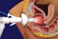
A speculum is a medical tool for investigating body orifices, with a form dependent on the orifice for which it is designed. In old texts, the speculum may also be referred to as a diopter or dioptra. Like an endoscope, a speculum allows a view inside the body; endoscopes, however, tend to have optics while a speculum is intended for direct vision.

<span class="mw-page-title-main">Cervicitis</span> Inflammation of the uterine cervix

Cervicitis is inflammation of the uterine cervix. Cervicitis in women has many features in common with urethritis in men and many cases are caused by sexually transmitted infections. Non-infectious causes of cervicitis can include intrauterine devices, contraceptive diaphragms, and allergic reactions to spermicides or latex condoms. Cervicitis affects over half of all women during their adult life.

<span class="mw-page-title-main">Pelvic floor dysfunction</span> Medical condition

Pelvic floor dysfunction is a term used for a variety of disorders that occur when pelvic floor muscles and ligaments are impaired. The condition affects up to 50 percent of women who have given birth. Although this condition predominantly affects women, up to 16 percent of men are affected as well. Symptoms can include pelvic pain, pressure, pain during sex, urinary incontinence (UI), overactive bladder, bowel incontinence, incomplete emptying of feces, constipation, myofascial pelvic pain and pelvic organ prolapse. When pelvic organ prolapse occurs, there may be visible organ protrusion or a lump felt in the vagina or anus. Research carried out in the UK has shown that symptoms can restrict everyday life for women. However, many people found it difficult to talk about it and to seek care, as they experienced embarrassment and stigma.

<span class="mw-page-title-main">Vaginal bleeding</span> Medical condition

Vaginal bleeding is any expulsion of blood from the vagina. This bleeding may originate from the uterus, vaginal wall, or cervix. Generally, it is either part of a normal menstrual cycle or is caused by hormonal or other problems of the reproductive system, such as abnormal uterine bleeding.

<span class="mw-page-title-main">Vaginal discharge</span> Medical condition

Vaginal discharge is a mixture of liquid, cells, and bacteria that lubricate and protect the vagina. This mixture is constantly produced by the cells of the vagina and cervix, and it exits the body through the vaginal opening. The composition, amount, and quality of discharge varies between individuals and can vary throughout the menstrual cycle and throughout the stages of sexual and reproductive development. Normal vaginal discharge may have a thin, watery consistency or a thick, sticky consistency, and it may be clear or white in color. Normal vaginal discharge may be large in volume but typically does not have a strong odor, nor is it typically associated with itching or pain. While most discharge is considered physiologic or represents normal functioning of the body, some changes in discharge can reflect infection or other pathological processes. Infections that may cause changes in vaginal discharge include vaginal yeast infections, bacterial vaginosis, and sexually transmitted infections. The characteristics of abnormal vaginal discharge vary depending on the cause, but common features include a change in color, a foul odor, and associated symptoms such as itching, burning, pelvic pain, or pain during sexual intercourse.

<span class="mw-page-title-main">Uterine prolapse</span> Medical condition

Uterine prolapse is a form of pelvic organ prolapse in which the uterus and a portion of the upper vagina protrude into the vaginal canal and, in severe cases, through the opening of the vagina. It is most often caused by injury or damage to structures that hold the uterus in place within the pelvic cavity. Symptoms may include vaginal fullness, pain with sexual intercourse, difficulty urinating, and urinary incontinence. Risk factors include older age, pregnancy, vaginal childbirth, obesity, chronic constipation, and chronic cough. Prevalence, based on physical exam alone, is estimated to be approximately 14%.

<span class="mw-page-title-main">Endometrial biopsy</span> Diagnostic medical procedure

The endometrial biopsy is a medical procedure that involves taking a tissue sample of the lining of the uterus. The tissue subsequently undergoes a histologic evaluation which aids the physician in forming a diagnosis.

<span class="mw-page-title-main">Hematometra</span> Medical condition

Hematometra is a medical condition involving collection or retention of blood in the uterus. It is most commonly caused by an imperforate hymen or a transverse vaginal septum.

Neuroendocrine carcinoma of the cervix is best defined separately:Neuroendocrine: Of, relating to, or involving the interaction between the nervous system and the hormones of the endocrine glands.Carcinoma: An invasive malignant tumor derived from epithelial tissue that tends to metastasize to other areas of the body.

<span class="mw-page-title-main">Fertility testing</span>

Fertility testing is the process by which fertility is assessed, both generally and also to find the "fertile window" in the menstrual cycle. General health affects fertility, and STI testing is an important related field.

A well-woman examination is an exam offered to women to review elements of their reproductive health. The exam includes a breast examination, a pelvic examination and a Pap smear but may include other procedures. Hospitals employ strict policies relating to the provision of consent by the patient, the availability of chaperones at the examination, and the absence of other parties.

<span class="mw-page-title-main">Postcoital bleeding</span> Medical condition

Postcoital bleeding (PCB) is non-menstrual vaginal bleeding that occurs during or after sexual intercourse. Though some causes are with associated pain, it is typically painless and frequently associated with intermenstrual bleeding.

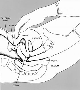
A rectovaginal examination is a type of gynecological examination used to supplement a pelvic examination. In the rectovaginal examination, a doctor or other health care provider places one finger in the vagina and another in the rectum to assess the rectovaginal septum. The examiner will look for any scarring or masses that may indicate cancer or another disease. Typically, a rectovaginal examination is performed to assess pelvic pain, rectal symptoms, or a pelvic mass. It can also provide a sample for fecal occult blood testing.

Pediatric gynaecology or pediatric gynecology is the medical practice dealing with the health of the vagina, vulva, uterus, and ovaries of infants, children, and adolescents. Its counterpart is pediatric andrology, which deals with medical issues specific to the penis and testes.

<span class="mw-page-title-main">Squamous cell carcinoma of the vagina</span> Medical condition

Squamous cell carcinoma of the vagina is a potentially invasive type of cancer that forms in the tissues of the vagina. Though uncommonly diagnosed, squamous cell cancer of the vagina (SCCV) is the most common type of vaginal cancer, accounting for 80-90% of cases as well as 2% of all gynecological cancers. SCCV forms in squamous cells, which are the thin, flat cells lining the vagina. SCCV initially spreads superficially within the vaginal wall and can slowly spread to invade other vaginal tissues. Because of its slow growth, this cancer may cause no symptoms, or it may present with signs like irregular bleeding, pain, or a vaginal mass. This carcinoma can metastasize to the lungs or less frequently to the liver, bone, or other sites. SCCV has many risk factors in common with cervical cancer and is similarly strongly associated with infection with oncogenic strains of human papillomavirus (HPV). Diagnosis of SCCV is done by pelvic exam and biopsy of the tissue. Treatment and prognosis will depend on the stage, location, and characteristics of the cancer.

References

  1. "Examination of the Female Pelvis". Archived from the original on 2001-11-15. Retrieved 2007-12-09.
  2. 1 2 ACOG Practice Advisory on Annual Pelvic Examination Recommendations (30 June 2014). "The Utility of and Indications for Routine Pelvic Examination". American Congress of Obstetricians and Gynecologists. Archived from the original on 2 August 2014.
  3. 1 2 3 4 Hoffman BL, Bradshaw KD, Cunningham FG, Halvorson LM, Schaffer JI, Schorge JO (2012). Williams gynecology (2nd ed.). New York: McGraw-Hill Medical. pp. 2–6. ISBN   978-0-07-171672-7. OCLC   779244257.
  4. Emery JD, Paraiso MF (2014-10-07). Office-based gynecologic surgical procedures. Springer. ISBN   978-1-4939-1414-2. OCLC   893426765.
  5. "Current Procedural Terminology - Pelvic examination under anesthesia (other than local) - Classes". NCBO BioPortal. Retrieved 2018-03-02.
  6. 1 2 3 4 5 Bickley LS, Szilagyi PG, Bates B (2013). Bates' guide to physical examination and history-taking (11th ed.). Philadelphia: Wolters Kluwer Health/Lippincott Williams & Wilkins. ISBN   978-1-60913-762-5. OCLC   801810287.
  7. 1 2 3 4 5 Qaseem A, Humphrey LL, Harris R, Starkey M, Denberg TD (July 2014). "Screening pelvic examination in adult women: a clinical practice guideline from the American College of Physicians". Annals of Internal Medicine. 161 (1): 67–72. doi:10.7326/M14-0701. PMID   24979451. S2CID   12370761.[Free text]
  8. "Don't perform pelvic exams on asymptomatic nonpregnant women, unless necessary for guideline-appropriate screening for cervical cancer". Choosing Wisely. American Academy of Family Physicians. August 8, 2018. Retrieved 5 December 2019.
  9. Berek and Novak's Gynecology (15th ed.). Philadelphia, PA: Lippincott, Williams, and Wilkins. 2012. p. 11. ISBN   978-1-4511-1433-1.
  10. 1 2 3 "ACOG Committee Opinion No. 754 Summary: The Utility of and Indications for Routine Pelvic Examination". Obstetrics and Gynecology. 132 (4): 1080–1083. October 2018. doi:10.1097/aog.0000000000002896. PMID   30247359. S2CID   52342353.
  11. "Recommendation: Gynecological Conditions: Periodic Screening With the Pelvic Examination | United States Preventive Services Taskforce". www.uspreventiveservicestaskforce.org. Retrieved 2024-02-07.
  12. "Stanford 25: Pelvic Examination". YouTube . 2017-09-27.
  13. 1 2 3 4 5 "Trauma-Informed Pelvic Exam - Contraceptive Pearl". Reproductive Health Access Project. Retrieved 2019-12-05.
  14. Allen RH, Singh R (June 2018). "Society of Family Planning clinical guidelines pain control in surgical abortion part 1 - local anesthesia and minimal sedation". Contraception. 97 (6): 471–477. doi: 10.1016/j.contraception.2018.01.014 . PMID   29407363.
  15. Yanikkerem E, Ozdemir M, Bingol H, Tatar A, Karadeniz G (October 2009). "Women's attitudes and expectations regarding gynaecological examination". Midwifery. 25 (5): 500–8. doi:10.1016/j.midw.2007.08.006. PMC   2801597 . PMID   18086509.
  16. "Sexual Trauma: Information for Women's Medical Providers". www.ptsd.va.gov. National Center for PTSD. Retrieved 2019-12-20.
  17. Hammoud, Maya M.; Spector-Bagdady, Kayte; O'Reilly, Meg; Major, Carol; Baecher-Lind, Laura (December 2019). "Consent for the Pelvic Examination Under Anesthesia by Medical Students: Recommendations by the Association of Professors of Gynecology and Obstetrics". Obstetrics & Gynecology. 134 (6): 1303–1307. doi:10.1097/AOG.0000000000003560. ISSN   0029-7844. PMC   6882529 . PMID   31764743.
  18. 1 2 3 4 "Pelvic exam - About - Mayo Clinic". www.mayoclinic.org. Retrieved 2018-01-04.
  19. "Your First Gynecologic Visit" (PDF). American College of Obstetricians and Gynecologists. May 2017.
  20. 1 2 3 4 5 6 7 8 9 10 11 12 13 14 15 16 17 18 19 20 21 Damico D (2016). Health & physical assessment in nursing. Boston: Pearson. p. 665. ISBN   978-0-13-387640-6.
  21. "Pelvic exam - MayoClinic.com" . Retrieved 2007-12-09.
  22. "Your First Gynecologic Visit" (PDF). American College of Obstetricians and Gynecologists. May 2017.
  23. 1 2 3 4 5 6 Berek, Jonathan S., ed. (2020). Berek & Novak's gynecology (16th ed.). Philadelphia: Wolters Kluwer. ISBN   978-1-4963-8033-3.
  24. Bickley, Lynn S., ed. (2021). Bates' guide to physical examination and history taking (13th ed.). Philadelphia Baltimore New York: Wolters Kluwer. ISBN   978-1-4963-9817-8.
  25. 1 2 3 4 5 6 7 Cunningham, F. Gary; Leveno, Kenneth J.; Dashe, Jodi S.; Hoffman, Barbara L.; Spong, Catherine Y.; Casey, Brian M. (2022). "10. Prenatal care". William's Obstetrics (27th ed.). McGraw Hill. ISBN   978-1-260-46273-9.