Uterine artery embolization

Last updated
Uterine artery embolization
Gray1170.png
Arteries of the female reproductive tract (posterior view): uterine artery, ovarian artery and vaginal arteries.
Specialty Interventional radiology
Illustration of uterine fibroids with examples of their possible locations Fibroid locations.jpg
Illustration of uterine fibroids with examples of their possible locations

Uterine artery embolization is a procedure in which an interventional radiologist uses a catheter to deliver small particles that block the blood supply to the uterine body. The procedure is primarily done for the treatment of uterine fibroids and adenomyosis. [1] [2] Since uterine fibroids are the most common indication, it is also often referred to as uterine fibroid embolization. Compared to surgical treatment for fibroids such as a hysterectomy, in which a woman's uterus is removed, uterine artery embolization may be beneficial in women who wish to retain their uterus. Other reasons for uterine artery embolization are postpartum hemorrhage and uterine arteriovenous malformations. [3]

Contents

Medical uses

Uterine fibroids are the most common type of benign uterine tumor and are composed of smooth muscle. [4] They often cause bulk-related symptoms, which can be characterized by back pain, heaviness in the pelvic area, abdominal bloating. [5] Uterine artery embolization may be done to treat bothersome bulk-related symptoms as well as abnormal or heavy uterine bleeding due to uterine fibroids. Fibroid size, number, and location are three potential predictors of a successful outcome. [6] [7] [8] Specifically, studies have demonstrated that submucosal (directly underneath the uterine lining) fibroids demonstrated the largest reduction in size while subserosal (outer layer of the uterus) had the smallest reduction. [9]

Uterine artery embolization may also be appropriate for the treatment of adenomyosis, which is when the lining of the uterus aberrantly grows into the muscle of the uterus. [10] Symptoms of adenomyosis include heavy or prolonged menstrual bleeding and painful menstrual periods. [11]

Uterine artery embolization can also be used to control heavy uterine bleeding for reasons other than fibroids, such as postpartum obstetrical hemorrhage. [12] Many women who experience postpartum hemorrhage may be successfully treated with medication or uterine balloon tamponade. [13] However, in cases where women continue to bleed, uterine artery embolization may be an appropriate option.

A less common indication for uterine artery embolization is for the treatment of uterine arteriovenous malformations which can be a cause of abnormal uterine bleeding or life-threatening bleeding. Roughly half of women with uterine arteriovenous malformations are born with them while the remaining form following surgical interventions or may be due to uterine tumors. [3]

Contra-indications

Prior to undergoing UAE, the patient should be evaluated for the following absolute contra-indications to the procedure: a viable pregnancy, a current infection that is not being treated, or gynecologic malignancy (except for cases where UAE is being used as a procedure in addition to treatment for the cancer). [14] Relative contra-indications for the procedure include a severe contrast allergy since contrast is necessary to visualize the arteries during the procedure, kidney impairment since contrast may cause damage to the kidneys, or coagulopathy (blood disorder that causes prolonged or excessive bleeding). [15] However, all of the stated relative contra-indications can be managed with appropriate pre-operative planning.

Risks/Complications

The rate of serious complications is comparable to that of myomectomy or hysterectomy. The advantage of somewhat faster recovery time is offset by a higher rate of minor complications and an increased likelihood of requiring surgical intervention within two to five years of the initial procedure. [16] An analysis of 15,000 women found that those who had myomectomy required fewer additional procedures, including hysterectomies, to treat fibroids over the next five years than those who had uterine artery embolization. [17] [18]

Complications include the following:

Technique

After access to the femoral or radial artery is established through needle puncture, interventional radiologists use image guidance to perform the procedure. The screen in front of the doctors provides a live image of the tools that are being used throughout the procedure as they navigate to the artery that they would like to embolize (block off). Administration of embolizing agents that are used to block off the arteries can also be seen in real-time. DVbFd-lXUAAxgFI.jpg
After access to the femoral or radial artery is established through needle puncture, interventional radiologists use image guidance to perform the procedure. The screen in front of the doctors provides a live image of the tools that are being used throughout the procedure as they navigate to the artery that they would like to embolize (block off). Administration of embolizing agents that are used to block off the arteries can also be seen in real-time.

Pre-operative preparation

Prior to a uterine artery embolization, patients should undergo a clinic visit with their gynecologist, have a recent Pap smear, and an endometrial biopsy in cases where abnormal uterine bleeding is a presenting symptom. [27] A clinic visit can then be made with the interventional radiologist performing the uterine artery embolization so that a thorough history and physical exam can be taken. Recent diagnostic imaging such as a pelvic magnetic resonance imaging (MRI) should also be reviewed by the interventional radiologist to rule out possible malignancy, evaluate uterine anatomy, and discuss the likelihood of fibroid passage with the patient. [27]

Procedure

The procedure is performed by an interventional radiologist under conscious sedation. [25] Access is commonly through the radial [3] or femoral artery via the wrist or groin, [25] respectively. After anesthetizing the skin over the artery of choice, the artery is accessed by a needle puncture using the Seldinger technique. [25] Under fluoroscopic guidance, a catheter is then introduced into the artery and used to select the uterine vessels for subsequent embolization. Once at the level of the uterine artery an angiogram with contrast is performed to confirm placement of the catheter, and the embolizing agent (spheres or beads) is released. As more embolizing agent is administered, blood flow will slow down significantly. Over time, the decreased blood flow causes the fibroid to shrink. Both the left and right uterine arteries are embolized since unilateral UAEs have a high risk of failure. [28] The procedure can be performed in a hospital or surgical center. More recently, there has been support for UAE as an outpatient procedure, but many doctors choose an overnight admission for pain control. [29] Follow-up for the procedure may very based on institution, but can include a clinic appointment at 1 to 3 months following the procedure and an MRI to see if the fibroids have shrunk from the preoperative MRI. [9]

Recovery

The vast majority of women who undergo UAE experience elimination of abnormal uterine bleeding and improvement in bulk symptoms. [15] Additionally, patient satisfaction following the procedure is about 80%. [14] One drawback of UAE is that it appears to require more repeat procedures than if surgery was done initially. [30] However, long-term patient satisfaction outcomes of UAE are similar to that of surgery and a short-term benefit is the reduction in hospital stay with UAE. [30] [15]

Currently the number of studies that compare pregnancy rates between UAE and myomectomy are limited. However, a 2020 systematic review assessing pregnancy outcomes after UAE for fibroids demonstrated that pregnancy rates between UAE and myomectomy are comparable. Additionally, they found that rates of pregnancy-related complications in women who underwent UAE were similar to that of the general population. [31] Despite these findings, there is still a lack of randomized control trials that directly compare the outcomes of myomectomy and UAE for fibroids, so future studies are needed to determine which procedure yields better results.

For women with adenomyosis, the data regarding outcomes is limited. However, studies have demonstrated that about 83% of women with adenomyosis experienced an improvement in their symptoms. Additionally, the rate of improvement in symptoms increased to about 93% in women who had both adenomyosis and fibroids.

Regarding cost, the American Journal of Gynecology reports that uterine artery embolization costs 12% less than hysterectomy and 8% less than myomectomy. [32]

History

UAE was used for the first time in 1979 to control bleeding in a woman with postpartum hemorrhage that did not improve after surgical treatment. [33] Since then studies have shown that UAE is a safe and effective procedure for postpartum hemorrhage with control of bleeding in greater than 90% of women. [34] The initial use of UAE for patients with fibroids was to limit bleeding during myomectomy. [35] During the 1990s, doctors began expanding the indications for UAE and started using it for the treatment of the fibroids specifically. [36] Previously, the primary treatment methods for fibroids were myomectomy or hysterectomy. Compared to surgery, UAE can be advantageous because blood loss is typically minimal, surgery and general anesthesia is avoided, recovery is shorter, and women can retain their uterus (relative to hysterectomy). [37] UAE is thought to treat fibroids by selectively decreasing blood flow to the tumor since it is highly vascular, which causes improvement in abnormal bleeding and the bulk symptoms that are often experienced with fibroids.

Related Research Articles

<span class="mw-page-title-main">Hysterectomy</span> Surgical removal of the uterus

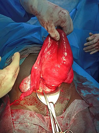
Hysterectomy is the surgical removal of the uterus and cervix. Supracervical hysterectomy refers to removal of the uterus while the cervix is spared. These procedures may also involve removal of the ovaries (oophorectomy), fallopian tubes (salpingectomy), and other surrounding structures. The term “partial” or “total” hysterectomy are lay-terms that incorrectly describe the addition or omission of oophorectomy at the time of hysterectomy. These procedures are usually performed by a gynecologist. Removal of the uterus renders the patient unable to bear children and has surgical risks as well as long-term effects, so the surgery is normally recommended only when other treatment options are not available or have failed. It is the second most commonly performed gynecological surgical procedure, after cesarean section, in the United States. Nearly 68 percent were performed for conditions such as endometriosis, irregular bleeding, and uterine fibroids. It is expected that the frequency of hysterectomies for non-malignant indications will continue to fall given the development of alternative treatment options.

<span class="mw-page-title-main">Interventional radiology</span> Medical subspecialty

Interventional radiology (IR) is a medical specialty that performs various minimally-invasive procedures using medical imaging guidance, such as x-ray fluoroscopy, computed tomography, magnetic resonance imaging, or ultrasound. IR performs both diagnostic and therapeutic procedures through very small incisions or body orifices. Diagnostic IR procedures are those intended to help make a diagnosis or guide further medical treatment, and include image-guided biopsy of a tumor or injection of an imaging contrast agent into a hollow structure, such as a blood vessel or a duct. By contrast, therapeutic IR procedures provide direct treatment—they include catheter-based medicine delivery, medical device placement, and angioplasty of narrowed structures.

Heavy menstrual bleeding (HMB), previously known as menorrhagia or hematomunia, is a menstrual period with excessively heavy flow. It is a type of abnormal uterine bleeding (AUB).

Abnormal uterine bleeding (AUB), also known as (AVB) or as atypical vaginal bleeding, is vaginal bleeding from the uterus that is abnormally frequent, lasts excessively long, is heavier than normal, or is irregular. The term dysfunctional uterine bleeding was used when no underlying cause was present. Vaginal bleeding during pregnancy is excluded. Iron deficiency anemia may occur and quality of life may be negatively affected.

<span class="mw-page-title-main">Embolization</span> Passage and lodging of an embolus within the bloodstream

Embolization refers to the passage and lodging of an embolus within the bloodstream. It may be of natural origin (pathological), in which sense it is also called embolism, for example a pulmonary embolism; or it may be artificially induced (therapeutic), as a hemostatic treatment for bleeding or as a treatment for some types of cancer by deliberately blocking blood vessels to starve the tumor cells.

<span class="mw-page-title-main">Placental abruption</span> Medical condition

Placental abruption is when the placenta separates early from the uterus, in other words separates before childbirth. It occurs most commonly around 25 weeks of pregnancy. Symptoms may include vaginal bleeding, lower abdominal pain, and dangerously low blood pressure. Complications for the mother can include disseminated intravascular coagulopathy and kidney failure. Complications for the baby can include fetal distress, low birthweight, preterm delivery, and stillbirth.

<span class="mw-page-title-main">Adenomyosis</span> Extension of endometrial tissue into the myometrium

Adenomyosis is a medical condition characterized by the growth of cells that proliferate on the inside of the uterus (endometrium) atypically located among the cells of the uterine wall (myometrium), as a result, thickening of the uterus occurs. As well as being misplaced in patients with this condition, endometrial tissue is completely functional. The tissue thickens, sheds and bleeds during every menstrual cycle.

<span class="mw-page-title-main">Vaginal bleeding</span> Medical condition

Vaginal bleeding is any expulsion of blood from the vagina. This bleeding may originate from the uterus, vaginal wall, or cervix. Generally, it is either part of a normal menstrual cycle or is caused by hormonal or other problems of the reproductive system, such as abnormal uterine bleeding.

<span class="mw-page-title-main">Uterine fibroid</span> Medical condition with benign tumors of uterus

Uterine fibroids, also known as uterine leiomyomas or fibroids, are benign smooth muscle tumors of the uterus. Most women with fibroids have no symptoms while others may have painful or heavy periods. If large enough, they may push on the bladder, causing a frequent need to urinate. They may also cause pain during penetrative sex or lower back pain. A woman can have one uterine fibroid or many. Occasionally, fibroids may make it difficult to become pregnant, although this is uncommon.

<span class="mw-page-title-main">Uterine myomectomy</span> Surgical removal of uterine fibroid

Myomectomy, sometimes also called fibroidectomy, refers to the surgical removal of uterine leiomyomas, also known as fibroids. In contrast to a hysterectomy, the uterus remains preserved and the woman retains her reproductive potential. It still may impact hormonal regulation and the menstrual cycle.

Adenomyoma is a tumor (-oma) including components derived from glands (adeno-) and muscle (-my-). It is a type of complex and mixed tumor, and several variants have been described in the medical literature. Uterine adenomyoma, the localized form of uterine adenomyosis, is a tumor composed of endometrial gland tissue and smooth muscle in the myometrium. Adenomyomas containing endometrial glands are also found outside of the uterus, most commonly on the uterine adnexa but can also develop at distant sites outside of the pelvis. Gallbladder adenomyoma, the localized form of adenomyomatosis, is a polypoid tumor in the gallbladder composed of hyperplastic mucosal epithelium and muscularis propria.

<span class="mw-page-title-main">Placenta accreta spectrum</span> Medical condition

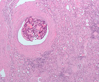
Placenta accreta occurs when all or part of the placenta attaches abnormally to the myometrium. Three grades of abnormal placental attachment are defined according to the depth of attachment and invasion into the muscular layers of the uterus:

  1. Accreta – chorionic villi attached to the myometrium, rather than being restricted within the decidua basalis.
  2. Increta – chorionic villi invaded into the myometrium.
  3. Percreta – chorionic villi invaded through the perimetrium.
<span class="mw-page-title-main">Uterine artery</span>

The uterine artery is an artery that supplies blood to the uterus in females.

<span class="mw-page-title-main">Uterine atony</span> Loss of tone in the uterine musculature

Uterine atony is the failure of the uterus to contract adequately following delivery. Contraction of the uterine muscles during labor compresses the blood vessels and slows flow, which helps prevent hemorrhage and facilitates coagulation. Therefore, a lack of uterine muscle contraction can lead to an acute hemorrhage, as the vasculature is not being sufficiently compressed. Uterine atony is the most common cause of postpartum hemorrhage, which is an emergency and potential cause of fatality. Across the globe, postpartum hemorrhage is among the top five causes of maternal death. Recognition of the warning signs of uterine atony in the setting of extensive postpartum bleeding should initiate interventions aimed at regaining stable uterine contraction.

<span class="mw-page-title-main">Postpartum bleeding</span> Loss of blood following childbirth

Postpartum bleeding or postpartum hemorrhage (PPH) is often defined as the loss of more than 500 ml or 1,000 ml of blood following childbirth. Some have added the requirement that there also be signs or symptoms of low blood volume for the condition to exist. Signs and symptoms may initially include: an increased heart rate, feeling faint upon standing, and an increased breathing rate. As more blood is lost, the patient may feel cold, blood pressure may drop, and they may become restless or unconscious. The condition can occur up to six weeks following delivery.

Reproductive surgery is surgery in the field of reproductive medicine. It can be used for contraception, e.g. in vasectomy, wherein the vasa deferentia of a male are severed, but is also used plentifully in assisted reproductive technology. Reproductive surgery is generally divided into three categories: surgery for infertility, in vitro fertilization, and fertility preservation.

<span class="mw-page-title-main">Interstitial pregnancy</span> Medical condition

An interstitial pregnancy is a uterine but ectopic pregnancy; the pregnancy is located outside the uterine cavity in that part of the fallopian tube that penetrates the muscular layer of the uterus. The term cornual pregnancy is sometimes used as a synonym, but remains ambiguous as it is also applied to indicate the presence of a pregnancy located within the cavity in one of the two upper "horns" of a bicornuate uterus. Interstitial pregnancies have a higher mortality than ectopics in general.

Interventional oncology is a subspecialty field of interventional radiology that deals with the diagnosis and treatment of cancer and cancer-related problems using targeted minimally invasive procedures performed under image guidance. Interventional oncology has developed to a separate pillar of modern oncology and it employs X-ray, ultrasound, computed tomography (CT) or magnetic resonance imaging (MRI) to help guide miniaturized instruments to allow targeted and precise treatment of solid tumours located in various organs of the human body, including but not limited to the liver, kidneys, lungs, and bones. Interventional oncology treatments are routinely carried out by interventional radiologists in appropriate settings and facilities.

<span class="mw-page-title-main">Prostatic artery embolization</span> Non-surgical technique for treatment of benign prostatic hypertrophy

Prostatic artery embolization is a non-surgical technique for treatment of benign prostatic hypertrophy (BPH).

Anna-Maria Belli, MD, FCIRSE is a British interventional radiologist known for her work in vascular interventional radiology and for holding leadership positions in interventional radiology societies in Britain and Europe.

References

  1. Siskin GP, Tublin ME, Stainken BF, et al. (2001). "Uterine artery embolization for the treatment of adenomyosis: clinical response and evaluation with MR imaging". American Journal of Roentgenology. 177 (2): 297–302. doi:10.2214/ajr.177.2.1770297. PMID   11461849.
  2. Chen C, Liu P, Lu J, et al. [Uterine arterial embolization in the treatment of adenomyosis]. Zhonghua Fu Chan Ke Za Zhi 2002; 37:77.
  3. 1 2 3 Keefe, Nicole A.; Haskal, Ziv J; Park, Auh Whan; Angle, John F., eds. (2018). IR Playbook. doi:10.1007/978-3-319-71300-7. ISBN   978-3-319-71299-4. S2CID   46964078.
  4. Donnez, Jacques; Dolmans, Marie-Madeleine (2016-07-27). "Uterine fibroid management: from the present to the future". Human Reproduction Update. 22 (6): 665–686. doi:10.1093/humupd/dmw023. ISSN   1355-4786. PMC   5853598 . PMID   27466209.
  5. Gonsalves, Carin (December 2008). "Uterine Artery Embolization for Treatment of Symptomatic Fibroids". Seminars in Interventional Radiology. 25 (4): 369–377. doi:10.1055/s-0028-1103001. ISSN   0739-9529. PMC   3036525 . PMID   21326578.
  6. Spies JB, Roth AR, Jha RC, et al. (2002). "Leiomyomata treated with uterine artery embolization: factors associated with successful symptom and imaging outcome". Radiology. 222 (1): 45–52. doi:10.1148/radiol.2221010661. PMID   11756703.
  7. Pelage JP, Walker WJ, Le Dref O, et al. (2001). "Treatment of uterine fibroids". Lancet. 357 (9267): 1530. doi:10.1016/s0140-6736(00)04683-3. PMC   5267678 . PMID   11383541.
  8. Katsumori T, Nakajima K, Mihara T (2003). "Is a large fibroid a high-risk factor for uterine artery embolization?". American Journal of Roentgenology. 181 (5): 1309–1314. doi:10.2214/ajr.181.5.1811309. PMID   14573425.
  9. 1 2 Young, Michael; Coffey, William; Mikhail, Lyree N. (2023), "Uterine Fibroid Embolization", StatPearls, Treasure Island (FL): StatPearls Publishing, PMID   30085558 , retrieved 2023-11-01
  10. Garcia, Lydia; Isaacson, Keith (July 2011). "Adenomyosis: Review of the Literature". Journal of Minimally Invasive Gynecology. 18 (4): 428–437. doi:10.1016/j.jmig.2011.04.004. ISSN   1553-4650. PMID   21622029.
  11. Gunther, Rutger; Walker, Christopher (2023), "Adenomyosis", StatPearls, Treasure Island (FL): StatPearls Publishing, PMID   30969690 , retrieved 2023-10-29
  12. Management of severe postpartum haemorrhage by uterine artery embolization
  13. Knight, Marian; Acosta, Colleen; Brocklehurst, Peter; Cheshire, Anna; Fitzpatrick, Kathryn; Hinton, Lisa; Jokinen, Mervi; Kemp, Bryn; Kurinczuk, Jennifer J.; Lewis, Gwyneth; Lindquist, Anthea; Locock, Louise; Nair, Manisha; Patel, Nishma; Quigley, Maria (2016-06-30). "Beyond maternal death: Improving the quality of maternal care through national studies of near-miss maternal morbidity". Programme Grants for Applied Research. 4 (9): 1–180. doi: 10.3310/pgfar04090 . ISSN   2050-4330. PMID   27386616.
  14. 1 2 Dariushnia, Sean R.; Nikolic, Boris; Stokes, LeAnn S.; Spies, James B. (November 2014). "Quality Improvement Guidelines for Uterine Artery Embolization for Symptomatic Leiomyomata". Journal of Vascular and Interventional Radiology. 25 (11): 1737–1747. doi:10.1016/j.jvir.2014.08.029. ISSN   1051-0443. PMID   25442136.
  15. 1 2 3 Kohi, Maureen P.; Spies, James B. (March 2018). "Updates on Uterine Artery Embolization". Seminars in Interventional Radiology. 35 (1): 048–055. doi:10.1055/s-0038-1636521. ISSN   0739-9529. PMC   5886769 . PMID   29628616.
  16. Gupta, Janesh K.; Sinha, Anju; Lumsden, M. A.; Hickey, Martha (2014-12-26). "Uterine artery embolization for symptomatic uterine fibroids". The Cochrane Database of Systematic Reviews (12): CD005073. doi:10.1002/14651858.CD005073.pub4. ISSN   1469-493X. PMID   25541260.
  17. Amoah, Alison; Quinn, Stephen D. (1 June 2023). "Uterine‐preserving treatments or hysterectomy reintervention after myomectomy or uterine artery embolisation: A retrospective cohort study of long‐term outcomes". BJOG: An International Journal of Obstetrics & Gynaecology. 130 (7): 823–831. doi: 10.1111/1471-0528.17412 . ISSN   1470-0328. PMID   36706789.
  18. "Surgical removal of fibroids has better long-term outcomes than blocking blood vessels". NIHR Evidence. 22 September 2023.
  19. Vashisht A, Studd JW, Carey AH (2000). "Fibroid Embolisation: A Technique Not Without Significant Complications". British Journal of Obstetrics and Gynaecology. 107 (9): 1166–1170. doi:10.1111/j.1471-0528.2000.tb11119.x. PMID   11002964. S2CID   12959753.
  20. Schirf, Brian E.; Vogelzang, Robert L.; Chrisman, Howard B. (June 2006). "Complications of Uterine Fibroid Embolization". Seminars in Interventional Radiology. 23 (2): 143–149. doi:10.1055/s-2006-941444. ISSN   0739-9529. PMC   3036365 . PMID   21326757.
  21. de Block S, de Bries C, Prinssen HM (2003). "Fatal Sepss after Uterine Artery Embolization with Microspheres". Journal of Vascular and Interventional Radiology. 14 (6): 779–783. doi:10.1097/01.rvi.0000079988.80153.61. PMID   12817046.
  22. Shlansky-Goldberg, Richard D.; Coryell, Lee; Stavropoulos, S. William; Trerotola, Scott O.; Mondschein, Jeffrey; Beshara, Mathew; Butts, Samantha F.; Sondheimer, Steven; Tureck, Richard W.; Rosen, Mark (November 2011). "Outcomes Following Fibroid Expulsion after Uterine Artery Embolization". Journal of Vascular and Interventional Radiology. 22 (11): 1586–1593. doi:10.1016/j.jvir.2011.08.004. ISSN   1051-0443. PMID   22024118.
  23. Common AA, Mocarski E, Kolin A (2001). "Leiomyosarcoma". Journal of Vascular and Interventional Radiology. 12 (12): 1449–1452. doi:10.1016/s1051-0443(07)61708-4. PMID   11742024.
  24. Dietz DM, Stahfeld KR, Bansal SK (2004). "Buttock Necrosis After Uterine Artery Embolization". Obstetrics & Gynecology. 104 (Supplement): 1159–1161. doi:10.1097/01.AOG.0000141567.25541.26. PMID   15516436. S2CID   39409507.
  25. 1 2 3 4 5 6 Rand, Thomas; Patel, Rafiuddin; Magerle, Wolfgang; Uberoi, Raman (December 2020). "CIRSE standards of practice on gynaecological and obstetric haemorrhage". CVIR Endovascular. 3 (1): 85. doi: 10.1186/s42155-020-00174-7 . ISSN   2520-8934. PMC   7695782 . PMID   33245432.
  26. Walker WJ, Pelage JP, Sutton C (2002). "Fibroid Embolization". Clinical Radiology. 57 (5): 325–331. doi:10.1053/crad.2002.0945. PMID   12014926.
  27. 1 2 Keung, Jonathan J.; Spies, James B.; Caridi, Theresa M. (2018-01-01). "Uterine artery embolization: A review of current concepts". Best Practice & Research Clinical Obstetrics & Gynaecology. Alternatives to Hysterectomy. 46: 66–73. doi:10.1016/j.bpobgyn.2017.09.003. ISSN   1521-6934. PMID   29128204.
  28. Ravina, J.H.; Ciraru-Vigneron, N.; Bouret, J.M.; Herbreteau, D.; Houdart, E.; Aymard, A.; Merland, J.J. (September 1995). "Arterial embolisation to treat uterine myomata". The Lancet. 346 (8976): 671–672. doi:10.1016/s0140-6736(95)92282-2. ISSN   0140-6736. PMID   7544859. S2CID   24270648.
  29. Guyer, A; Raggio, T; Sor, M; Usvyat, L; Latif, W; Miller, G; Koh, E; Rosenblatt, M; Suchin, C (February 2017). "Safety of uterine artery embolization performed as an outpatient procedure: retrospective analysis of 876 patients across a network of 26 outpatient interventional radiology practices". Journal of Vascular and Interventional Radiology. 28 (2): S47. doi: 10.1016/j.jvir.2016.12.697 . ISSN   1051-0443.
  30. 1 2 Gupta, JK; Sinha, A; Lumsden, MA; Hickey, M (26 December 2014). "Uterine artery embolization for symptomatic uterine fibroids". The Cochrane Database of Systematic Reviews. 12 (12): CD005073. doi:10.1002/14651858.CD005073.pub4. PMID   25541260.
  31. Ghanaati, Hossein; Sanaati, Mina; Shakiba, Madjid; Bakhshandeh, Hooman; Ghavami, Nafiseh; Aro, Solmaz; Jalali, Amir Hossein; Firouznia, Kavous (2020-08-01). "Pregnancy and its Outcomes in Patients After Uterine Fibroid Embolization: A Systematic Review and Meta-Analysis". CardioVascular and Interventional Radiology. 43 (8): 1122–1133. doi:10.1007/s00270-020-02521-6. ISSN   1432-086X. PMID   32458009. S2CID   218898884.
  32. Al-Fozan, Haya; Dufort, Joanne; Kaplow, Marilyn; Valenti, David; Tulandi, Togas (November 2002). "Cost analysis of myomectomy, hysterectomy, and uterine artery embolization". American Journal of Obstetrics and Gynecology. 187 (5): 1401–1404. doi:10.1067/mob.2002.127374. PMID   12439538.
  33. Heaston, Dk; Mineau, De; Brown, Bj; Miller, Fj (1979-07-01). "Transcatheter arterial embolization for control of persistent massive puerperal hemorrhage after bilateral surgical hypogastric artery ligation". American Journal of Roentgenology. 133 (1): 152–154. doi:10.2214/ajr.133.1.152. ISSN   0361-803X. PMID   110056.
  34. Liu, ZhiRong; Wang, Yaxuan; Yan, Jingxin; Li, Juan; Liu, XinLian; Zhang, LuShun; Cheng, Li (2020-02-16). "Uterine artery embolization versus hysterectomy in the treatment of refractory postpartum hemorrhage: a systematic review and meta-analysis". The Journal of Maternal-Fetal & Neonatal Medicine. 33 (4): 693–705. doi:10.1080/14767058.2018.1497599. ISSN   1476-7058. PMID   30354858. S2CID   53027764.
  35. Ravina, J.H.; Ciraru-Vigneron, N.; Bouret, J.M.; Herbreteau, D.; Houdart, E.; Aymard, A.; Merland, J.J. (September 1995). "Arterial embolisation to treat uterine myomata". The Lancet. 346 (8976): 671–672. doi:10.1016/s0140-6736(95)92282-2. ISSN   0140-6736. PMID   7544859. S2CID   24270648.
  36. Manyonda, Isaac; Belli, Anna-Maria; Lumsden, Mary-Ann; Moss, Jonathan; McKinnon, William; Middleton, Lee J.; Cheed, Versha; Wu, Olivia; Sirkeci, Fusun; Daniels, Jane P.; McPherson, Klim (2020-07-30). "Uterine-Artery Embolization or Myomectomy for Uterine Fibroids". New England Journal of Medicine. 383 (5): 440–451. doi: 10.1056/NEJMoa1914735 . ISSN   0028-4793. PMID   32726530.
  37. Smith, S. J. (2000-06-15). "Uterine fibroid embolization". American Family Physician. 61 (12): 3601–3607, 3611–3612. ISSN   0002-838X. PMID   10892632.