Columella (auditory system)

Last updated
Columella (highlighted) in the skull of the extinct therapsid Dicynodon. Columella in Dicynodon.jpg
Columella (highlighted) in the skull of the extinct therapsid Dicynodon.

In the auditory system, the columella contributes to hearing in amphibians, reptiles and birds. The columella form thin, bony structures in the interior of the skull and serve the purpose of transmitting sounds from the eardrum. It is an evolutionary homolog of the stapes, one of the auditory ossicles in mammals.

Contents

In many species, the extracolumella is a cartilaginous structure that grows in association with the columella. During development, the columella is derived from the dorsal end of the hyoid arch. [1]

Evolution

The evolution of the columella is closely related to the evolution of the jaw joint. It is an ancestral homolog of the stapes, and is derived from the hyomandibular bone of fishes. [2]

As the columella is derived from the hyomandibula, many of its functional relationships remain the same. The columella resides in the air-filled tympanic cavity of the middle ear. The footplate, or proximal end of the columella, rests in the oval window. Sound is conducted through the oval window to the interior of the otic capsule. [2] This motion ultimately stimulates sensory cells in the inner ear. [3]

Depiction of the evolution of the ossicles of the ear. Columella (Co) and extra-columella (E) evolve into the stapes and extra-stapes in embryonic mammals (7). EB1911 Reptiles - Evolution of the Ossicular Chain of the Ear.jpg
Depiction of the evolution of the ossicles of the ear. Columella (Co) and extra-columella (E) evolve into the stapes and extra-stapes in embryonic mammals (7).

In the transition of tetrapods from sea to land, the earliest appearance of functional columella appeared in temnospondyls. [5]

Extracolumella

Crocodilians evolved to lift the head and body off the ground, isolating the head from ground vibrations. Under selective pressure to detect airborne sound vibrations, the columella in crocodilians have become more slender and reduce their mass. The extracolumella, a cartilaginous outgrowth on the distal end of the columella, couples the columella to the tympanum to conduct sound from the exterior air. [6]

Birds and modern crocodilians have evolved a trifurcated columella, which forms a Y-shaped support structure on the surface of the tympanic membrane. [7] In birds, this is thought to increase the surface area of the columellar footplate, thus lowering the threshold of hearing and improving the detection of airborne sound waves. [7] [3]

Anatomy in amphibians

Frogs

In frogs, the extracolumella is simple and club-shaped. [3]

Anatomy in reptiles

In reptiles, the columella function to transduce sound through the middle ear as part of the auditory pathway. The columella is relatively straight and moves in a piston-like motion in response to vibration. [3] Due to the rigid bony structure, the columella primarily responds to low-frequency vibrations transmitted through the ground. [2]

Crocodilians

In crocodilians, the columella arises from a proximal and a distal component which develop into the columella and extracolumella, respectively. It is typically trifurcated, with three finger-like projections supporting it against the tympanic membrane. [3] The extracolumella remains cartilaginous while the columella ossifies during development. [8] The connection between the columella and extracolumella remains flexible over the animal's lifetime. [7]

Snakes

Mounted skull of a python with disarticulated upper and lower jaw joints. In snakes, the columella would be attached directly to the quadrate bone (c). Python Daudin, 1803.jpg
Mounted skull of a python with disarticulated upper and lower jaw joints. In snakes, the columella would be attached directly to the quadrate bone (c).

Snakes have lost a tympanic membrane, and hence a distal attachment for the columella. The columella is instead connected to the quadrate bone of the jaw. Thus, snakes are able to detect and localize ground vibrations through the lower jaw, rather than the sides of the head. [3]

Worm lizards

In Amphisbaenia, the extracolumella is particularly lengthened and firmly connects with a layer of skin over dentary bone of the lower jaw. This connection appears to facilitate detection of airborne vibrations in the facial area. [6] The embedding in the skin often occurs at a specially enlarged labial scale. As a result, the amphisbaenian is able to detect substrate vibrations as it burrows through the ground while protecting the internal ear from damage. [9] Amphisbaenians otherwise lack an external ear structure, likely due to selective pressure to protect the middle and inner ears from damage as the animal burrows. [10]

Birds

In birds, the columella is anchored to the conical tympanic membrane at an acute angle, rather than a 90-degree angle relative to the plane of the tympanic membrane. This is thought to provide a lever advantage in conducting airborne sound from the distal to the proximal end of the columella. [6]

Development in chickens

In chick embryos, the primordial columella arises from a mesenchymal condensation. Chondrification of the columella occurs earlier than the extracolumella. During endochondral ossification, the columella ossifies from two origins of periosteum: the shaft and the footplate. [11]

Homology in mammals

Within mammals and other synapsids the columella has evolved into the stapes, a homologous bone within the newly evolved inner ear. As the tympanic cavity evolved to reduce in size, the columella shortened in length. The stirrup-shaped articular processes of the columella inspired a new name for this auditory ossicle, the stapes. The auditory ossicles continue to function in conducting transmitting sound through the auditory pathway; however, they have lost their function in conducting low frequency ground vibrations.

Later-arising reptiles with columella likely evolved stronger limbs and a more crawling posture, which removed the body from the ground and prevented the transmission of ground-conducted sounds. The skin over the ear evolved into the eardrum, which allowed for the detection of high-frequency airborne vibrations. In mammals, the newly specialized ossicles function to transduce and amplify these vibrations along the auditory pathway. [2]

Artificial columella

In humans, artificially made columella may be produced as autografts from cortical bone. These prostheses are used as replacements for the stapes in ear surgery to correct for hearing problems (such as cholesteatoma or re-perforation). [12] [13]

Related Research Articles

<span class="mw-page-title-main">Inner ear</span> Innermost part of the vertebrate ear

The inner ear is the innermost part of the vertebrate ear. In vertebrates, the inner ear is mainly responsible for sound detection and balance. In mammals, it consists of the bony labyrinth, a hollow cavity in the temporal bone of the skull with a system of passages comprising two main functional parts:

<span class="mw-page-title-main">Middle ear</span> Portion of the ear internal to the eardrum, and external to the oval window of the inner ear

The middle ear is the portion of the ear medial to the eardrum, and distal to the oval window of the cochlea.

The ossicles are three bones in either middle ear that are among the smallest bones in the human body. They serve to transmit sounds from the air to the fluid-filled labyrinth (cochlea). The absence of the auditory ossicles would constitute a moderate-to-severe hearing loss. The term "ossicle" literally means "tiny bone". Though the term may refer to any small bone throughout the body, it typically refers to the malleus, incus, and stapes of the middle ear.

<span class="mw-page-title-main">Incus</span> Bone in the middle ear

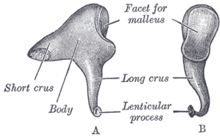
The incus or anvil is a bone in the middle ear. The anvil-shaped small bone is one of three ossicles in the middle ear. The incus receives vibrations from the malleus, to which it is connected laterally, and transmits these to the stapes medially. The incus is so-called because of its resemblance to an anvil.

<i>Stapes</i> Bone in the middle ear

The stapes or stirrup is a bone in the middle ear of humans and other animals which is involved in the conduction of sound vibrations to the inner ear. This bone is connected to the oval window by its annular ligament, which allows the footplate to transmit sound energy through the oval window into the inner ear. The stapes is the smallest and lightest bone in the human body, and is so-called because of its resemblance to a stirrup.

<span class="mw-page-title-main">Oval window</span> Membrane-covered opening in the ear

The oval window is a membrane-covered opening from the middle ear to the cochlea of the inner ear.

<span class="mw-page-title-main">Cochlea</span> Snail-shaped part of inner ear involved in hearing

The cochlea is the part of the inner ear involved in hearing. It is a spiral-shaped cavity in the bony labyrinth, in humans making 2.75 turns around its axis, the modiolus. A core component of the cochlea is the organ of Corti, the sensory organ of hearing, which is distributed along the partition separating the fluid chambers in the coiled tapered tube of the cochlea.

The angular is a large bone in the lower jaw (mandible) of amphibians and reptiles, which is connected to all other lower jaw bones: the dentary, the splenial, the suprangular, and the articular. It is homologous to the tympanic bone in mammals, due to the incorporation of several jaw bones into the mammalian middle ear early in mammal evolution.

<span class="mw-page-title-main">Ear</span> Organ of hearing and balance

An ear is the organ that enables hearing and body balance using the vestibular system. In mammals the ear is usually described as having three parts: the outer ear, the middle ear and the inner ear. The outer ear consists of the pinna and the ear canal. Since the outer ear is the only visible portion of the ear in most animals, the word "ear" often refers to the external part alone. The middle ear includes the tympanic cavity and the three ossicles. The inner ear sits in the bony labyrinth, and contains structures which are key to several senses: the semicircular canals, which enable balance and eye tracking when moving; the utricle and saccule, which enable balance when stationary; and the cochlea, which enables hearing. The ear is a self cleaning organ through its relationship with earwax and the ear canals. The ears of vertebrates are placed somewhat symmetrically on either side of the head, an arrangement that aids sound localization.

<span class="mw-page-title-main">Tympanoplasty</span> Surgical operation on the ear

Tympanoplasty is the surgical operation performed to reconstruct hearing mechanism of middle ear.

<span class="mw-page-title-main">Stapedius muscle</span> Muscle in the human ear

The stapedius is the smallest skeletal muscle in the human body. At just over one millimeter in length, its purpose is to stabilize the smallest bone in the body, the stapes or stirrup bone of the middle ear.

<span class="mw-page-title-main">Tensor tympani muscle</span> Muscle of the middle ear

The tensor tympani is a muscle within the middle ear, located in the bony canal above the bony part of the auditory tube, and connects to the malleus bone. Its role is to dampen loud sounds, such as those produced from chewing, shouting, or thunder. Because its reaction time is not fast enough, the muscle cannot protect against hearing damage caused by sudden loud sounds, like explosions or gunshots.

<span class="mw-page-title-main">Tympanic cavity</span> Small cavity surrounding the bones of the middle ear

The tympanic cavity is a small cavity surrounding the bones of the middle ear. Within it sit the ossicles, three small bones that transmit vibrations used in the detection of sound.

<span class="mw-page-title-main">Round window</span>

The round window is one of the two openings from the middle ear into the inner ear. It is sealed by the secondary tympanic membrane, which vibrates with opposite phase to vibrations entering the inner ear through the oval window. It allows fluid in the cochlea to move, which in turn ensures that hair cells of the basilar membrane will be stimulated and that audition will occur.

<span class="mw-page-title-main">Tympanum (anatomy)</span> External hearing structure in animals

The tympanum is an external hearing structure in animals such as mammals, birds, some reptiles, some amphibians and some insects.

<span class="mw-page-title-main">Evolution of mammalian auditory ossicles</span> Middle ear bones evolved from jaw bones

The evolution of mammalian auditory ossicles was an evolutionary process that resulted in the formation of the bones of the mammalian middle ear. These bones, or ossicles, are a defining characteristic of all mammals. The event is well-documented and important as a demonstration of transitional forms and exaptation, the re-purposing of existing structures during evolution.

<span class="mw-page-title-main">Hearing</span> Sensory perception of sound by living organisms

Hearing, or auditory perception, is the ability to perceive sounds through an organ, such as an ear, by detecting vibrations as periodic changes in the pressure of a surrounding medium. The academic field concerned with hearing is auditory science.

Cochlea is Latin for “snail, shell or screw” and originates from the Greek word κοχλίας kokhlias. The modern definition, the auditory portion of the inner ear, originated in the late 17th century. Within the mammalian cochlea exists the organ of Corti, which contains hair cells that are responsible for translating the vibrations it receives from surrounding fluid-filled ducts into electrical impulses that are sent to the brain to process sound.

<span class="mw-page-title-main">Malleus</span> Hammer-shaped small bone of the middle ear

The malleus, or hammer, is a hammer-shaped small bone or ossicle of the middle ear. It connects with the incus, and is attached to the inner surface of the eardrum. The word is Latin for 'hammer' or 'mallet'. It transmits the sound vibrations from the eardrum to the incus (anvil).

A middle ear implant is a hearing device that is surgically implanted into the middle ear. They help people with conductive, sensorineural or mixed hearing loss to hear. 

References

  1. Goodrich ES (July 1915). "Memoirs: The Chorda Tympani and Middle Ear in Reptiles, Birds, and Mammals". Journal of Cell Science. 61 (2): 137–160. S2CID   27277400.
  2. 1 2 3 4 Homberger DG, Walker WF (2004). Vertebrate dissection (9th ed.). Belmont, CA: Thomson Brooks/Cole. ISBN   0-03-022522-1. OCLC   53074665.
  3. 1 2 3 4 5 6 Christensen-Dalsgaard J, Manley GA (October 2013). "The malleable middle ear: an underappreciated player in the evolution of hearing in vertebrates.". In Köppl C, Manley GA, Popper AN, Fay RR (eds.). Insights from comparative hearing research. Springer Handbook of Auditory Research. Vol. 49. New York, NY.: Springer. pp. 157–191. doi:10.1007/2506_2013_33. ISBN   978-1-4614-9077-7.
  4. Olson EC (August 1966). "The middle ear--morphological types in amphibians and reptiles". American Zoologist. 6 (3): 399–419. doi: 10.1093/icb/6.3.399 . PMID   5949350.
  5. Manley GA (May 2010). "An evolutionary perspective on middle ears". Hearing Research. 263 (1–2): 3–8. doi:10.1016/j.heares.2009.09.004. PMID   19786082. S2CID   25664943.
  6. 1 2 3 Saunders J (2000). The Middle Ear of Reptiles and Birds. Springer. ISBN   978-1-4612-7036-2.
  7. 1 2 3 Claes R (2018). Understanding functioning and evolution of bird middle ear mechanics: a functional morphological analysis (PDF) (Ph.D. thesis). University of Antwerp.
  8. Frank GH, Smit AL (1974). "The Early Ontogeny of the Columella Auris of Crocodilus Niloticus and its Bearing on Problems Concerning the Upper End of the Reptilian Hyoid Arch". African Zoology. 9 (1): 59–87. doi:10.1080/00445096.1974.11448520.
  9. Evans, Susan Evans (2016). "The Lepidosaurian Ear: Variations on a Theme". Evolution of the Vertebrate Ear. Springer Handbook of Auditory Research. Vol. 59. pp. 245–284. doi:10.1007/978-3-319-46661-3_9. ISBN   978-3-319-46659-0.
  10. Gans, Carl (1972). "The ear and hearing in Amphisbaenia (Reptilia)". Journal of Experimental Zoology. 179 (1): 17–34. doi:10.1002/jez.1401790103.
  11. Wood JL, Hughes AJ, Mercer KJ, Chapman SC (February 2010). "Analysis of chick (Gallus gallus) middle ear columella formation". BMC Developmental Biology. 10 (1): 16. doi: 10.1186/1471-213X-10-16 . PMC   2834582 . PMID   20158901.
  12. Kylén P, Albrektsson T, Ekvall L, Hellkvist H, Tjellström A (1987). "Survival of the cortical bone columella in ear surgery". Acta Oto-Laryngologica. 104 (1–2): 158–65. doi:10.3109/00016488709109062. PMID   3310512.
  13. Rönnblom A, Gladiné K, Niklasson A, von Unge M, Dirckx J, Tano K (December 2019). "A New, Promising Experimental Ossicular Prosthesis: A Human Temporal Bone Study With Laser Doppler Vibrometry". Otology & Neurotology. 41 (4): 537–544. doi: 10.1097/MAO.0000000000002556 . PMC   7208281 . PMID   31821265.