Premaxilla

Last updated
Premaxilla
Spinosaurus skull en.svg
Skull of Spinosaurus aegyptiacus , premaxilla in orange
Gray199.png
Human premaxilla and its sutures
Details
Precursor median nasal prominence
Identifiers
TA98 A02.1.12.031
TA2 833
FMA 77231
Anatomical terminology

The premaxilla (or praemaxilla) is one of a pair of small cranial bones at the very tip of the upper jaw of many animals, usually, but not always, bearing teeth. In humans, they are fused with the maxilla. The "premaxilla" of therian mammals has been usually termed as the incisive bone. Other terms used for this structure include premaxillary bone or os premaxillare, intermaxillary bone or os intermaxillare, and Goethe's bone.

Contents

Human anatomy

Incisive bone
Gray160.png
The bony palate and alveolar arch. (Premaxilla is not labeled, but region is visible.)
Details
Identifiers
Latin os incisivum
TA98 A02.1.12.031
TA2 833
FMA 77231
Anatomical terms of bone

In human anatomy, the premaxilla is referred to as the incisive bone (os incisivum) and is the part of the maxilla which bears the incisor teeth, and encompasses the anterior nasal spine and alar region. In the nasal cavity, the premaxillary element projects higher than the maxillary element behind. The palatal portion of the premaxilla is a bony plate with a generally transverse orientation. The incisive foramen is bound anteriorly and laterally by the premaxilla and posteriorly by the palatine process of the maxilla. [1]

It is formed from the fusion of a pair of small cranial bones at the very tip of the jaws of many animals, usually bearing teeth, but not always. They are connected to the maxilla and the nasals. While Johann Wolfgang von Goethe was not the first one to discover the incisive bone in humans, he was the first to prove its presence across mammals. Hence, the incisive bone is also known as Goethe's bone. [2]

Incisive bone and premaxilla

Incisive bone is a term used for mammals, and it has been generally thought to be homologous to premaxilla in non-mammalian animals. However, there are counterarguments. According to them, the incisive bone is a novel character first acquired in therian mammals as a composition of premaxilla derived from medial nasal prominence and septomaxilla derived from maxillary prominence. In the incisive bones, only the palatine process corresponds to the premaxilla, while the other parts are the septomaxilla. Based on this, the incisive bone is not completely homologous to the non-mammalian premaxilla. This was hypothesized by Ernst Gaupp in 1905 [3] and demonstrated by developmental biological- and paleontological experiments in 2021. [4] This issue is still under debate.

Embryology

In the embryo, the nasal region develops from neural crest cells which start their migration down to the face during the fourth week of gestation. A pair of symmetrical nasal placodes (thickenings in the epithelium) are each divided into medial and lateral processes by the nasal pits. The medial processes become the septum, philtrum, and premaxilla. [5]

The first ossification centers in the area of the future premaxilla appear during the seventh week above the germ of the second incisor on the outer surface of the nasal capsule. After eleven weeks an accessory ossification center develops into the alar region of the premaxilla. Then a premaxillary process grow upwards to fuse with the frontal process of the maxilla; and later expands posteriorly to fuse with the alveolar process of the maxilla. The boundary between the premaxilla and the maxilla remains discernible after birth and a suture is often observable up to five years of age. [1]

It is also common in non-mammals, such as chickens, that premaxilla is derived from medial nasal prominence. However, experiments using mice have shown a different result. The bone that has been called the "premaxilla" (incisive bone) in mice consists of two parts: most of the bone covering the face originates from the maxillary prominence, and only a part of the palate originates from the medial nasal prominence. [4] This may be due to the replacement of most of the incisive bone with septomaxilla in the therian mammal, as following section. In any case, the development and evolution of this region is complex and needs to be considered carefully.

In bilateral cleft lip and palate, the growth pattern of the premaxilla differs significantly from the normal case; in utero growth is excessive and directed more horizontally, resulting in a protrusive premaxilla at birth. [6]

Evolutionary variation

Forming the oral edge of the upper jaw in most jawed vertebrates, the premaxillary bones comprise only the central part in more primitive forms. They are fused in blowfishes and absent in cartilaginous fishes such as sturgeons. [7]

Reptiles and most non-mammalian therapsids have a large, paired, intramembranous bone behind the premaxilla called the septomaxilla. Because this bone is vestigial in Acristatherium (a Cretaceous eutherian) this species is believed to be the oldest known therian mammal. Intriguingly the septomaxilla is still present in monotremes. [8] [9]

However, embryonic and fossil studies in 2021 suggest that the incisive bone, which has been called "premaxilla" in therian mammals, has been largely replaced by septomaxilla; and that only a palatal part of the incisive bone remains a vestige of premaxilla. [4] If this theory is accurate, the bones that have been called "premaxilla" in therian mammals are not entirely homologous to the original premaxilla of other vertebrates.

The differences in the size and composition in the premaxilla of various families of bats is used for classification. [10]

The premaxillae of squamates are fused; this feature can be used to distinguish fossil squamates from relatives. [11]

History

In 1573, Volcher Coiter was the first to illustrate the incisive suture in humans. Pierre Marie Auguste Broussonet and Félix Vicq-d'Azyr were the first to describe the incisive bone as a separate bone within the skull in 1779 and 1780, respectively. [2]

In the 1790s, Johann Wolfgang von Goethe began studying zoology, and formed the impression that all animals are similar, being bodies composed of vertebrae and their permutations. The human skull is one example of a metamorphosed vertebra, and within it, the intermaxillary bone rests as evidence linking the species to other animals. [12]

Related Research Articles

<span class="mw-page-title-main">Maxilla</span> Upper jaw bone

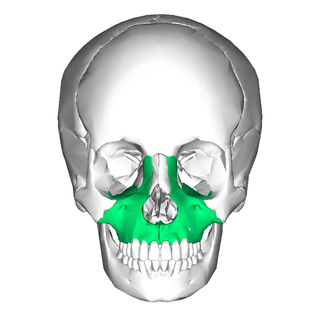
In vertebrates, the maxilla is the upper fixed bone of the jaw formed from the fusion of two maxillary bones. In humans, the upper jaw includes the hard palate in the front of the mouth. The two maxillary bones are fused at the intermaxillary suture, forming the anterior nasal spine. This is similar to the mandible, which is also a fusion of two mandibular bones at the mandibular symphysis. The mandible is the movable part of the jaw.

<span class="mw-page-title-main">Jaw</span> Opposable articulated structure at the entrance of the mouth

The jaws are a pair of opposable articulated structures at the entrance of the mouth, typically used for grasping and manipulating food. The term jaws is also broadly applied to the whole of the structures constituting the vault of the mouth and serving to open and close it and is part of the body plan of humans and most animals.

<span class="mw-page-title-main">Zygomatic bone</span> Facial bone

In the human skull, the zygomatic bone, also called cheekbone or malar bone, is a paired irregular bone, situated at the upper and lateral part of the face and forms part of the lateral wall and floor of the orbit, of the temporal fossa and the infratemporal fossa. It presents a malar and a temporal surface; four processes, and four borders.

<span class="mw-page-title-main">Palatine bone</span> Bone of the facial skeleton

In anatomy, the palatine bones are two irregular bones of the facial skeleton in many animal species, located above the uvula in the throat. Together with the maxilla, they comprise the hard palate.

<span class="mw-page-title-main">Vomer</span> Unpaired facial bone of the skull

The vomer is one of the unpaired facial bones of the skull. It is located in the midsagittal line, and articulates with the sphenoid, the ethmoid, the left and right palatine bones, and the left and right maxillary bones. The vomer forms the inferior part of the nasal septum in humans, with the superior part formed by the perpendicular plate of the ethmoid bone. The name is derived from the Latin word for a ploughshare and the shape of the bone.

<i>Galesaurus</i> Extinct genus of cynodonts from the Triassic of South Africa

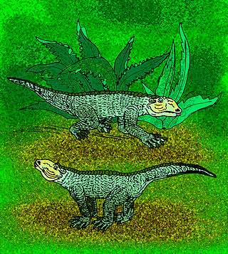
Galesaurus is an extinct genus of carnivorous cynodont therapsid that lived between the Induan and the Olenekian stages of the Early Triassic in what is now South Africa. It was incorrectly classified as a dinosaur by Sir Richard Owen in 1859.

<span class="mw-page-title-main">Incisive foramen</span> Opening of the incisive canals on the hard palate immediately behind the incisor teeth

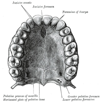
In the human mouth, the incisive foramen is the opening of the incisive canals on the hard palate immediately behind the incisor teeth. It gives passage to blood vessels and nerves. The incisive foramen is situated within the incisive fossa of the maxilla.

<span class="mw-page-title-main">Maxillary prominence</span>

Continuous with the dorsal end of the first pharyngeal arch, and growing forward from its cephalic border, is a triangular process, the maxillary prominence, the ventral extremity of which is separated from the mandibular arch by a ">"-shaped notch.

<span class="mw-page-title-main">Palatine process of maxilla</span> Thick, horizontal process of the maxilla

In human anatomy of the mouth, the palatine process of maxilla, is a thick, horizontal process of the maxilla. It forms the anterior three quarters of the hard palate, the horizontal plate of the palatine bone making up the rest. It is the most important bone in the midface. It provides structural support for the viscerocranium.

<i>Chimaerasuchus</i> Extinct genus of reptiles

Chimaerasuchus is an extinct genus of Chinese crocodyliform from the Early Cretaceous Wulong Formation. The four teeth in the very tip of its short snout gave it a "bucktoothed" appearance. Due its multicusped teeth and marked heterodonty, it is believed to have been an herbivore. Chimaerasuchus was originally discovered in the 1960s but not identified as a crocodyliform until 1995, instead thought to possibly be a multituberculate mammal. It is highly unusual, as only two other crocodyliforms have displayed any characteristics resembling its adaptations to herbivory.

<span class="mw-page-title-main">Frontonasal process</span>

The frontonasal process, or frontonasal prominence is one of the five swellings that develop to form the face. The frontonasal process is unpaired, and the others are the paired maxillary prominences, and the paired mandibular prominences. During the fourth week of embryonic development, an area of thickened ectoderm develops, on each side of the frontonasal process called the nasal placodes or olfactory placodes, and appear immediately under the forebrain.

<span class="mw-page-title-main">Primary palate</span>

Around the 5th week, the intermaxillary segment arises as a result of fusion of the two medial nasal processes and the frontonasal process within the embryo. The intermaxillary segment gives rise to the primary palate. The primary palate will form the premaxillary portion of the maxilla. This small portion is anterior to the incisive foramen and will contain the maxillary incisors.

<span class="mw-page-title-main">Human nose</span> Feature of the human face

The human nose is the first organ of the respiratory system. It is also the principal organ in the olfactory system. The shape of the nose is determined by the nasal bones and the nasal cartilages, including the nasal septum, which separates the nostrils and divides the nasal cavity into two.

<i>Xixiasaurus</i> Genus of dinosaur

Xixiasaurus is a genus of troodontid dinosaur that lived during the Late Cretaceous Period in what is now China. The only known specimen was discovered in Xixia County, Henan Province, in central China, and became the holotype of the new genus and species Xixiasaurus henanensis in 2010. The names refer to the areas of discovery, and can be translated as "Henan Xixia lizard". The specimen consists of an almost complete skull, part of the lower jaw, and teeth, as well as a partial right forelimb.

<i>Herpetoskylax</i> Extinct genus of therapsids

Herpetoskylax is an extinct genus of biarmosuchians which existed in South Africa. The type species is Herpetoskylax hopsoni. It lived in the Late Permian Period.

<i>Yonghesuchus</i> Extinct genus of reptiles

Yonghesuchus is an extinct genus of Late Triassic archosaur reptile. Remains have been found from the early Late Triassic Tongchuan Formation in Shanxi, China. It is named after Yonghe County, the county where fossils were found. Currently only one species, Y. sangbiensis, is known. The specific name refers to Sangbi Creek, as fossils were found in one of its banks.

Ovoo gurvel is an extinct varanid lizard from the Late Cretaceous of Mongolia. It is one of the smallest and earliest monitor lizards. It was described in 2008. Ovoo possesses a pair of small bones in its skull that are not seen in any other lizard.

The face and neck development of the human embryo refers to the development of the structures from the third to eighth week that give rise to the future head and neck. They consist of three layers, the ectoderm, mesoderm and endoderm, which form the mesenchyme, neural crest and neural placodes. The paraxial mesoderm forms structures named somites and somitomeres that contribute to the development of the floor of the brain and voluntary muscles of the craniofacial region. The lateral plate mesoderm consists of the laryngeal cartilages. The three tissue layers give rise to the pharyngeal apparatus, formed by six pairs of pharyngeal arches, a set of pharyngeal pouches and pharyngeal grooves, which are the most typical feature in development of the head and neck. The formation of each region of the face and neck is due to the migration of the neural crest cells which come from the ectoderm. These cells determine the future structure to develop in each pharyngeal arch. Eventually, they also form the neurectoderm, which forms the forebrain, midbrain and hindbrain, cartilage, bone, dentin, tendon, dermis, pia mater and arachnoid mater, sensory neurons, and glandular stroma.

<i>Arisierpeton</i> Extinct genus of synapsids

Arisierpeton is an extinct genus of synapsids from the Early Permian Garber Formation of Richards Spur, Oklahoma. It contains a single species, Arisierpeton simplex.

<i>Nyaphulia</i> Extinct genus of dicynodonts

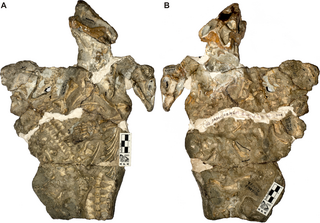
Nyaphulia is an extinct genus of dicynodont therapsid from the middle Permian of South Africa, containing only the type species N. oelofseni. The generic name is in honour of John Nyaphuli of the National Museum of Bloemfontein, who contributed extensively to South African palaeontology and discovered the holotype specimen of Nyaphulia in 1982. Nyaphulia was initially named as a second species of the basal dicynodont Eodicynodon by Professor Bruce Rubidge in 1990 as E. oelofseni, named after his mentor in palaeontology and geology Dr. Burger Oelofsen.

References

  1. 1 2 Lang, Johannes (1995). Clinical anatomy of the masticatory apparatus peripharyngeal spaces. Thieme. ISBN   978-3-13-799101-4.
  2. 1 2 Barteczko, K; Jacob, M (March 2004). "A re-evaluation of the premaxillary bone in humans". Anatomy and Embryology. 207 (6): 417–437. doi:10.1007/s00429-003-0366-x. PMID   14760532. S2CID   13069026.
  3. Gaupp, E. (1905). "Neue Deutungen auf dem Gebiete der Lehre vom Säugetierschädel". Anat. Anz. 27: 273–310.
  4. 1 2 3 Higashiyama, Hiroki; Koyabu, Daisuke; Hirasawa, Tatsuya; Werneburg, Ingmar; Kuratani, Shigeru; Kurihara, Hiroki (November 2, 2021). "Mammalian face as an evolutionary novelty". PNAS. 118 (44): e2111876118. doi:10.1073/pnas.2111876118. PMC   8673075 . PMID   34716275.
  5. "Nasal Anatomy". Medscape. June 2011. Retrieved 3 December 2011.
  6. Vergervik, Karin (1983). "Growth Characteristics of the Premaxilla and Orthodontic Treatment Principles in Bilateral Cleft Lip and Palate" (PDF). Cleft Palate Journal. 20 (4). Retrieved 3 December 2011.
  7. "Premaxilla". ZipCodeZoo. Archived from the original on 21 January 2012. Retrieved 3 December 2011.
  8. Hu, Yaoming; Meng, Jin; Li, Chuankui; Wang, Yuanqing (January 22, 2010). "New basal eutherian mammal from the Early Cretaceous Jehol biota, Liaoning, China". Proc Biol Sci. 277 (1679): 229–236. doi:10.1098/rspb.2009.0203. PMC   2842663 . PMID   19419990.
  9. Wible, John R.; Miao, Desui; Hopson, James A. (March 1990). "The septomaxilla of fossil and recent synapsids and the problem of the septomaxilla of monotremes and armadillos". Zoological Journal of the Linnean Society. 98 (3): 203–228. doi:10.1111/j.1096-3642.1990.tb01207.x.
  10. Myers, P.; Espinosa, R.; Parr, C. S.; Jones, T.; Hammond, G. S.; Dewey, T. A. (2006). "Premaxillae of bats". Animal Diversity Web. Archived from the original on 3 January 2012. Retrieved 3 December 2011.
  11. Brownstein, Chase D.; Meyer, Dalton L.; Fabbri, Matteo; Bhullar, Bhart-Anjan S.; Gauthier, Jacques A. (2022-11-29). "Evolutionary origins of the prolonged extant squamate radiation". Nature Communications. 13 (1): 7087. doi:10.1038/s41467-022-34217-5. ISSN   2041-1723. PMC   9708687 .
  12. Gillispie, Charles Coulston (1960). The Edge of Objectivity: An Essay in the History of Scientific Ideas. Princeton, NJ: Princeton University Press. p. 194. ISBN   0-691-02350-6.