Medical illustration

Last updated
Medical illustration
Wikipedia medical illustration thoracic outlet syndrome brachial plexus anatomy with labels.jpg
An illustration of the relevant neurovascular anatomy in anterior supraclavicular neurosurgical approach to the brachial plexus and subclavian vessels for thoracic outlet syndrome.
Sobo 1909 589.png
Anatomical illustration of a dissection of the skull showing meninges. A 1909 illustration from Sobotta's Atlas and Text-book of Human Anatomy.
Identifiers
MeSH D008488
Anatomical terminology

Medical illustrations can be illustrations or animations that are created to visually represent medical or biological subjects that can be difficult to explain only using words

Contents

History

Medical illustrations have been made possibly since the beginning of medicine [1] in any case for hundreds (or thousands) of years. Many illuminated manuscripts and Arabic scholarly treatises of the medieval period contained illustrations representing various anatomical systems (circulatory, nervous, urogenital), pathologies, or treatment methodologies. Many of these illustrations can look odd to modern eyes, since they reflect early reliance on classical scholarship (especially Galen) rather than direct observation, and the representation of internal structures can be fanciful. An early high-water mark was the 1543 CE publication of Andreas Vesalius's De Humani Corporis Fabrica Libri Septum, which contained more than 600 exquisite woodcut illustrations based on careful observation of human dissection.

Since the time of the Leonardo da Vinci and his depictions of the human form, there have been great advancements in the art of representing the human body. The art has evolved over time from illustration to digital imaging using the technological advancements of the digital age. Berengario da Carpi was the first known anatomist to include medical illustration within his textbooks. Gray's Anatomy, originally published in 1858, is one well-known human anatomy textbook that showcases a variety of anatomy depiction techniques. [2]

In 1895, Konrad Roentgen, a German physicist discovered the X-Ray. Internal imaging became a reality after the invention of the X-Ray. Since then internal imaging has progressed to include ultrasonic, CT, and MRI imaging. [2]

As a profession, medical illustration has a more recent history. In the late 1890s, Max Brödel, a talented artist from Leipzig, was brought to The Johns Hopkins School of Medicine in Baltimore to illustrate for Harvey Cushing, William Halsted, Howard Kelly, and other notable clinicians. In addition to being an extraordinary artist, he created new techniques, such as carbon dust, that were especially suitable to his subject matter and then-current printing technologies. In 1911 he presided over the creation of the first academic department of medical illustration, which continues to this day. His graduates spread out across the world, and founded a number of the academic programs listed below under "Education".

Notable medical illustrators include Max Brödel and Dr. Frank H. Netter. For an online inventory of scientific illustrators including currently already more than 1000 medical illustrators active 1450-1950 and specializing in anatomy, dermatology and embryology, see the Stuttgart Database of Scientific Illustrators 1450–1950

Medical illustration is used in the history of medicine. [3]

Profession

Medical illustrators not only produce such material but can also function as consultants and administrators within the field of biocommunication. A certified medical illustrator continues to obtain extensive training in medicine, science, and art techniques throughout his or her career.

The Association of Medical Illustrators is an international organization founded in 1945, and incorporated in Illinois. Its members are primarily artists who create material designed to facilitate the recording and dissemination of medical and bioscientific knowledge through visual communication media. Members are involved not only in the creation of such material, but also serve in consultant, advisory, educational and administrative capacities in all aspects of bioscientific communications and related areas of visual education.

The professional objectives of the AMI are to promote the study and advancement of medical illustration and allied fields of visual communication, and to promote understanding and cooperation with the medical profession and related health science professions.

The AMI publishes an annual Medical Illustration Source Book which is distributed to creative and marketing professionals that regularly hire medical/scientific image makers for editorial, publishing, educational and advertising projects. There is a companion Source Book with searchable illustration, animation and multimedia portfolios from hundreds of artists in the field.

The obvious abilities necessary are to be able to visualize the subject-matter, some degree of originality in style of drawing and the refined skill of colour discrimination. [4]

Education

Medical illustrators in the profession either have a master's degree from an accredited graduate program in medical illustration or degrees in science, such as biology, or the arts. The Association of Medical Illustrators is a sponsor member of CAAHEP (Commission on Accreditation of Allied Health Education Programs), the organization that grants accreditation to the graduate programs in medical illustration upon recommendation of ARC-MI (Accreditation Review Committee for the Medical Illustrator) which is a standing committee of the AMI and a Committee on Accreditation of CAAHEP. Currently there are four accredited Programs in North America:

The Department of Art as Applied to Medicine on the East Baltimore Campus of the Johns Hopkins Medical Institutions was the first program of its kind in the world. Endowed in 1911, the program has been in existence for over 90 years. In 1959, the Johns Hopkins University approved a two-year graduate program leading to the university-wide degree of Master of Arts in Medical and Biological Illustration. The academic calendar, faculty and student affairs are administered by The Johns Hopkins University School of Medicine. The program has been fully accredited since 1970, currently accredited by the Commission on Accreditation of Allied Health Education Programs (CAAHEP).

The Biomedical Visualization Program at the University of Illinois at Chicago's College of Applied Health Sciences is the second oldest school of medical illustration in the western hemisphere, founded in 1921 by Thomas Smith Jones (Jones also was co-founder of the Association of Medical Illustrators). The UIC program is located in the national healthcare and pharmaceutical hub of Chicago, and offers a market-based curriculum that includes the highest ends of technology (including the renowned Virtual Reality Medical Laboratory and a rigorous animation curriculum). Biomedical Visualization is located on the UIC Medical Center campus, home of the largest medical school in the United States. The UIC program blends the more traditional aspects of medical illustration and the emerging markets of digital, pharmaceutical, and "edutainment" industries. UIC previously offered an extensive study in the field of anaplastology (facial and somatic prosthetics) and medical sculpture, though it is no longer available in the current curriculum. [5] A two-year Master of Science (MS) in Biomedical Visualization degree is awarded, and the program is accredited by the Commission on Accreditation of Allied Health Education Programs (CAAHEP).

The Medical Illustration Graduate Program at Augusta University (formerly the Medical College of Georgia), in Augusta, Georgia is fully accredited by CAAHEP. Graduates receive a Master of Science in Medical Illustration. The first Master of Science degree in Medical Illustration at MCG was awarded in 1951. The program emphasizes anatomical and surgical illustration for print and electronic publication, as well as for projection and broadcast distribution. Because of the importance of good drawing skills, the students learn a variety of traditional illustration techniques during the first year. In addition, computer technologies and digital techniques, used to prepare both vector and raster images for print and motion media, are well and extensively integrated into the curriculum.

The Biomedical Communications Program at the University of Toronto. This program was begun in 1945 by Maria Wishart, a student of Max Brödel's. Faculty and graduates of the program contributed the drawings for Grant's Atlas of Anatomy, a renowned guide to dissection, structure, and function for medical students. The current two-year professional Master's program, offered through the Institute of Medical Science, emphasizes a research-based approach to the creation and evaluation of visual material for health promotion, medical education, and the process of scientific discovery.

The Biomedical Communication Graduate Program at the University of Texas Southwestern Medical Center at Dallas. The University of Texas Southwestern Medical Center was the first school in the world to offer a graduate degree in medical illustration in 1945. Lewis Boyd Waters, who studied under Max Brodel at Johns Hopkins in the 1920s, was a founding member of the medical school and was responsible for starting the master's program. Waters died in 1969 and was later succeeded by several of his students who continued and expanded the program. The program was designed to be an interdisciplinary program that provides opportunities for development of the skills and knowledge needed in the application of communications arts and technology to the health sciences. The program closed in 2012.

Review

Medical illustrators create medical illustrations using traditional and digital techniques which can appear in medical textbooks, medical advertisements, professional journals [6] , instructional videotapes and films, animations, web-based media [7] , computer-assisted learning programs, exhibits, lecture presentations [8] , and patient education [9] . Medical illustrators also work in three dimensions, creating anatomical teaching models, patient simulators, games, and facial prosthetics. Medical illustrations can also be used in court cases to assist the jury with conceptualizing the extent of people’s injuries [10] .

The traditional tools of medical illustration are slowly being replaced and supplemented by a range of unique modern artistic practices. Three-dimensional renders and endoscopic cameras are replacing carbon dust and cadavers.

Additional images

See also

Related Research Articles

<span class="mw-page-title-main">Biomedical engineering</span> Application of engineering principles and design concepts to medicine and biology

Biomedical engineering (BME) or medical engineering is the application of engineering principles and design concepts to medicine and biology for healthcare applications. BME is also traditionally logical sciences to advance health care treatment, including diagnosis, monitoring, and therapy. Also included under the scope of a biomedical engineer is the management of current medical equipment in hospitals while adhering to relevant industry standards. This involves procurement, routine testing, preventive maintenance, and making equipment recommendations, a role also known as a Biomedical Equipment Technician (BMET) or as a clinical engineer.

<span class="mw-page-title-main">Ponce Health Sciences University</span> Private university in Ponce, Puerto Rico

The Ponce Health Sciences University (PHSU), formerly Ponce School of Medicine & Health Sciences, is a private, for-profit university in Ponce, Puerto Rico and St. Louis, Missouri. It awards graduate degrees in Medicine (MD), Clinical Psychology (PsyD and PhD), Biomedical Sciences (PhD), Medical Sciences (MS), and Public Health (MPH and DrPH). The university has 360 students in its medical school and, as of 11 February 2019, was authorized to increase the student body at the medical school to 600 which, when fully in place, will make it the largest private medical school in Puerto Rico and one of the largest under the American flag.

<span class="mw-page-title-main">Perfusionist</span> Healthcare professional who uses the cardiopulmonary bypass machine

A cardiovascular perfusionist, clinical perfusionist or perfusiologist, and occasionally a cardiopulmonary bypass doctor or clinical perfusion scientist, is a healthcare professional who operates the cardiopulmonary bypass machine during cardiac surgery and other surgeries that require cardiopulmonary bypass to manage the patient's physiological status. As a member of the cardiovascular surgical team, the perfusionist also known as the clinical perfusionist helps maintain blood flow to the body's tissues as well as regulate levels of oxygen and carbon dioxide in the blood, using a heart–lung machine.

<span class="mw-page-title-main">University of Tennessee Health Science Center</span> Health Sciences division of the University of Tennessee

The University of Tennessee Health Science Center (UTHSC) is a public medical school in Memphis, Tennessee. It includes the Colleges of Health Professions, Dentistry, Graduate Health Sciences, Medicine, Nursing, and Pharmacy. Since 1911, the University of Tennessee Health Science Center has educated nearly 57,000 health care professionals. As of 2010, U.S. News & World Report ranked the College of Pharmacy 17th among American pharmacy schools.

<span class="mw-page-title-main">Des Moines University</span> Medical school in West Des Moines, Iowa

Des Moines University (DMU) is a private medical school in West Des Moines, Iowa. Founded in 1898, Des Moines University is the second oldest osteopathic medical school and the fifteenth largest medical school in the United States. DMU's three colleges—the College of Osteopathic Medicine, College of Podiatric Medicine and Surgery, and College of Health Sciences—offer nine academic degrees, including master's and doctorate degrees.

<span class="mw-page-title-main">University of Puerto Rico School of Medicine</span> Medical school of the University of Puerto Rico

The University of Puerto Rico, Medical Sciences Campus, School of Medicine is located in the University of Puerto Rico, Medical Sciences Campus in San Juan, Puerto Rico. It's the only medical school in the University of Puerto Rico System. It is accredited by the Liaison Committee on Medical Education (LCME). Its students are predominantly Puerto Rican residents. However, anyone is allowed to apply to this university. It is also considered the top bilingual medical school in the world due to its requirement for each admitted student to fully master both English and Spanish.

The University of North Texas Health Science Center is a public academic health science center in Fort Worth, Texas. It is part of the University of North Texas System and was founded in 1966 as the Texas College of Osteopathic Medicine, with its first cohort admitted in 1970. UNT Health Science Center consists of six schools with a total enrollment of 2,329 students (2020–21).

A biomedical scientist is a scientist trained in biology, particularly in the context of medical laboratory sciences or laboratory medicine. These scientists work to gain knowledge on the main principles of how the human body works and to find new ways to cure or treat disease by developing advanced diagnostic tools or new therapeutic strategies. The research of biomedical scientists is referred to as biomedical research.

The School of Engineering and Applied Science (SEAS) at the George Washington University in Washington, D.C. is a technical school which specializes in engineering, technology, communications, and transportation. The school is located on the main campus of the George Washington University and offers both undergraduate and graduate programs.

<span class="mw-page-title-main">Touro University California</span> Health professions graduate school

Touro University California is a private graduate school focused primarily on health professions and located on Mare Island in Vallejo, California. It is part of the Touro College and University System and is jointly administered with its sister campus Touro University Nevada.

<span class="mw-page-title-main">Max Brödel</span> German medical illustrator

Max Brödel was a medical illustrator. Born in Leipzig, Germany, he began his artistic career after graduating from the Leipzig Academy of Fine Arts, working for Carl Ludwig. Under Ludwig's instruction, Brödel gained a basic knowledge of medicine and became recognized for his detailed medical illustrations. In the late 1890s, he was brought to the Johns Hopkins School of Medicine in Baltimore to illustrate for Harvey Cushing, William Halsted, Howard Kelly, and other notable clinicians. In addition to being a prolific medical illustrator, he developed new artistic techniques such as the carbon dust technique that helped the advancement of the quality and accuracy of medical illustrations for physicians. In 1911, he presided over the creation of the first Department of Art as Applied to Medicine; located at the Johns Hopkins School of Medicine, it continues to train medical illustrators to this day. His graduates spread out across the world, and have founded a number of other academic programs.

Commission on Accreditation of Allied Health Education Programs (CAAHEP) is an accreditation agency for postsecondary education programs in 30 health science fields.

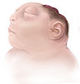
A medical animation is a short educational film, usually based around a physiological or surgical topic, that is rendered using 3D computer graphics. While it may be intended for an array of audiences, the medical animation is most commonly utilized as an instructional tool for medical professionals or their patients.

Certified anesthesiologist assistants (CAAs) are highly trained master’s degree level non-physician anesthesia care providers. CAAs are integral members of the anesthesia care team as described by the American Society of Anesthesiologists (ASA). This designation must be disambiguated from the Certified Clinical Anesthesia Assistant (CCAA) designation conferred by the Canadian Society of Respiratory Therapists. All CAAs possess a baccalaureate degree, and complete an intensive didactic and clinical program at a postgraduate level. CAAs are trained in the delivery and maintenance of all types of anesthesia care as well as advanced patient monitoring techniques. The goal of CAA education is to guide the transformation of student applicants into competent clinicians.

Audrey Juliet Arnott (1901–1974) was a medical illustrator who worked with the neurosurgeon Hugh Cairns at the London Hospital and followed him to Oxford when he was appointed Nuffield Professor of Surgery in 1939. She founded the Medical Artists Association of Great Britain from her home in Wolvercote in 1949.

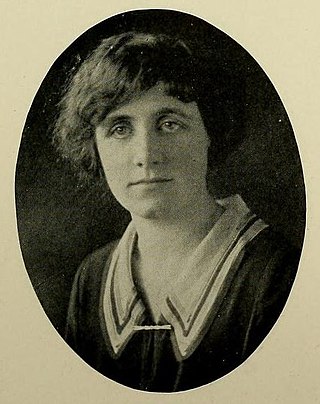
Maria Torrence Wishart was a Canadian medical illustrator and the founder of the University of Toronto's Art as Applied to Medicine program. She was educated at Johns Hopkins School of Medicine under Max Brödel, and in 1925 returned to Canada to found the Department of Medical Art Service in the Faculty of Medicine.

California Health Sciences University (CHSU) is a private, for-profit university located in Clovis, in the U.S. state of California. Founded in 2012, the school operates three academic programs, two of which offer doctoral degrees (in pharmacy and osteopathic medicine), and the third offers a masters degree in science. Graduates of the College of Pharmacy (COP) will receive the Doctor of Pharmacy (Pharm D) degree, graduates of the College of Osteopathic Medicine (COM) will receive the Doctor of Osteopathic Medicine (DO) degree, and graduates of the College of Biosciences and Health Professions (CBHP) will receive the Masters of Science in Biomedical Sciences (MSBS) degree. The College of Osteopathic Medicine is fully pre-accredited by the American Osteopathic Association's (AOA) Commission on Osteopathic College Accreditation (COCA). The college is accredited by the Western Association of Schools and Colleges (WASC) Senior College and University Commission and has approval to operate from the Bureau of Private Postsecondary Education (BPPE).

<span class="mw-page-title-main">Mildred Codding</span> American medical illustrator

Mildred B. Codding was an American medical illustrator. Her illustrations are featured in numerous textbooks and academic journal articles.

<span class="mw-page-title-main">Elizabeth H. Brödel</span> American scientific illustrator

Elizabeth Huntington Brödel, also seen as Elizabeth H. Broedel, was an American medical illustrator and daughter of medical illustrator Max Brödel.

The Visible Embryo Project (VEP) is a multi-institutional, multidisciplinary research project originally created in the early 1990s as a collaboration between the Developmental Anatomy Center at the National Museum of Health and Medicine and the Biomedical Visualization Laboratory (BVL) at the University of Illinois at Chicago, "to develop software strategies for the development of distributed biostructural databases using cutting-edge technologies for high-performance computing and communications (HPCC), and to implement these tools in the creation of a large-scale digital archive of multidimensional data on normal and abnormal human development." This project related to BVL's other research in the areas of health informatics, educational multimedia, and biomedical imaging science. Over the following decades, the list of VEP collaborators grew to include over a dozen universities, national laboratories, and companies around the world.

References

Notes

  1. FM. Corl1, MR. Garland and EK. Fishman - Role of Computer Technology in Medical Illustration AJR December 2000 vol. 175 no. 6 1519-1524 Retrieved 2012-12-20
  2. 1 2 Tsafrir, Jenni; Ohry, Avi (18 July 2008). "Medical illustration: from caves to cyberspace‡". Health Information & Libraries Journal. 18 (2): 99–109. doi:10.1046/j.1471-1842.2001.d01-16.x. ISSN   1471-1834.
  3. Camillia Matuk (2006). "Seeing the Body: The Divergence of Ancient Chinese and Western Medical Illustration" (PDF). Journal of Biocommunication. 32 (1).
  4. nih.gov Archived 2011-10-26 at the Wayback Machine Retrieved 2012-12-20
  5. "MS in Biomedical Visualization < University of Illinois at Chicago". catalog.uic.edu. Retrieved 2021-02-08.
  6. Hajar, Rachel (2011). "Medical illustration: Art in medical education". Heart Views. 12 (2): 83–91. doi: 10.4103/1995-705X.86023 . ISSN   1995-705X. PMC   3221200 . PMID   22121469.
  7. Bernhardt, Butch (2017-08-17). "Wood Poles Still Best Alternative for Most Uses". Natural Gas & Electricity. 34 (2): 9–14. doi:10.1002/gas.22002. ISSN   1545-7893.
  8. Sutton, Halley (2015-12-15). "Bring a complex texture of life experiences to campus with transfer students". Recruiting & Retaining Adult Learners. 18 (4): 1–5. doi:10.1002/nsr.30113. ISSN   2155-644X.
  9. "Custom Illustration Specialist - Affordable Illustrator Agency London – Maa Illustrations". www.maaillustrations.com. Retrieved 2024-03-16.
  10. "Utilizing Medical Illustrations as Demonstrative Aids for Trial". Academy of Forensic Nursing | AFN | Home. Retrieved 2024-03-16.