Microtia

Last updated
Microtia
Microtia lvl3.jpg
Unilateral grade III microtia (more often affecting the right ear).
Specialty Medical genetics   OOjs UI icon edit-ltr-progressive.svg

Microtia is a congenital deformity where the auricle (external ear) is underdeveloped. A completely undeveloped auricle is referred to as anotia. Because microtia and anotia have the same origin, it can be referred to as microtia-anotia. [1] Microtia can be unilateral (one side only) or bilateral (affecting both sides). Microtia occurs in 1 out of about 8,000–10,000 births. In unilateral microtia, the right ear is most commonly affected. It may occur as a complication of taking Accutane (isotretinoin) during pregnancy. [2]

Contents

Classification

Bilateral grade III microtia in a 9-year-old boy Various types 2 (detail B).jpg
Bilateral grade III microtia in a 9-year-old boy

According to the Altman-classification, there are four grades of microtia: [3]

Causes and risk factors

The etiology of microtia in children remains uncertain but there are some cases that associate the cause of microtia with genetic defects in multiple or single genes, altitude, and gestational diabetes. [3] Risk factors gathered from studies include infants born underweight, male sex, women gravidity and parity, and medication use while pregnant. [4] [3] [5] [6] Genetic inheritance has not been fully studied but in the few studies available, it has shown to occur during the early stages of pregnancy. [3]

Microtia is also feature of many conditions and syndromes: [7]

Microtia in a 10-year-old girl with Goldenhar syndrome Apert Azerad 2.jpg
Microtia in a 10-year-old girl with Goldenhar syndrome
Microtia in a 50-year-old woman with Saethre-Chotzen syndrome Davis Oxycephalus 8.jpg
Microtia in a 50-year-old woman with Saethre-Chotzen syndrome

Diagnosis

At birth, lower grade microtia is difficult to visually diagnose with a physical exam. While higher grade microtia can be visually diagnosed due to noticeable abnormalities. [3] Infants that have noticeable abnormalities are closely monitored by physicians and hearing specialists. [8]

Treatment

The goal of medical intervention is to provide the best form and function to the underdeveloped ear. [9]

Hearing

Typically, testing is first done to determine the quality of hearing. This can be done as early as in the first two weeks with a BAER test (Brain Stem Auditory Response Test). [10] [11] At age 5–6, CT or CAT scans of the middle ear can be done to elucidate its development and clarify which patients are appropriate candidates for surgery to improve hearing. For younger individuals, this is done under sedation.

The hearing loss associated with congenital aural atresia is a conductive hearing loss—hearing loss caused by inefficient conduction of sound to the inner ear. Essentially, children with aural atresia have hearing loss because the sound cannot travel into the (usually) healthy inner ear—there is no ear canal, no eardrum, and the small ear bones (malleus/hammer, incus/anvil, and stapes/stirrup) are underdeveloped. "Usually" is in parentheses because rarely, a child with atresia also has a malformation of the inner ear leading to a sensorineural hearing loss (as many as 19% in one study). [12] Sensorineural hearing loss is caused by a problem in the inner ear, the cochlea. Sensorineural hearing loss is not correctable by surgery, but properly fitted and adjusted hearing amplification (hearing aids) generally provide excellent rehabilitation for this hearing loss. If the hearing loss is severe to profound in both ears, the child may be a candidate for a cochlear implant (beyond the scope of this discussion).

Unilateral sensorineural hearing loss was not generally considered a serious disability by the medical establishment before the nineties; it was thought that the afflicted person was able to adjust to it from birth. In general, there are exceptional advantages to gain from an intervention to enable hearing in the microtic ear, especially in bilateral microtia. Children with untreated unilateral sensorineural hearing loss are more likely to have to repeat a grade in school and/or need supplemental services (e.g., FM system – see below) than their peers. [13] [14]

Children with unilateral sensorineural hearing loss often require years of speech therapy in order to learn how to enunciate and understand spoken language. What is truly unclear, and the subject of an ongoing research study, is the effect of unilateral conductive hearing loss (in children with unilateral aural atresia) on scholastic performance. If atresia surgery or some form of amplification is not used, special steps should be taken to ensure that the child is accessing and understanding all of the verbal information presented in school settings. Recommendations for improving a child's hearing in the academic setting include preferential seating in class, an FM system (the teacher wears a microphone, and the sound is transmitted to a speaker at the child's desk or to an ear bud or hearing aid the child wears), a bone-anchored hearing aid (BAHA), or conventional hearing aids. Age for BAHA implantation depends on whether the child is in Europe (18 months) or the US (age 5). Until then it is possible to fit a BAHA on a softband [15] [16]

It is important to note that not all children with aural atresia are candidates for atresia repair. Candidacy for atresia surgery is based on the hearing test (audiogram) and CT scan imaging. If a canal is built where one does not exist, minor complications can arise from the body's natural tendency to heal an open wound closed. Repairing aural atresia is a very detailed and complicated surgical procedure which requires an expert in atresia repair. [17] While complications from this surgery can arise, the risk of complications is greatly reduced when using a highly experienced otologist. Atresia patients who opt for surgery will temporarily have the canal packed with gelatin sponge and silicone sheeting to prevent closure. The timing of ear canal reconstruction (canalplasty) depends on the type of external ear (Microtia) repair desired by the patient and family. Two surgical teams in the USA are currently able to reconstruct the canal at the same time as the external ear in a single surgical stage (one stage ear reconstruction).

In cases where a later surgical reconstruction of the external ear of the child might be possible, positioning of the BAHA implant is critical. It may be necessary to position the implant further back than usual to enable successful reconstructive surgery – but not so far as to compromise hearing performance. If the reconstruction is ultimately successful, it is easy to remove the percutaneous BAHA abutment. If the surgery is unsuccessful, the abutment can be replaced and the implant re-activated to restore hearing.

External ear

The age when outer ear surgery can be attempted depends upon the technique chosen. The earliest is 7 for Rib Cartilage Grafts. However, some surgeons recommend waiting until a later age, such as 8–10 when the ear is closer to adult size. External ear prostheses have been made for children as young as 5.

For auricular reconstruction, there are several different options:

  1. Rib Cartilage Graft Reconstruction: This surgery may be performed by specialists in the technique. [18] [19] [20] [21] [22] It involves sculpting the patient's own rib cartilage into the form of an ear. Because the cartilage is the patient's own living tissue, the reconstructed ear continues to grow as the child does. In order to be sure that the rib cage is large enough to provide the necessary donor tissue, some surgeons wait until the patient is 8 years of age; [21] [22] however, some surgeons with more experience with this technique may begin the surgery on a child aged six. [19] [20] [23] The major advantage of this surgery is that the patient's own tissue is used for the reconstruction. This surgery varies from two to four stages depending on the surgeon's preferred method. A novel one stage ear reconstruction technique is performed by a few select surgeons. One team is able to reconstruct the entire external ear and ear canal in one operation.
  2. Reconstruct the ear using a polyethylene plastic implant (also called Medpor): This is a 1–2 stage surgery that can start at age 3 and can be done as an outpatient without hospitalization. Using the porous framework, which allows the patient's tissue to grow into the material and the patient's own tissue flap, a new ear is constructed in a single surgery. A small second surgery is performed in 3–6 months if needed for minor adjustments. Medpor was developed by John Reinisch. [24] This surgery should only be performed by experts in the techniques involved. [25] The use of porous polyethylene implants for ear reconstruction was initiated in the 1980s by Alexander Berghaus. [26]
  3. Ear Prosthesis: An auricular (ear) prosthesis is custom made by an anaplastologist to mirror the other ear. [27] Prosthetic ears can appear very realistic. They require a few minutes of daily care. They are typically made of silicone, which is colored to match the surrounding skin and can be attached using either adhesive or with titanium screws inserted into the skull to which the prosthetic is attached with a magnetic or bar/clip type system. These screws are the same as the BAHA (bone anchored hearing aid) screws and can be placed simultaneously. The biggest advantage over any surgery is having a prosthetic ear that allows the affected ear to appear as normal as possible to the natural ear. The biggest disadvantage is the daily care involved and knowing that the prosthesis is not real.[ citation needed ] In 2022, success of transplantation of a 3D bioprinted auricle made from the microtia patient's own cells was reported, [28] also achieving a first in 3D bioprinting for transplants. [29]

Aural atresia is the underdevelopment of the middle ear and canal and usually occurs in conjunction with microtia. Atresia occurs because patients with microtia may not have an external opening to the ear canal, though. However, the cochlea and other inner ear structures are usually present. The grade of microtia usually correlates to the degree of development of the middle ear. [11] [17] Microtia is usually isolated, but may occur in conjunction with hemifacial microsomia, Goldenhar Syndrome or Treacher-Collins Syndrome. [30] It is also occasionally associated with kidney abnormalities (rarely life-threatening), and jaw problems, and more rarely, heart defects and vertebral deformities. [20]

Notable cases

Related Research Articles

<span class="mw-page-title-main">Outer ear</span> Outer part of ear

The outer ear, external ear, or auris externa is the external part of the ear, which consists of the auricle and the ear canal. It gathers sound energy and focuses it on the eardrum.

<span class="mw-page-title-main">Anotia</span> Medical condition

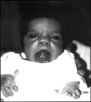
Anotia describes a rare congenital deformity that involves the complete absence of the auricle, the outer projected portion of the ear, and narrowing or absence of the ear canal. This contrasts with microtia, in which a small part of the auricle is present. Anotia and microtia may occur unilaterally or bilaterally. This deformity results in conductive hearing loss, deafness.

<span class="mw-page-title-main">Ear</span> Organ of hearing and balance

An ear is the organ that enables hearing and body balance using the vestibular system. In mammals, the ear is usually described as having three parts: the outer ear, the middle ear and the inner ear. The outer ear consists of the pinna and the ear canal. Since the outer ear is the only visible portion of the ear in most animals, the word "ear" often refers to the external part alone. The middle ear includes the tympanic cavity and the three ossicles. The inner ear sits in the bony labyrinth, and contains structures which are key to several senses: the semicircular canals, which enable balance and eye tracking when moving; the utricle and saccule, which enable balance when stationary; and the cochlea, which enables hearing. The ear is a self cleaning organ through its relationship with earwax and the ear canals. The ears of vertebrates are placed somewhat symmetrically on either side of the head, an arrangement that aids sound localization.

<span class="mw-page-title-main">Auricle (anatomy)</span> Visible part of the ear that is outside the head

The auricle or auricula is the visible part of the ear that is outside the head. It is also called the pinna, a term that is used more in zoology.

<span class="mw-page-title-main">Treacher Collins syndrome</span> Human genetic disorder

Treacher Collins syndrome (TCS) is a genetic disorder characterized by deformities of the ears, eyes, cheekbones, and chin. The degree to which a person is affected, however, may vary from mild to severe. Complications may include breathing problems, problems seeing, cleft palate, and hearing loss. Those affected generally have normal intelligence.

<span class="mw-page-title-main">Conductive hearing loss</span> Medical condition

Conductive hearing loss (CHL) occurs when there is a problem transferring sound waves anywhere along the pathway through the outer ear, tympanic membrane (eardrum), or middle ear (ossicles). If a conductive hearing loss occurs in conjunction with a sensorineural hearing loss, it is referred to as a mixed hearing loss. Depending upon the severity and nature of the conductive loss, this type of hearing impairment can often be treated with surgical intervention or pharmaceuticals to partially or, in some cases, fully restore hearing acuity to within normal range. However, cases of permanent or chronic conductive hearing loss may require other treatment modalities such as hearing aid devices to improve detection of sound and speech perception.

Otoplasty is a procedure for correcting the deformities and defects of the auricle, whether these defects are congenital conditions or caused by trauma. Otoplastic surgeons may reshape, move, or augment the cartilaginous support framework of the auricle to correct these defects.

Unilateral hearing loss (UHL) is a type of hearing impairment where there is normal hearing in one ear and impaired hearing in the other ear.

Atresia is a condition in which an orifice or passage in the body is closed or absent.

<span class="mw-page-title-main">Hemifacial microsomia</span> Birth defect which affects the lower half of the face

Hemifacial microsomia (HFM) is a congenital disorder that affects the development of the lower half of the face, most commonly the ears, the mouth and the mandible. It usually occurs on one side of the face, but both sides are sometimes affected. If severe, it may result in difficulties in breathing due to obstruction of the trachea—sometimes even requiring a tracheotomy. With an incidence in the range of 1:3500 to 1:4500, it is the second most common birth defect of the face, after cleft lip and cleft palate. HFM shares many similarities with Treacher Collins syndrome.

<span class="mw-page-title-main">Bone-anchored hearing aid</span>

A bone-anchored hearing aid (BAHA) is a type of hearing aid based on bone conduction. It is primarily suited for people who have conductive hearing losses, unilateral hearing loss, single-sided deafness and people with mixed hearing losses who cannot otherwise wear 'in the ear' or 'behind the ear' hearing aids. They are more expensive than conventional hearing aids, and their placement involves invasive surgery which carries a risk of complications, although when complications do occur, they are usually minor.

<span class="mw-page-title-main">Robinow syndrome</span> Rare genetic disorder characterized by a fetal face

Robinow syndrome is an extremely rare genetic disorder characterized by short-limbed dwarfism, abnormalities in the head, face, and external genitalia, as well as vertebral segmentation. The disorder was first described in 1969 by human geneticist Meinhard Robinow, along with physicians Frederic N. Silverman and Hugo D. Smith, in the American Journal of Diseases of Children. By 2002, over 100 cases had been documented and introduced into medical literature.

Hearing loss with craniofacial syndromes is a common occurrence. Many of these multianomaly disorders involve structural malformations of the outer or middle ear, making a significant hearing loss highly likely.

Amastia refers to a rare clinical anomaly in which both internal breast tissue and the visible nipple are absent on one or both sides. It affects both men and women. Amastia can be either isolated or comorbid with other syndromes, such as ectodermal dysplasia, Syndactyly and lipoatrophic diabetes. This abnormality can be classified into various types, and each could result from different pathologies. Amastia differs from amazia and athelia. Amazia is the absence of one or both mammary glands but the nipples remain present, and athelia is the absence of one or both nipples, but the mammary gland remains.

Burt Brent is a retired reconstructive plastic surgeon best known for his work in reconstructing the absent outer ear. He built upon the techniques of his mentor, Dr. Radford Tanzer of the Mary Hitchcock Clinic at Dartmouth Medical School and repaired ear defects in 1,800 patients, most of them children born with ear deformities such as microtia. He also reconstructed ears lost or due to some form of trauma.

A facial cleft is an opening or gap in the face, or a malformation of a part of the face. Facial clefts is a collective term for all sorts of clefts. All structures like bone, soft tissue, skin etc. can be affected. Facial clefts are extremely rare congenital anomalies. There are many variations of a type of clefting and classifications are needed to describe and classify all types of clefting. Facial clefts hardly ever occur isolated; most of the time there is an overlap of adjacent facial clefts.

<span class="mw-page-title-main">Cryptotia</span> Medical condition

Cryptotia is the condition where an ear appears to have its upper portion buried underneath the side of the head. The condition also involves underdeveloped scapha and antihelical crura. Cryptotia is also known as buried ear or hidden ear.

Cartilage conduction is a pathway by which sound signals are transmitted to the inner ear. In 2004, Hiroshi Hosoi discovered this pathway and named “cartilage conduction”. Hearing by cartilage conduction is distinct from conventional sound-conduction pathways, such as air or bone, because it is realized by touching a transducer on the aural cartilage and does not involve the vibration of the skull bone. Therefore, cartilage conduction is referred to as the “third auditory pathway”.

<span class="mw-page-title-main">Auricular splint</span>

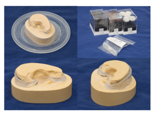
An auricular splint (AS) or ear splint is a custom-made medical device that is used to maintain auricular projection and dimensions following second stage auricular reconstruction. The AS is made from ethylene-vinyl acetate (EVA), which is typically used to make custom-made mouthguards and was developed by a team from Great Ormond Street Hospital in the United Kingdom.

<span class="mw-page-title-main">Mandibulofacial dysostosis-microcephaly syndrome</span> Medical condition

Mandibulofacial dysostosis with microcephaly syndrome, also known as growth delay-intellectual disability-mandibulofacial dysostosis-microcephaly-cleft palate syndrome, mandibulofacial dysostosis, guion-almeida type, or simply as MFDM syndrome is a rare genetic disorder which is characterized by developmental delays, intellectual disabilities, and craniofacial dysmorphisms.

References

  1. Online Mendelian Inheritance in Man (OMIM): Microtia-Anotia - 600674
  2. Pretest self assessment and review for the USMLE, pediatrics, 12th edition, question 84, general pediatrics
  3. 1 2 3 4 5 Luquetti, Daniela V.; Heike, Carrie L.; Hing, Anne V.; Cunningham, Michael L.; Cox, Timothy C. (January 2012). "Microtia: Epidemiology & Genetics". American Journal of Medical Genetics. Part A. 158A (1): 124–139. doi:10.1002/ajmg.a.34352. ISSN   1552-4825. PMC   3482263 . PMID   22106030.
  4. Guo, Rui et al. “Epidemiological Analysis and the Nomogram for Possible Risk Factors for Severe Microtia.” The Journal of craniofacial surgery vol. 32,2 (2021): e184-e189. doi:10.1097/SCS.0000000000007068
  5. Forrester, Mathias B.; Merz, Ruth D. (December 2005). "Descriptive epidemiology of anotia and microtia, Hawaii, 1986-2002". Congenital Anomalies. 45 (4): 119–124. doi: 10.1111/j.1741-4520.2005.00080.x . ISSN   0914-3505. PMID   16359491.
  6. Zhang, Qing-guo; Zhang, Jiao; Yu, Pei; Shen, Hao (October 2009). "Environmental and genetic factors associated with congenital microtia: a case-control study in Jiangsu, China, 2004 to 2007". Plastic and Reconstructive Surgery. 124 (4): 1157–1164. doi:10.1097/PRS.0b013e3181b454d8. ISSN   1529-4242. PMID   19935299. S2CID   3388083.
  7. "Microtia (Concept Id: C0152423)". www.ncbi.nlm.nih.gov. Retrieved 2023-11-18.
  8. Bly, Randall A.; Bhrany, Amit D.; Murakami, Craig S.; Sie, Kathleen C.Y. (November 2016). "Microtia Reconstruction". Facial Plastic Surgery Clinics of North America. 24 (4): 577–591. doi:10.1016/j.fsc.2016.06.011. ISSN   1064-7406. PMC   5950715 . PMID   27712823.
  9. Thomas, Daniel J. (August 2016). "Could 3D bioprinted tissues offer future hope for microtia treatment?". International Journal of Surgery (London, England). 32: 43–44. doi: 10.1016/j.ijsu.2016.06.036 . ISSN   1743-9159. PMID   27353851.
  10. De la Cruz A, Kesser BW (1999). "Management of the Unilateral Atretic Ear". In Pensak M (ed.). Controversies in Otolaryngology–Head and Neck Surgery. New York: Thieme Medical Publishers. pp. 381–385.
  11. 1 2 Kountakis SE, Helidonis E, Jahrsdoerfer RA (1995). "Microtia grade as an indicator of middle ear development in aural atresia". Arch Otolaryngol Head Neck Surg. 121 (8): 885–6. doi:10.1001/archotol.1995.01890080053010. PMID   7619415.
  12. Vrabec JT, Lin JW (2010). "Inner Ear Anomalies in Congenital Aural Atresia". Otology & Neurotology. 31 (9): 1421–1426. doi:10.1097/mao.0b013e3181f7ab62. PMID   21113986. S2CID   23454415.
  13. Bess FH, Tharpe AM (1986). "Case History Data on Unilaterally Hearing-Impaired Children". Ear and Hearing. 7 (1): 14–19. doi:10.1097/00003446-198602000-00004. PMID   3949096. S2CID   11839568.
  14. Bess FH, Tharpe AM (1988). "Performance and Management of Children with Unilateral Sensorineural Hearing Loss". Cand Audiol Supple. 30: 75–9. PMID   3067327.
  15. Nicholson N, Christensen L, Dornhoffer J, Martin P, Smith-Olinde L (2011). "Verification of speech spectrum audibility for pediatric baha softband users with craniofacial anomalies". The Cleft Palate-Craniofacial Journal. 48 (1): 56–65. doi:10.1597/08-178. PMID   20180710. S2CID   46151637.
  16. Verhagen CV, Hol MK, Coppens-Schellekens W, Snik AF, Cremers CW (2008). "The Baha Softband. A new treatment for young children with bilateral congenital aural atresia". International Journal of Pediatric Otorhinolaryngology. 72 (10): 1455–9. doi:10.1016/j.ijporl.2008.06.009. PMID   18667244.
  17. 1 2 Jahrsdoerfer RA, Kesser BW (1995). "Issues on Aural Atresia for the facial Plastic Surgeon". Facial Plastic Surgery. 11 (4): 274–277. doi:10.1055/s-2008-1064543. PMID   9046615. S2CID   34448986.
  18. Tanzer RC (1959). "Total Reconstruction of the External Ear". Plastic & Reconstructive Surgery. 23 (1): 1–15. doi:10.1097/00006534-195901000-00001. PMID   13633474.
  19. 1 2 Brent B (1999). "Technical Advances with Autogenous Rib Cartilage Grafts—A Personal Review of 1,200 Cases". Plastic & Reconstructive Surgery. 104 (2): 319–334. doi:10.1097/00006534-199908000-00001. PMID   10654675.
  20. 1 2 3 Brent B (1992). "Auricular Repair with Autogenous Rib Cartilage Grafts: Two Decades of Experience with 600 Cases". Plastic & Reconstructive Surgery. 90 (3): 355–374. doi:10.1097/00006534-199209000-00001. PMID   1513882. S2CID   24327009.
  21. 1 2 Firmin F (1992). "Microtie Reconstruction par la Technique de Brent". Annals Chirurgie Plastica Esthetica. 1: 119.
  22. 1 2 Nagata S (1994). "Modification of the Stages in Total Reconstruction of the Auricle: Part I. Grafting the Three-Dimensional Costal Cartilage Framework for Lobule-Type Microtia". Plastic & Reconstructive Surgery. 93 (2): 221–30. doi:10.1097/00006534-199402000-00001. PMID   8310014. S2CID   39161797.
  23. Brent B (2000). "The Team Approach to Treating the Microtia-Atresia Patient". Otolaryngologic Clinics of North America. 33 (6): 1353–65, viii. doi:10.1016/s0030-6665(05)70286-3. PMID   11449792.
  24. "MEDPOR Reconstruction For Mictrotia". Cedars-Sinai Medical Center. Retrieved 19 October 2011.
  25. Reinisch JF, Lewin S (2009). "Ear reconstruction using a porous polyethylene framework and temporoparietal fascia flap". Facial Plast Surg. 25 (3): 181–9. doi:10.1055/s-0029-1239448. PMID   19809950. S2CID   260135290.
  26. Alexander Berghaus. Implantate für die rekonstruktive Chirurgie der Nase und des Ohres. Sammelwerk=Laryngo-Rhino-Otologie. Vol.86. 2007. Page 67–76. DOI=10.1055/s-2007-966301
  27. Tanner PB; Mobley SR. (2006). "External Auricular and Facial Prosthetics: A Collaborative Effort of the Reconstructive Surgeon and Anaplastologist. Auricular Surgery: Aesthetic and Reconstructive". Facial Plast Surg Clin North Am. 14 (2): 137–45, vi–vii. doi:10.1016/j.fsc.2006.01.003. PMID   16750771.
  28. "A Multicenter, Single Arm, Prospective, Open-Label, Staged Study of the Safety and Efficacy of the AuriNovo Construct for Auricular Reconstruction in Subjects With Unilateral Microtia". clinicaltrials.gov. 15 October 2021. Retrieved 19 July 2022.
  29. Rabin, Roni Caryn (2 June 2022). "Doctors Transplant Ear of Human Cells, Made by 3-D Printer". The New York Times. Retrieved 19 July 2022.
  30. Huston Katsanis S, Cutting GR (July 2004). "Treacher Collins Syndrome". GeneReviews. PMID   20301704.

Further reading