Formula
where (P+M+A+T) is the sum of all cells in phase as prophase, metaphase, anaphase and telophase, respectively and N is total number of cells.
Mitotic index is defined as the ratio between the number of a population's cells undergoing mitosis to its total number of cells.
The mitotic index is a measure of cellular proliferation. [1]
It is defined as the percentage of cells undergoing mitosis in a given population of cells. Mitosis is the division of somatic cells into two daughter cells. Durations of the cell cycle and mitosis vary in different cell types. An elevated mitotic index indicates more cells are dividing. In cancer cells, the mitotic index may be elevated compared to normal growth of tissues or cellular repair of the site of an injury. [2] The mitotic index is therefore an important prognostic factor predicting both overall survival and response to chemotherapy in most types of cancer. It may lose much of its predictive value for elderly populations. For example, a low mitotic index loses any prognostic value for women over 70 years old with breast cancer. [3]
The mitotic index is the number of cells undergoing mitosis divided by the total number of cells. [4]
Counting the total number of cells is of course laborious. In a clinical setting, and where the intention is only to compare observations rather than to state an index, informal alternatives may be used: for example "12 mitotic figures are noted per 10 high power [microscopic] fields" in contrast with "4 mitotic figures noted per 50 high power fields." (Mitotic figures are cells recognisably in mitotic configuration.)
where (P+M+A+T) is the sum of all cells in phase as prophase, metaphase, anaphase and telophase, respectively and N is total number of cells.
The fastest rate of mitosis happens in the zygote, embryo and infant stage for humans and animals because mitosis is essential for embryological development. Mitosis is also required at a higher rate to grow and repair tissue. Some examples include human lymph nodes and bone marrow. Also, skin, hair, and the cells lining the intestines (epithelial cells) have high rates of mitosis. That's because those tissues constantly need to be repaired (by the cells being replaced) or growing. Plants have higher rates of mitosis at the cells of the shoot and root tips.

The cell cycle, or cell-division cycle, is the series of events that take place in a cell that causes it to divide into two daughter cells. These events include the duplication of its DNA and some of its organelles, and subsequently the partitioning of its cytoplasm, chromosomes and other components into two daughter cells in a process called cell division.

Mitosis is a part of the cell cycle in which replicated chromosomes are separated into two new nuclei. Cell division by mitosis is an equational division which gives rise to genetically identical cells in which the total number of chromosomes is maintained. Mitosis is preceded by the S phase of interphase and is followed by telophase and cytokinesis, which divide the cytoplasm, organelles, and cell membrane of one cell into two new cells containing roughly equal shares of these cellular components. The different stages of mitosis altogether define the mitotic phase of a cell cycle—the division of the mother cell into two daughter cells genetically identical to each other.

Cell division is the process by which a parent cell divides into two daughter cells. Cell division usually occurs as part of a larger cell cycle in which the cell grows and replicates its chromosome(s) before dividing. In eukaryotes, there are two distinct types of cell division: a vegetative division (mitosis), producing daughter cells genetically identical to the parent cell, and a cell division that produces haploid gametes for sexual reproduction (meiosis), reducing the number of chromosomes from two of each type in the diploid parent cell to one of each type in the daughter cells. Mitosis is a part of the cell cycle, in which, replicated chromosomes are separated into two new nuclei. Cell division gives rise to genetically identical cells in which the total number of chromosomes is maintained. In general, mitosis is preceded by the S stage of interphase and is followed by telophase and cytokinesis; which divides the cytoplasm, organelles, and cell membrane of one cell into two new cells containing roughly equal shares of these cellular components. The different stages of mitosis all together define the M phase of an animal cell cycle—the division of the mother cell into two genetically identical daughter cells. To ensure proper progression through the cell cycle, DNA damage is detected and repaired at various checkpoints throughout the cycle. These checkpoints can halt progression through the cell cycle by inhibiting certain cyclin-CDK complexes. Meiosis undergoes two divisions resulting in four haploid daughter cells. Homologous chromosomes are separated in the first division of meiosis, such that each daughter cell has one copy of each chromosome. These chromosomes have already been replicated and have two sister chromatids which are then separated during the second division of meiosis. Both of these cell division cycles are used in the process of sexual reproduction at some point in their life cycle. Both are believed to be present in the last eukaryotic common ancestor.

Carcinoma is a malignancy that develops from epithelial cells. Specifically, a carcinoma is a cancer that begins in a tissue that lines the inner or outer surfaces of the body, and that arises from cells originating in the endodermal, mesodermal or ectodermal germ layer during embryogenesis.
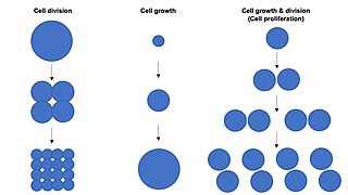
Cell growth refers to an increase in the total mass of a cell, including both cytoplasmic, nuclear and organelle volume. Cell growth occurs when the overall rate of cellular biosynthesis is greater than the overall rate of cellular degradation.

An ovarian follicle is a roughly spheroid cellular aggregation set found in the ovaries. It secretes hormones that influence stages of the menstrual cycle. At the time of puberty, women have approximately 200,000 to 300,000 follicles, each with the potential to release an egg cell (ovum) at ovulation for fertilization. These eggs are developed once every menstrual cycle with around 450–500 being ovulated during a woman's reproductive lifetime.
Carcinogenesis, also called oncogenesis or tumorigenesis, is the formation of a cancer, whereby normal cells are transformed into cancer cells. The process is characterized by changes at the cellular, genetic, and epigenetic levels and abnormal cell division. Cell division is a physiological process that occurs in almost all tissues and under a variety of circumstances. Normally, the balance between proliferation and programmed cell death, in the form of apoptosis, is maintained to ensure the integrity of tissues and organs. According to the prevailing accepted theory of carcinogenesis, the somatic mutation theory, mutations in DNA and epimutations that lead to cancer disrupt these orderly processes by interfering with the programming regulating the processes, upsetting the normal balance between proliferation and cell death. This results in uncontrolled cell division and the evolution of those cells by natural selection in the body. Only certain mutations lead to cancer whereas the majority of mutations do not.

Cyclin B is a member of the cyclin family. Cyclin B is a mitotic cyclin. The amount of cyclin B and the activity of the cyclin B-Cdk complex rise through the cell cycle until mitosis, where they fall abruptly due to degradation of cyclin B. The complex of Cdk and cyclin B is called maturation promoting factor or mitosis promoting factor (MPF).
In cell biology, contact inhibition refers to two different but closely related phenomena: contact inhibition of locomotion (CIL) and contact inhibition of proliferation (CIP). CIL refers to the avoidance behavior exhibited by fibroblast-like cells when in contact with one another. In most cases, when two cells contact each other, they attempt to alter their locomotion in a different direction to avoid future collision. When collision is unavoidable, a different phenomenon occurs whereby growth of the cells of the culture itself eventually stops in a cell-density dependent manner.

Aurora kinase A also known as serine/threonine-protein kinase 6 is an enzyme that in humans is encoded by the AURKA gene.

Antigen Kiel 67, also known as Ki-67 or MKI67, is a protein that in humans is encoded by the MKI67 gene.
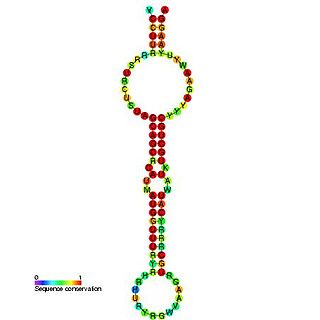
The miR-15 microRNA precursor family is made up of small non-coding RNA genes that regulate gene expression. The family includes the related mir-15a and mir-15b sequences, as well as miR-16-1, miR-16-2, miR-195 and miR-497. These six highly conserved miRNAs are clustered on three separate chromosomes. In humans miR-15a and miR-16 are clustered within 0.5 kilobases at chromosome position 13q14. This region has been found to be the most commonly affected in chronic lymphocytic leukaemia (CLL), with deletions of the entire region in more than half of cases. Both miR-15a and miR-16 are thus frequently deleted or down-regulated in CLL samples with 13q14 deletions; occurring in more than two thirds of CLL cases. The expression of miR-15a is associated with survival in triple negative breast cancer.

A mitotic inhibitor, microtubule inhibitor, or tubulin inhibitor, is a drug that inhibits mitosis, or cell division, and is used in treating cancer, gout, and nail fungus. These drugs disrupt microtubules, which are structures that pull the chromosomes apart when a cell divides. Mitotic inhibitors are used in cancer treatment, because cancer cells are able to grow through continuous division that eventually spread through the body (metastasize). Thus, cancer cells are more sensitive to inhibition of mitosis than normal cells. Mitotic inhibitors are also used in cytogenetics, where they stop cell division at a stage where chromosomes can be easily examined.
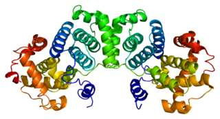
G2/mitotic-specific cyclin-B1 is a protein that in humans is encoded by the CCNB1 gene.

SCL-interrupting locus protein is a protein that in humans is encoded by the STIL gene. STIL is present in many different cell types and is essential for centriole biogenesis. This gene encodes a cytoplasmic protein implicated in regulation of the mitotic spindle checkpoint, a regulatory pathway that monitors chromosome segregation during cell division to ensure the proper distribution of chromosomes to daughter cells. The protein is phosphorylated in mitosis and in response to activation of the spindle checkpoint, and disappears when cells transition to G1 phase. It interacts with a mitotic regulator, and its expression is required to efficiently activate the spindle checkpoint.
Proliferation, as one of the hallmarks and most fundamental biological processes in tumors, is associated with tumor progression, response to therapy, and cancer patient survival. Consequently, the evaluation of a tumor proliferative index has clinical significance in characterizing many solid tumors and hematologic malignancies. This has led investigators to develop different technologies to evaluate the proliferation index in tumor samples. The most commonly used methods in evaluating a proliferative index include mitotic indexing, thymidine-labeling index, bromodeoxyuridine assay, the determination of fraction of cells in various phases of cell cycle, and the immunohistochemical evaluation of cell cycle-associated proteins.
Breast cancer classification divides breast cancer into categories according to different schemes criteria and serving a different purpose. The major categories are the histopathological type, the grade of the tumor, the stage of the tumor, and the expression of proteins and genes. As knowledge of cancer cell biology develops these classifications are updated.

Mitotic catastrophe has been defined as either a cellular mechanism to prevent potentially cancerous cells from proliferating or as a mode of cellular death that occurs following improper cell cycle progression or entrance. Mitotic catastrophe can be induced by prolonged activation of the spindle assembly checkpoint, errors in mitosis, or DNA damage and operates to prevent genomic instability. It is a mechanism that is being researched as a potential therapeutic target in cancers, and numerous approved therapeutics induce mitotic catastrophe.
Amitosis, also known as karyostenotic, direct cell division, or binary fission, represents a mode of asexual cell division predominantly observed in prokaryotes. This process is distinct from other cell division mechanisms such as mitosis and meiosis, primarily because it bypasses the complexities associated with the mitotic apparatus, such as spindle formation. Additionally, amitosis does not involve the condensation of chromatin into distinct chromosomes before the division of the cell occurs, simplifying the process of cellular replication.
A somatic mutation is a change in the DNA sequence of a somatic cell of a multicellular organism with dedicated reproductive cells; that is, any mutation that occurs in a cell other than a gamete, germ cell, or gametocyte. Unlike germline mutations, which can be passed on to the descendants of an organism, somatic mutations are not usually transmitted to descendants. This distinction is blurred in plants, which lack a dedicated germline, and in those animals that can reproduce asexually through mechanisms such as budding, as in members of the cnidarian genus Hydra.
https://heimduo.org/which-tissues-have-the-highest-rate-of-mitosis/
| | This cell biology article is a stub. You can help Wikipedia by expanding it. |
