Mitrofanoff procedure

Last updated
Mitrofanoff procedure
Mitrofanoff.jpg
The appendix is used in the Mitrofanoff procedure. The appendix is used to connect the umbilicus to the urinary bladder. The blood supply of the appendix is preserved. [1]
Other namesMitrofanoff appendicovesicostomy

The Mitrofanoff procedure, also known as the Mitrofanoff appendicovesicostomy, is a surgical procedure in which the appendix is used to create a conduit, or channel, between the skin surface and the urinary bladder. [2] The small opening on the skin surface, or the stoma, is typically located either in the navel or nearby the navel on the right lower side of the abdomen. [3] Originally developed by Professor Paul Mitrofanoff in 1980, the procedure represents an alternative to urethral catheterization [4] and is sometimes used by people with urethral damage or by those with severe autonomic dysreflexia. [5] An intermittent catheter, or a catheter that is inserted and then removed after use, is typically passed through the channel every 3–4 hours [6] and the urine is drained into a toilet or a bottle. [6] As the bladder fills, rising pressure compresses the channel against the bladder wall, creating a one-way valve that prevents leakage of urine between catheterizations. [3]

Contents

Description of procedure

A surgeon creates a small channel using the appendix or in the absence of the appendix, a piece of small bowel. [3] When bowel is used instead of appendix, it is called a Monti procedure. [7] One end of the channel is sewn to the skin, creating an opening on the surface called a stoma. [3] The other end of the channel is sewn to the bladder and a flap valve of tissue is created to prevent leakage from the stoma between catheterizations. [3] Sometimes, the bladder is enlarged with bowel tissue to enable greater urine storage in an additional procedure called bladder augmentation. [3] The Mitrofanoff procedure is different from an indwelling catheter placement because the catheter is removed from the channel between urine drainage events. [5] Some people with Mitrofanoff channels can also void urethrally, while others catheterize exclusively.

Relation to MACE

The Malone antegrade continence enema (MACE), used to treat fecal incontinence, is like the Mitrofanoff procedure as it uses the Mitrofanoff principle and, thus, can be considered an analogous procedure. [8] As fecal and urinary incontinence frequently co-exist, a MACE is often created at the same time as a continent catheterizable urinary conduit. [8]

Relation to Monti procedure

If the appendix is not available, due to appendectomy, or unusable for another reason, the Monti procedure is done. [9]

Relation to bladder augmentation and neobladder construction

If the bladder is not sufficiently large, some people may need a bladder augmentation at the same time as a Mitrofanoff. [10] Augmentation enlarges the bladder, making it possible to hold more urine and prevent backflow into the kidneys. [10] This is usually done with one's own bowel tissue and typically bowel tissue produces mucus. [10] Hence, regular washouts are usually required. [11] Because bowel tissue aids in absorption, its use for an augmentation may result in metabolic imbalance and result in the need to monitor vitamin B12, bicarbonate, and chloride. [11] If bowel tissue is used to create an entirely new bladder, the resulting reservoir to hold urine is called a neobladder. [12] Neobladders are usually created in instances of bladder removal. [11]

History

The concept of clean intermittent catheterization via the urethra was widely introduced by Jack Lapides when he published a seminal paper on the subject in 1972. [13] Clean intermittent catheterization provides an alternative to the sterile technique and allows individuals to self-catheterize after washing their hands, without the need for medical professionals and sterile equipment. [13] In 1980, Professor Paul Mitrofanoff described a "trans-appendicular continent cystostomy," the technique that would later be named for him. [14] Mitrofanoff's concept revolutionized clean intermittent catheterization because it allows urine to be drained via a route other than the urethra. [14] However, the Mitrofanoff procedure was slow to be adopted until a pediatric resident named Marc Cendron translated Mitrofanoff's French language paper for the well-known pediatric urologist Dr. John Duckett Jr. [15] in Philadelphia. [14] The Mitrofanoff procedure is sometimes performed along with bladder neck closure, [16] but Duckett advised against the closure of the bladder neck. [14] Today, the Mitrofanoff procedure can be performed robotically or using laparoscopic techniques and it paved the way for the creation of other urinary conduits using fallopian tubes, ureters, and segments of bowel, as in the Monti procedure. [14]

Indications

The Mitrofanoff procedure is typically performed as an alternative for people who experience painful urethral catheterization and has been particularly useful for females. [5] It is also used in people with neurogenic bladder dysfunction, urethral trauma, and spinal cord injuries. [5] The procedure is sometimes recommended for those with spinal cord injuries who have severe autonomic dysreflexia. [5] Wheelchair users who cannot use a toilet independently or who struggle to catheterize independently may get a Mitrofanoff to gain greater control over their care. [17] For people who would otherwise leak via the urethra, the Mitrofanoff channel can provide continence and enable them to stop using diapers. [17] Other conditions for which the procedure may be appropriate include urethral cancer, congenital absence of a urethra, Prune Belly syndrome, sacral agenesis, and traumatic loss of urethra from a gunshot. [18] Appropriate candidates are prepared to commit to a lifetime of followup care. [18]

Contraindications

People who have high pressure bladders, meaning their bladder pressure increases significantly with small increments of fluid, are not good candidates for the procedure due to the risk of damage to the kidneys. [19] Also contraindicated are those who cannot hold large volumes of urine without refluxing into the kidneys. [3] The procedure is not recommended for people with poor hand function, especially those without access to reliable assistance. [13]

Presurgical testing

People who have been determined to be candidates for a Mitrofanoff surgery will need to undergo a variety of presurgical tests. [20] Testing often includes blood tests such as a complete blood count, a basic metabolic panel, and an assessment of cystatin c. [20] Additionally, urodynamic testing and a kidney bladder ultrasound are typically performed to assess characteristics of the urinary tract prior to surgery. [20] The urodynamics test should be done within 2 years prior to the procedure and the ultrasound within 1 year. [20] Prior to surgery, the bowels are typically cleared with a routine called bowel prep. [20] Bowel prep can be performed at home the 1–2 days before surgery or in some instances, occurs in a hospital before the operation. [20] Bowel prep may require magnesium citrate drink to empty the colon. [21] Bowel prep is done to reduce infection risk. [22]

Recovery from surgery and life with a Mitrofanoff

Hospitalization and learning to use the channel

The Mitrofanoff procedure is a major surgery and typically requires inpatient hospitalization for 5–7 days. [23] Initially, eating and drinking by mouth is not permitted and fluids are delivered intravenously for a few days. [24] Progression to a regular diet can be accomplished, starting with the consumption of clear fluids. [24] After the surgery, a tube is placed in the newly created channel to keep it open while it heals. [25] A tube is typically also placed in the urethra or through a suprapubic opening to ensure full urine drainage and to rest the bladder during recovery. [25] The tubes are generally removed and the channel is ready to use with intermittent catheters in 4–6 weeks, [25] provided that a medical professional first instructs on how to catheterize. [23] Depending on one's neurological status, a person with a Mitrofanoff may or may not feel the sensation to urinate. [26] Full recovery time varies from person to person and ranges from 3–12 months. [27]

Types of catheters

There are three major types of intermittent catheters used with a Mitrofanoff. [28] Intermittent catheters are used to drain the bladder at regular intervals. [28] The three types are:

Dietary considerations

People with Mitrofanoff channels should eat a balanced diet that is high in fiber, including a recommended 5 servings of fruits and vegetables daily. [29] Drinking plenty of fluids is recommended to flush out the kidneys and bladder and to lessen the chance of urinary tract infection. [29] Recommended fluid intake is 2-3 liters of water a day, resulting in the production of healthy urine that is light in color. [29] Most people with Mitrofanoff channels can drink alcohol in moderation provided that they follow a regular catheterization schedule to avoid overfilling the bladder. [29]

Sex and pregnancy

Sexual activity is typically avoided for six weeks postoperatively. It is possible to have a healthy pregnancy after Mitrofanoff surgery, but the pregnancy should be monitored closely by a urologist. A cesarean section may be considered. In individuals with a conduit made from bowel tissue, a standard pregnancy test will not be accurate in most instances; pregnancy can instead be confirmed by a blood test. Male fertility is typically unaffected. [25] [5]

Exercise and physical activity

For 2–3 weeks after surgery, extra care should be taken to prevent trauma to the surgical area. [30] For this time period, physical education and activities such as bicycle riding are not advisable. [30] It is possible to play sports with a Mitrofanoff, but high contact sports such as rugby are typically not advised. [25] Light exercise following surgery can facilitate recovery; though specific exercise recommendations generally require consultation with a medical professional. [29]

Bladder washouts

Bladder washouts are performed to prevent build up of mucus and debris that can lead to urinary tract infection and increase the chance of a bladder stone. [31] Bladder stones can stop the bladder from emptying completely during catheterization and cause infection. [26] Those with an augmented bladder are more likely than those with a native bladder to require washouts. [26] A washout, also called an irrigation, [31] is performed by pushing saline or sterile water into the channel using a syringe connected to a catheter. [31] The water is pulled back out when the syringe is withdrawn and the process is repeated until the mucus is cleared. [31] Certain foods and drinks such as dairy products and soft drinks (soda) can increase mucus production. [31] The frequency of bladder washouts is dictated by medical advice. [31]

Annual follow up testing

People with Mitrofanoff channels can expect a lifetime of annual testing to evaluate their urological health. [5] These tests may include:

Risks and complications

Every surgery has some risks. Some possible complications are:

Related Research Articles

<span class="mw-page-title-main">Urology</span> Medical specialty

Urology, also known as genitourinary surgery, is the branch of medicine that focuses on surgical and medical diseases of the urinary-tract system and the reproductive organs. Organs under the domain of urology include the kidneys, adrenal glands, ureters, urinary bladder, urethra, and the male reproductive organs.

<span class="mw-page-title-main">Urethra</span> Tube that connects the urinary bladder to the external urethral orifice

The urethra is a tube that connects the urinary bladder to the urinary meatus for the removal of urine from the body of both females and males. In human females and other primates, the urethra connects to the urinary meatus above the vagina, whereas in marsupials, the female's urethra empties into the urogenital sinus.

<span class="mw-page-title-main">Cystoscopy</span> Medical procedure; endoscopy of the urinary bladder via the urethra

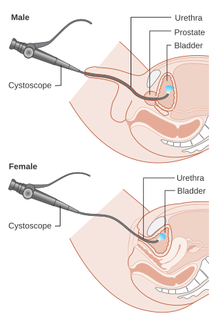
Cystoscopy is endoscopy of the urinary bladder via the urethra. It is carried out with a cystoscope.

<span class="mw-page-title-main">Urinary catheterization</span> Insertion of a catheter through the urethra to drain urine

In urinary catheterization, a latex, polyurethane, or silicone tube known as a urinary catheter is inserted into the bladder through the urethra to allow urine to drain from the bladder for collection. It may also be used to inject liquids used for treatment or diagnosis of bladder conditions. A clinician, often a nurse, usually performs the procedure, but self-catheterization is also possible. A catheter may be in place for long periods of time or removed after each use.

<span class="mw-page-title-main">Urethral stricture</span> Medical condition

A urethral stricture is a narrowing of the urethra, the tube connected to the bladder that allows the passing of urine. The narrowing reduces the flow of urine and makes it more difficult or even painful to empty the bladder.

<span class="mw-page-title-main">Urinary retention</span> Inability to completely empty the bladder

Urinary retention is an inability to completely empty the bladder. Onset can be sudden or gradual. When of sudden onset, symptoms include an inability to urinate and lower abdominal pain. When of gradual onset, symptoms may include loss of bladder control, mild lower abdominal pain, and a weak urine stream. Those with long-term problems are at risk of urinary tract infections.

<span class="mw-page-title-main">Foley catheter</span> Medical device

In urology, a Foley catheter is a brand name for one of many brands of urinary catheters (UC). Foleys and their namesakes are indwelling UC, often referred to as an IDCs or the alternative type being an in/out catheters. The foley UC was named after Frederic Foley, who produced the original design in 1929. The UC is a flexible tube if it is indwelling and stays put, or rigid if it is in/out, that a clinician, or the client themselves, often in the case of in/out UC, passes it through the urethra and into the bladder to drain urine.

<span class="mw-page-title-main">Urostomy</span>

A urostomy is a surgical procedure that creates a stoma for the urinary system. A urostomy is made to avail for urinary diversion in cases where drainage of urine through the bladder and urethra is not possible, e.g. after extensive surgery or in case of obstruction.

An Indiana pouch is a surgically-created urinary diversion used to create a way for the body to store and eliminate urine for patients who have had their urinary bladders removed as a result of bladder cancer, pelvic exenteration, bladder exstrophy or who are not continent due to a congenital, neurogenic bladder. This particular urinary diversion results in a continent reservoir that the patient must catheterize to empty urine. This concept and technique was developed by Drs. Mike Mitchell, Randall Rowland, and Richard Bihrle at Indiana University.

A urethrotomy is an operation which involves incision of the urethra, especially for relief of a stricture. It is most often performed in the outpatient setting, with the patient (usually) being discharged from the hospital or surgery center within six hours from the procedure's inception.

<span class="mw-page-title-main">Posterior urethral valve</span> Medical condition

Posterior urethral valve (PUV) disorder is an obstructive developmental anomaly in the urethra and genitourinary system of male newborns. A posterior urethral valve is an obstructing membrane in the posterior male urethra as a result of abnormal in utero development. It is the most common cause of bladder outlet obstruction in male newborns. The disorder varies in degree, with mild cases presenting late due to milder symptoms. More severe cases can have renal and respiratory failure from lung underdevelopment as result of low amniotic fluid volumes, requiring intensive care and close monitoring. It occurs in about one in 8,000 babies.

In urology, voiding cystourethrography (VCUG) is a frequently performed technique for visualizing a person's urethra and urinary bladder while the person urinates (voids). It is used in the diagnosis of vesicoureteral reflux, among other disorders. The technique consists of catheterizing the person in order to fill the bladder with a radiocontrast agent, typically diatrizoic acid. Under fluoroscopy the radiologist watches the contrast enter the bladder and looks at the anatomy of the patient. If the contrast moves into the ureters and back into the kidneys, the radiologist makes the diagnosis of vesicoureteral reflux, and gives the degree of severity a score. The exam ends when the person voids while the radiologist is watching under fluoroscopy. Consumption of fluid promotes excretion of contrast media after the procedure. It is important to watch the contrast during voiding, because this is when the bladder has the most pressure, and it is most likely this is when reflux will occur. Despite this detailed description of the procedure, at least as of 2016 the technique had not been standardized across practices.

<span class="mw-page-title-main">Ureteral stent</span>

A ureteral stent, or ureteric stent, is a thin tube inserted into the ureter to prevent or treat obstruction of the urine flow from the kidney. The length of the stents used in adult patients varies between 24 and 30 cm. Additionally, stents come in differing diameters or gauges, to fit different size ureters. The stent is usually inserted with the aid of a cystoscope. One or both ends of the stent may be coiled to prevent it from moving out of place; this is called a JJ stent, double J stent or pig-tail stent.

Neurogenic bladder dysfunction, or neurogenic bladder, refers to urinary bladder problems due to disease or injury of the central nervous system or peripheral nerves involved in the control of urination. There are multiple types of neurogenic bladder depending on the underlying cause and the symptoms. Symptoms include overactive bladder, urinary urgency, frequency, incontinence or difficulty passing urine. A range of diseases or conditions can cause neurogenic bladder including spinal cord injury, multiple sclerosis, stroke, brain injury, spina bifida, peripheral nerve damage, Parkinson's disease, or other neurodegenerative diseases. Neurogenic bladder can be diagnosed through a history and physical as well as imaging and more specialized testing. Treatment depends on underlying disease as well as symptoms and can be managed with behavioral changes, medications, surgeries, or other procedures. The symptoms of neurogenic bladder, especially incontinence, can have a significant impact on quality of life.

Urethroplasty is the surgical repair of an injury or defect within the walls of the urethra. Trauma, iatrogenic injury and infections are the most common causes of urethral injury/defect requiring repair. Urethroplasty is regarded as the gold standard treatment for urethral strictures and offers better outcomes in terms of recurrence rates than dilatations and urethrotomies. It is probably the only useful modality of treatment for long and complex strictures though recurrence rates are higher for this difficult treatment group.

<span class="mw-page-title-main">Retrograde urethrogram</span> Medical imaging of the urethra

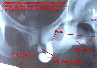
A retrograde urethrography is a routine radiologic procedure used to image the integrity of the urethra. Hence a retrograde urethrogram is essential for diagnosis of urethral injury, or urethral stricture.

<span class="mw-page-title-main">Urethrostomy</span>

Urethrostomy is a surgical procedure that creates a permanent opening in the urethra, commonly to remove obstructions to urine flow. The procedure is most often performed in male cats, where the opening is made in the perineum.

Bladder outlet obstruction occurs when urine is unable to flow from the kidneys through the ureters and out of the bladder through the urethra. Decreased flow of urine leads to swelling of the urinary tract, called hydronephrosis. This process of decreased flow of urine through the urinary tract can begin as early as during intrauterine life and it prevents normal development of fetal kidneys and fetal urine. Low levels of fetal urine leads to low amniotic fluid levels and incomplete lung maturation. Older children and adults can also experience bladder outlet obstruction; however, this process is usually reversible and isn't associated with as many poor outcomes as in infants with congenital bladder outlet obstruction.

Suprapubic aspiration is a procedure to take a urine sample. It involves putting a needle through the skin just above the pubic bone into the bladder. It is typically used as a method to collect urine in child less than 2 years of age who is not yet toilet trained in an effort to diagnose a urinary tract infection.

The genitourinary tract, or simply the urinary tract, consists of the kidneys, ureters, bladder, and the urethra. The kidney is the most frequently injured. Injuries to the kidney commonly occur after automobile or sports-related accidents. A blunt force is involved in 80-85% of injuries. Major decelerations can result in vascular injuries near the kidney's hilum. Gunshots and knife wounds and fractured ribs can result in penetrating injuries to the kidney.

References

  1. Breen, Micheál; Phelps, Andrew; Estrada, Carlos; Chow, Jeanne S. (2015-09-01). "The role of imaging in pediatric bladder augmentation". Pediatric Radiology. 45 (10): 1440–1447. doi:10.1007/s00247-015-3349-1. ISSN   1432-1998. PMID   25903841.
  2. Mingin GC, Baskin LS (2003). "Surgical management of the neurogenic bladder and bowel". Int Braz J Urol. 29 (1): 53–61. doi: 10.1590/S1677-55382003000100012 . PMID   15745470.
  3. 1 2 3 4 5 6 7 "Mitrofanoff Procedure". Columbia University Department of Urology. 2016-06-06. Retrieved 2020-03-28.
  4. "Mitrofanoff". Urostomy Association. Retrieved 2020-03-28.
  5. 1 2 3 4 5 6 7 8 9 10 11 12 "Mitrofanoff Procedure". Craig Hospital. Retrieved 2020-03-28.
  6. 1 2 "Mitrofanoff: Catheterization and care". www.aboutkidshealth.ca. Retrieved 2020-03-28.
  7. Levy, Mya E.; Elliott, Sean P. (2016). "Reconstructive techniques for creation of catheterizable channels: tunneled and nipple valve channels". Translational Andrology and Urology. 5 (1): 136–144. doi:10.3978/j.issn.2223-4683.2016.01.04. ISSN   2223-4691. PMC   4739983 . PMID   26904419.
  8. 1 2 Teichman JM, Rogenes VJ, Barber DB (1997). "The utility of the Malone antegrade continence enema for urologists". Tech Urol. 3 (1): 30–3. PMID   9170222.
  9. Shapiro E (1999). "Early Clinical Experience With Monti Procedure and Managing the Nonpalpable Testis in Children". Rev Urol. 1 (2): 94–6. PMC   1476142 . PMID   16985779.
  10. 1 2 3 "Bladder Surgery: Augmentation with Mitrofanoff and Cecostomy" (PDF). Phoenix Children's Hospital. n.d.
  11. 1 2 3 4 5 6 "The Mitrofanoff Procedure". Mitrofanoff Support. Retrieved 2020-04-10.
  12. "Neobladder reconstruction - Mayo Clinic". www.mayoclinic.org. Retrieved 2020-04-26.
  13. 1 2 3 "Neurogenic Bladder". ngbladder.org. Retrieved 2020-04-10.
  14. 1 2 3 4 5 Farrugia, Marie Klaire; Malone, Padraig S. (25 February 2010). "Educational Article: The Mitrofanoff Procedure". Journal of Pediatric Urology. 6 (4): 330–337. doi:10.1016/j.jpurol.2010.01.015. PMID   20188633 via Science Direct.
  15. "Deaths: Duckett, Csiszar, Jackson". almanac.upenn.edu. Retrieved 2020-04-24.
  16. De Troyer, Bart; Van Laecke, Erik; Groen, Luitzen A.; Everaert, Karel; Hoebeke, Piet (April 2011). "A comparative study between continent diversion and bladder neck closure versus continent diversion and bladder neck reconstruction in children". Journal of Pediatric Urology. 7 (2): 209–212. doi:10.1016/j.jpurol.2010.03.011.
  17. 1 2 "What Is the Mitrofanoff Procedure (Appendicovesicostomy)?". www.nationwidechildrens.org. Retrieved 2020-04-05.
  18. 1 2 Barratt, Rachel; Marsden, Theresa; Greenwell, Tamsin J. (1 January 2018). "The Mitrofanoff procedure: a continent revolution". Urology News. 22: 2 via www.urologynews.com.
  19. "What is bladder pressure assessment?". www.medscape.com. Retrieved 2020-04-10.
  20. 1 2 3 4 5 6 "Urology Reconstruction v1.0: Pre-Operative Plan" (PDF). Seattle Children's Hospital. n.d. Retrieved 24 April 2020.
  21. "Two Day Bowel Preparation for Your Urology Surgery or Procedure". Memorial Sloan Kettering Cancer Center. 28 November 2018.
  22. "Bowel Preparation: Before Your Surgery". myhealth.alberta.ca. Retrieved 2020-04-26.
  23. 1 2 "Mitrofanoff Procedure | Gillette Children's Specialty Healthcare". www.gillettechildrens.org. Retrieved 2020-04-05.
  24. 1 2 "Mitrofanoff" (PDF). McMaster Children's Hospital. 17 July 2017. Retrieved 12 April 2020.
  25. 1 2 3 4 5 6 "Having a Mitrofanoff (creation of a catheterizable urinary stoma)" (PDF). Queen Elizabeth Hospital Birmingham. Retrieved 5 April 2020.
  26. 1 2 3 "FAQs". Mitrofanoff Support. Retrieved 2020-04-05.
  27. "The Operation and Recovery". Mitrofanoff Support. Retrieved 2020-04-05.
  28. 1 2 3 4 5 "Useful information". Mitrofanoff Support. Retrieved 2020-04-20.
  29. 1 2 3 4 5 "Healthy Living". Mitrofanoff Support. Retrieved 2020-04-10.
  30. 1 2 3 "Postoperative Care Instructions for Mitrofanoff". CHOC Children's. Retrieved 2020-04-10.
  31. 1 2 3 4 5 6 "Bladder Washouts". Mitrofanoff Support. Retrieved 2020-04-10.
  32. 1 2 "Bladder stones - Symptoms and causes". Mayo Clinic. Retrieved 2020-04-10.
  33. "Bladder Control Medicines | NIDDK". National Institute of Diabetes and Digestive and Kidney Diseases. Retrieved 2020-04-10.