Mycoplasma orale

Last updated

Mycoplasma orale
Scientific classification Red Pencil Icon.png
Domain: Bacteria
Phylum: Mycoplasmatota
Class: Mollicutes
Order: Mycoplasmatales
Family: Mycoplasmataceae
Genus: Mycoplasma
Species:
M. orale
Binomial name
Mycoplasma orale
Taylor-Robinson et al. 1964

Mycoplasma orale is a small bacterium found in the class Mollicutes. [1] It belongs to the genus Mycoplasma , a well-known group of obligate intracellular parasites that inhabit humans. [2] It also is known to be an opportunistic pathogen in immunocompromised humans. [3] As with other Mycoplasma species, M. orale is not readily treated with many antibiotics due to its lack of a peptidoglycan cell wall. [4] Therefore, this species is relevant to the medical field as physicians face the task of treating patients infected with this microbe. It is characterized by a small physical size (0.1 micrometer in diameter on average), a small genome size, and a limited metabolism. [4] [5] [2] It is also known to frequently contaminate laboratory experiments. [5] This bacteria is very similar physiologically and morphologically to its sister species within the genus Mycoplasma; however, its recent discovery leaves many questions still unanswered about this microbe.

Contents

Discovery and isolation

Taylor-Robinson et al. identified and named M. orale in 1964 after isolating it from the oral cavity of a child at a Washington hospital and three servicemen stationed in North Carolina. [6] Taylor-Robinson et al. cultured the organism under anaerobic conditions on PPLO agar and broth. [6] The resulting colony growth had a "fried egg" appearance when cultured on solid PPLO agar. [6] PPLO is stands for "Pleuropneumonia-like organisms" and was originally designed for growth of parasitic bacteria found in the respiratory tract of cattle; it is now known that these bacteria were Mycoplasma species. [7] PPLO agar is used for isolation and growth of Mycoplasma and Ureaplasma species; it contains beef heart infusion, yeast extract, phenol red, sodium chloride, the antibiotic Polymyxin B, the antifungal Amphotericin B, the antibiotic Penicillin, deionized water, and horse serum. [7] Taylor-Robinson et al. confirmed M. orale was distinct from previously discovered Mycoplasma species with a serological test using haemagglutination of sheep erythrocytes, tannic acid and sonnicated Mycoplasma extracts. [6]

L.G. Tallgren et al. also isolated M.orale in 1974 from a sample of bone marrow taken from a three-year-old boy with Eosinophilic leukaemia. [8] The sample was obtained via a bone marrow biopsy performed at Aurora Hospital in Helsinki, Finland. [8] L.G. Tallgren et al. noted this organism had a typical Mycoplasma "fried egg" appearance when cultured on solid media. [8]

Taxonomic and phylogenetic classification

As determined by 16S rRNA sequencing, M. orale is a bacterium belonging to the genus Mycoplasma. [2] Its closest relative within the genus is M. salivarius, while its most distant is M. mycoides. [2] M. orale and M. salivarius have both been observed in the human oral cavity, evidencing their close proximity in the genus Mycoplasma. [5]

Of the over 100 documented species in the genus Mycoplasma, 14 are known pathogens to humans. [1] This comes as no surprise, as the class Mollicutes contains common commensals or pathogens of several different organisms. [1] Many species in the genus Mycoplasma are commonly found associated with pelvic or genital region infections including M. fermentans and M. hominis. [9] Other species in this genus are the causative agents of respiratory related infections; these species include the well-known Mycoplasma pneumoniae . [10] The family Mycoplastaceae includes the genera Mycoplasma and Ureaplasma. [11] Bacteria in the genus Ureaplasma are known commensals in humans and possess the enzyme Urease (catalyzes urea to carbon dioxide and ammonia). [11] Both Mycoplasma and Ureaplasma species can be grown on the same PPLO medium due to their similarities in metabolism and growth requirement. [7]

Parasitism and genomics

While the genome of M. orale itself has not been fully sequenced, information can be surmised from the sequence data of its close relatives. [12] [2] Members of the genus Mycoplasma are known for their incredibly small genomes, with an average size of 0.6 Mb. [2] [13] This is the smallest discovered self-replicating genome of all known prokaryotes. [2] This significantly reduced genome size is thought to be the result of the taxon's evolution into obligate parasites. [14] Mycoplasma species typically invade and adhere to host cells from which they obtain their nutrients, usually at the expense of the host. [5] All members of this genus, including M. orale, inhabit a wide range of mammalian hosts. [15] Though M. orale usually exists as a commensal in human oral cavities, it is an opputunistic pathogen and will cause illness in human hosts when conditions are right. [3] Due to their small genome size and parasitic lifecycle, they lack many non-essential biosynthetic pathways in their metabolism. [2] These include those for cell wall synthesis as well as purine synthesis. [2] These genomic characteristics make them a good model for the Minimal Genome Concept. [2] Mycoplasma members, including M. orale, also generally have a low G-C content compared to other bacteria. [16] Research has revealed that the 16S rRNA gene of M. orale is about 1,510 bp. [13]

Growth: metabolism and reproduction

As stated previously, M. orale is an obligate parasite found in humans and primates according to the GIDEON Guide of medically relevant bacteria. [15] This evolution into parasitism has resulted in the loss of many functional metabolic pathways. [9] Therefore, these organisms are unable to grow and reproduce in culture without the necessary nutrients and metabolites present in media. [17] M. orale is cultured using a special medium, the 1076b. SP4-Z MEDIUM. [12] This medium contains either glucose or arginine (but not both at the same time) as a carbon source; however, it does not contain yeast extract. [18] For exact information regarding all components of this medium see "DSMZ Medium" in External Links. [18] It is considered to be a mesophilic bacteria growing best at a temperature of 37 °C. [12] This is expected, as theoretically it should grow best at the basal temperatures of its host. This organism was also found to grow optimally at pH 6.0 and had phosphatase activity at .2 Ual/Mg of protein. [19] Unlike other Mycoplasmas, M. orale does not have the ability to ferment glucose. [6] It is also unable to aerobically reduce 2-3-5 triphenyl tetrazolium. [14] This demonstrated its close phylogenetic relationship to other Mycoplasma species which inhabit human hosts. [14] M. orale was observed to divide in two ways: by either binary fission or by forming mycelial filaments in colonies. [17] Binary fission was observed deeper within colony growth while mycelial growth was found to occur more toward the air interface. [17] Thus, it was inferred that mycelial growth is associated with aerobic conditions, while growth by binary fission is associated with a more anaerobic environment. [17]

Medical relevance

Mycoplasma orale is considered to be a non-pathogenic commensal, especially in immunocompetent individuals. [3] However, abscesses containing M. orale have been noted in patients who are immunocompromised. [3] A study was conducted on bacterial samples taken from a 33-year-old immunocompromised male. [3] This individual presented with fever, increased weight loss, and shoulder pain among other pathologies. [3] Samples were obtained from the patient and grown on media for testing. [3] 16S rRNA sequencing was completed and revealed the culprit of the infection, M. orale. [3] This revealed the ability of this organism to cause pathology in humans. [3] Infections with Mycoplasma members, including M. orale, can be difficult to treat with antibiotics due to their lack of a cell wall. [4] This lack of peptidoglycan confers resistance to any antibiotics that may target its synthesis, including the commonly used penicillin and its derivatives as well as vancomycin. [20] This presents a challenge to physicians as they try to treat their patients with bacterial Mycoplasma infections. [20]

Lab contamination

Mycoplama species are commonly found in labs as contaminants. Beginning in 1956 through the early 2000s, various organizations including Johns Hopkins, US Food and Drug Administration and the Deutsche Sammlung von Mikroorganismen und Zellkulturen (DSMZ) in Germany, among others, have reported varying degrees of Mycoplasma contamination ranging from 15%-70% of cell cultures. [5] Mycoplasma contaminations are problematic because they are difficult to prevent and negatively impact host cell growth. [5] Contamination is common due to their small physical size, lack of cell wall, and pleomorphism. [5] Pleomorphism is defined as an organism's ability to modify their shape and size as a response to changes in their environment. [21] Pleomorphism in Mycoplasma species allows bypassing of typical membrane filtration systems. [5] As previous stated, their lack of a cell wall prevents the use of antibiotics to inhibit unwanted cell growth on cultured media. [5] In addition to culture contamination, Mycoplasma contaminating cell cultures can have a negative effect on host cell growth by stealing nutrients from host cells. [5] M. orale has been found to inhibit host cell growth by outcompeting the host cell for arginine. [5] It is important to prevent contamination of Mycoplasma for reliable and accurate laboratory research results. [5]

Related Research Articles

<i>Mycoplasma genitalium</i> Species of bacterium

Mycoplasma genitalium is a sexually transmitted, small and pathogenic bacterium that lives on the mucous epithelial cells of the urinary and genital tracts in humans. Medical reports published in 2007 and 2015 state that Mgen is becoming increasingly common. Resistance to multiple antibiotics is becoming prevalent, including to azithromycin, which until recently was the most reliable treatment. The bacteria was first isolated from the urogenital tract of humans in 1981, and was eventually identified as a new species of Mycoplasma in 1983. It can cause negative health effects in men and women. It also increases the risk factor for HIV spread with higher occurrences in those previously treated with the azithromycin antibiotics.

<span class="mw-page-title-main">Infection</span> Invasion of an organisms body by pathogenic agents

An infection is the invasion of tissues by pathogens, their multiplication, and the reaction of host tissues to the infectious agent and the toxins they produce. An infectious disease, also known as a transmissible disease or communicable disease, is an illness resulting from an infection.

<i>Mycoplasma</i> Genus of bacteria

Mycoplasma is a genus of bacteria that, like the other members of the class Mollicutes, lack a cell wall around their cell membranes. Peptidoglycan (murein) is absent. This characteristic makes them naturally resistant to antibiotics that target cell wall synthesis. They can be parasitic or saprotrophic. Several species are pathogenic in humans, including M. pneumoniae, which is an important cause of "walking" pneumonia and other respiratory disorders, and M. genitalium, which is believed to be involved in pelvic inflammatory diseases. Mycoplasma species are among the smallest organisms yet discovered, can survive without oxygen, and come in various shapes. For example, M. genitalium is flask-shaped, while M. pneumoniae is more elongated, many Mycoplasma species are coccoid. Hundreds of Mycoplasma species infect animals.

Mycoplasma hominis is a species of bacteria in the genus Mycoplasma. M. hominis has the ability to penetrate the interior of human cells. Along with ureaplasmas, mycoplasmas are the smallest free-living organisms known.

Mycoplasma pneumoniae is a very small bacterium in the class Mollicutes. It is a human pathogen that causes the disease mycoplasma pneumonia, a form of atypical bacterial pneumonia related to cold agglutinin disease. M. pneumoniae is characterized by the absence of a peptidoglycan cell wall and resulting resistance to many antibacterial agents. The persistence of M. pneumoniae infections even after treatment is associated with its ability to mimic host cell surface composition.

<i>Campylobacter jejuni</i> Species of bacterium

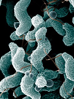
Campylobacter jejuni is one of the most common causes of food poisoning in Europe and in the US. The vast majority of cases occur as isolated events, not as part of recognized outbreaks. Active surveillance through the Foodborne Diseases Active Surveillance Network (FoodNet) indicates that about 20 cases are diagnosed each year for each 100,000 people in the US, while many more cases are undiagnosed or unreported; the CDC estimates a total of 1.5 million infections every year. The European Food Safety Authority reported 246,571 cases in 2018, and estimated approximately nine million cases of human campylobacteriosis per year in the European Union.

Ureaplasma urealyticum is a bacterium belonging to the genus Ureaplasma and the family Mycoplasmataceae in the order Mycoplasmatales. This family consists of the genera Mycoplasma and Ureaplasma. Its type strain is T960. There are two known biovars of this species; T960 and 27. These strains of bacterium are commonly found in the urogenital tracts of human beings, but overgrowth can lead to infections that cause the patient discomfort. Unlike most bacteria, Ureaplasma urealyticum lacks a cell wall making it unique in physiology and medical treatment.

Mollicutes is a class of bacteria distinguished by the absence of a cell wall. The word "Mollicutes" is derived from the Latin mollis, and cutis. Individuals are very small, typically only 0.2–0.3 μm in size and have a very small genome size. They vary in form, although most have sterols that make the cell membrane somewhat more rigid. Many are able to move about through gliding, but members of the genus Spiroplasma are helical and move by twisting. The best-known genus in the Mollicutes is Mycoplasma. Colonies show the typical "fried-egg" appearance.

Mycoplasmataceae is a family of bacteria in the order Mycoplasmatales. This family consists of the genera Mycoplasma and Ureaplasma.

<i>Aeromonas hydrophila</i> Species of heterotrophic, Gram-negative, bacterium

Aeromonas hydrophila is a heterotrophic, Gram-negative, rod-shaped bacterium mainly found in areas with a warm climate. This bacterium can be found in fresh or brackish water. It can survive in aerobic and anaerobic environments, and can digest materials such as gelatin and hemoglobin. A. hydrophila was isolated from humans and animals in the 1950s. It is the best known of the species of Aeromonas. It is resistant to most common antibiotics and cold temperatures and is oxidase- and indole-positive. Aeromonas hydrophila also has a symbiotic relationship as gut flora inside of certain leeches, such as Hirudo medicinalis.

Capnocytophaga canimorsus is a fastidious, slow-growing, Gram-negative rod of the genus Capnocytophaga. It is a commensal bacterium in the normal gingival flora of canine and feline species, but can cause illness in humans. Transmission may occur through bites, licks, or even close proximity with animals. C. canimorsus generally has low virulence in healthy individuals, but has been observed to cause severe, even grave, illness in persons with pre-existing conditions. The pathogenesis of C. canimorsus is still largely unknown, but increased clinical diagnoses have fostered an interest in the bacillus. Treatment with antibiotics is effective in most cases, but the most important yet basic diagnostic tool available to clinicians remains the knowledge of recent exposure to canines or felines.

<span class="mw-page-title-main">Bacteria</span> Domain of micro-organisms

Bacteria are ubiquitous, mostly free-living organisms often consisting of one biological cell. They constitute a large domain of prokaryotic microorganisms. Typically a few micrometres in length, bacteria were among the first life forms to appear on Earth, and are present in most of its habitats. Bacteria inhabit soil, water, acidic hot springs, radioactive waste, and the deep biosphere of Earth's crust. Bacteria are vital in many stages of the nutrient cycle by recycling nutrients such as the fixation of nitrogen from the atmosphere. The nutrient cycle includes the decomposition of dead bodies; bacteria are responsible for the putrefaction stage in this process. In the biological communities surrounding hydrothermal vents and cold seeps, extremophile bacteria provide the nutrients needed to sustain life by converting dissolved compounds, such as hydrogen sulphide and methane, to energy. Bacteria also live in symbiotic and parasitic relationships with plants and animals. Most bacteria have not been characterised and there are many species that cannot be grown in the laboratory. The study of bacteria is known as bacteriology, a branch of microbiology.

Ureaplasma parvum is a species of Ureaplasma, a genus of bacteria belonging to the family Mycoplasmataceae.

In biology, a pathogen in the oldest and broadest sense, is any organism or agent that can produce disease. A pathogen may also be referred to as an infectious agent, or simply a germ.

Parachlamydia acanthamoebae are bacterium that fall into the category of host-associated microorganisms. This bacterium lives within free-living amoebae that are an intricate part of their reproduction. Originally named Candidatus Parachlamydia acanthamoebae, its current scientific name was introduced shortly after. This species has shown to have over eighty percent 16S rRNA gene sequencing identity with the class Chlamydiia. Parachlamydia acanthamoebae has the same family as the genus Neochlamydia with which it shares many similarities.

Mycoplasma faucium is a species of bacteria in the genus Mycoplasma. This genus of bacteria lacks a cell wall around their cell membrane. Without a cell wall, they are unaffected by many common antibiotics such as penicillin or other beta-lactam antibiotics that target cell wall synthesis. Mycoplasma are the smallest bacterial cells yet discovered, can survive without oxygen and are typically about 0.1 µm in diameter.

Mycoplasma salivarium is a species of bacteria in the genus Mycoplasma. This genus of bacteria lacks a cell wall around their cell membrane. Without a cell wall, they are unaffected by many common antibiotics such as penicillin or other beta-lactam antibiotics that target cell wall synthesis. Mycoplasma are the smallest bacterial cells yet discovered, can survive without oxygen and are typically about 0. 1 μm in diameter. Mycoplasma salivarium is found in the mouths of 97% of the healthy population, and is generally considered to be a commensal organism and part of the normal oral flora.

Mycoplasma alligatoris is a species of bacteria in the genus Mycoplasma. It is classified in the family Mycoplasmataceae, order Mycoplasma, class Mollicutes, phylum Firmicutes and domain Bacteria. Many organisms of the genus Mycoplasma are known pathogens in humans and animal species. Mycoplasma alligatoris is known to elicit a fatal disease with inflammatory characteristics that can cause rapid death of alligators and caimans.

The exact role of Mycoplasma hominis in regards to a number of conditions related to pregnant women and their (unborn) offspring is controversial. This is mainly because many healthy adults have genitourinary colonization with Mycoplasma, published studies on pathogenicity have important design limitations and the organisms are very difficult to detect. The likelihood of colonization with M. hominis appears directly linked to the number of lifetime sexual partners Neonatal colonization does occur, but only through normal vaginal delivery. Caesarean section appears protective against colonization and is much less common. Neonatal colonization is transient.

<span class="mw-page-title-main">Uterine microbiome</span>

The uterine microbiome is the commensal, nonpathogenic, bacteria, viruses, yeasts/fungi present in a healthy uterus, amniotic fluid and endometrium and the specific environment which they inhabit. It has been only recently confirmed that the uterus and its tissues are not sterile. Due to improved 16S rRNA gene sequencing techniques, detection of bacteria that are present in low numbers is possible. Using this procedure that allows the detection of bacteria that cannot be cultured outside the body, studies of microbiota present in the uterus are expected to increase.

References

  1. 1 2 3 Tortoli, E. (2014). "Microbiological features and clinical relevance of new species of the genus Mycobacterium". Clinical Microbiology Reviews. 27 (4): 727–752. doi:10.1128/cmr.00035-14. PMC   4187642 . PMID   25278573.
  2. 1 2 3 4 5 6 7 8 9 10 Chambaud, I.; Heilig, R.; Ferris, S.; Barbe, V.; Samson, D.; Galisson, F.; Moszer, I.; Dybvig, K.; Wroblewski, H.; Viari, A.; Rocha, E.P.C.; Blanchard, A (2001). "The complete genome sequence of the murine respiratory pathogen Mycoplasma pulmonis". Nucleic Acids Research. 29 (10): 2145–2153. doi:10.1093/nar/29.10.2145. PMC   55444 . PMID   11353084.
  3. 1 2 3 4 5 6 7 8 9 Paessler, Michelle; Levinson, Arnold; Patel, Jean Baldus; Schuster, Mindy; Minda, Melanie; Nachamkin, Irving (2002-10-01). "Disseminated Mycoplasma orale infection in a patient with common variable immunodeficiency syndrome". Diagnostic Microbiology and Infectious Disease. 44 (2): 201–204. doi:10.1016/S0732-8893(02)00429-7. PMID   12458129.
  4. 1 2 3 Niederweis, M.; Danilchanka, O.; Huff, J.; Hoffman, C.; Engelhardt, H. (2010). "Mycobacterial outermembranes: in search of proteins". Trends in Microbiology. 18 (3): 109–116. doi:10.1016/j.tim.2009.12.005. PMC   2931330 . PMID   20060722.
  5. 1 2 3 4 5 6 7 8 9 10 11 12 Olarerin-George, A. & Hogenesch, J. (2015). "Assessing the prevalence of mycoplasma contamination in cell culture via a survey of NCBI's RNA-seq archive". Nucleic Acids Research. 43 (5): 2535–2542. doi:10.1093/nar/gkv136. PMC   4357728 . PMID   25712092.
  6. 1 2 3 4 5 Taylor-Robinson, D.; Canchola, J.; Chanock, R. M. (1964). "A Newly Identified Oral Mycoplasma (M. orale) and its Relationship to other Human Mycoplasmas". American Journal of Hygiene. 80 (1): 135–148. doi:10.1093/oxfordjournals.aje.a120454. PMID   14192765.
  7. 1 2 3 "PPLO (Mycoplasma) Media". Hardy Diagnostics.
  8. 1 2 3 Tallgren, L.G.; Wegelius, R.; Andersson, L.C. & Jansson, E. (1974). "Eosiniphilic Leukaemia—Recovery of Mycoplasma Orale from the Bone Marrow". Acta Medica Scandinavica. 195 (1–2): 87–92. doi:10.1111/j.0954-6820.1974.tb08102.x. PMID   4522224.
  9. 1 2 Razin, S.; Baron, S. (1996). "Mycoplasmas". Chapter 37: Mycoplasmas. In Medical Microbiology, 4th edition. Galveston, TX: University of Texas Medical Branch at Galveston. ISBN   9780963117212.
  10. "Mycoplasma pneumoniae". Center for Disease Control. Retrieved February 21, 2017.
  11. 1 2 Blanchard, A., Razin, S., Kenny, G. E., & Barile, M. F. (1988). "Characteristics of Ureaplasma urealyticum urease". Journal of Bacteriology. 170 (6): 2692–2697. doi:10.1128/jb.170.6.2692-2697.1988. PMC   211190 . PMID   3131306.{{cite journal}}: CS1 maint: multiple names: authors list (link)
  12. 1 2 3 "BacDrive: Mycoplasma orale".
  13. 1 2 "AY796060 Sequence Browser - StrainInfo". www.straininfo.net.
  14. 1 2 3 Fox, H.; Purcell, R. H.; Chanock, R. M. (1969). "Characterization of a Newly Identified Mycoplasma (Mycoplasma orale Type 3) from the Human Oropharynx". Journal of Bacteriology. 98 (1): 36–43. doi:10.1128/jb.98.1.36-43.1969. PMC   249900 . PMID   4976470.
  15. 1 2 Berger, Stephen (2014). GIDEON guide to medically important bacteria. Los Angeles, California: GIDEON Informatics Inc. ISBN   9781617558412.
  16. Boxer, Linda Minium; Korn, David (1979). "Structural and enzymological characterization of the homogeneous deoxyribonucleic acid polymerase from Mycoplasma orale". Biochemistry. 18 (21): 4742–4749. doi:10.1021/bi00588a039. ISSN   0006-2960.
  17. 1 2 3 4 NAKAMURA, M. & KAWAGUCHI, M. (1972). "Ultrastructure of Mycoplasma orale Serotype 1 in Agar Growth". Journal of General Microbiology. 70 (2): 305–314. doi: 10.1099/00221287-70-2-305 . PMID   5038879.
  18. 1 2 "DSMZ Microorganisms. 1076b. SP4-Z MEDIUM" (PDF). DSMZ. Retrieved February 21, 2017.
  19. Shibata, K.; Totsuka, M.; Watanabe, T. (1986). "Phosphatase Activity as a Criterion for Differentiation of Oral Mycoplasma". Journal of Clinical Microbiology. 23 (5): 970–972. doi:10.1128/jcm.23.5.970-972.1986. PMC   268765 . PMID   3011850.
  20. 1 2 Romaniuk, J. A. H.; Cegelski, L. (2015). "Bacterial cell wall composition and the influence of antibiotics by cell-wall and whole-cell NMR". Philosophical Transactions of the Royal Society B: Biological Sciences. 370 (1679): 20150024. doi:10.1098/rstb.2015.0024. PMC   4632600 . PMID   26370936.
  21. Joshi, Hiren M; Toleti, Rao S (2009-07-07). "Nutrition induced pleomorphism and budding mode of reproduction in Deinococcus radiodurans". BMC Research Notes. 2: 123. doi:10.1186/1756-0500-2-123. ISSN   1756-0500. PMC   2714317 . PMID   19583846.