N-localizer

Last updated
N-localizer
Photograph of Stereotactic Frame With 3 N-localizers.jpg
Three N-localizers attached to a stereotactic frame. [1]
Specialty neurosurgery, radiation oncology
Interventionstereotactic surgery, radiosurgery
Inventor(s) Russell A. Brown [2]

The N-localizer [3] is a device that enables guidance of stereotactic surgery or radiosurgery using tomographic images that are obtained via computed tomography (CT), [4] magnetic resonance imaging (MRI), [5] or positron emission tomography (PET). [6] The N-localizer comprises a diagonal rod that spans two vertical rods to form an N-shape (Figure 1) and permits calculation of the point where a tomographic image plane intersects the diagonal rod. Attaching three N-localizers to a stereotactic instrument allows calculation of three points where a tomographic image plane intersects three diagonal rods (Figure 2). These points determine the spatial orientation of the tomographic image plane relative to the stereotactic frame. [7]

Contents

The N-localizer is integrated with the Brown-Roberts-Wells (BRW), [8] Kelly-Goerss, [9] Leksell, [10] Cosman-Roberts-Wells (CRW), [11] Micromar-ETM03B, FiMe-BlueFrame, Macom, and Adeor-Zeppelin [12] stereotactic frames and with the Gamma Knife radiosurgery system. [13]

An alternative to the N-localizer is the Sturm-Pastyr localizer that comprises three rods wherein two diagonal rods form a V-shape and a third, vertical rod is positioned midway between the two diagonal rods (Figure 3). [14] The Sturm-Pastyr localizer is integrated with the Riechert-Mundinger and Zamorano-Dujovny stereotactic frames. [15]

Compared to the N-localizer, the Sturm-Pastyr localizer is less accurate and necessitates more elaborate calculations to determine the spatial orientation of the tomographic image plane relative to the stereotactic frame. [16] In contrast to the N-localizer that does not require specification of the pixel size in a tomographic image, [17] the Sturm-Pastyr localizer requires precise specification of the pixel size. [18]

Research conducted four decades after the introduction of the N-localizer [19] and Sturm-Pastyr localizer [20] has revealed computational techniques that improve the accuracy of both localizers.

Figures

Related Research Articles

<span class="mw-page-title-main">Neurosurgery</span> Medical specialty of disorders which affect any portion of the nervous system

Neurosurgery or neurological surgery, known in common parlance as brain surgery, is the medical specialty concerned with the surgical treatment of disorders which affect any portion of the nervous system including the brain, spinal cord and peripheral nervous system.

<span class="mw-page-title-main">Positron emission tomography</span> Medical imaging technique

Positron emission tomography (PET) is a functional imaging technique that uses radioactive substances known as radiotracers to visualize and measure changes in metabolic processes, and in other physiological activities including blood flow, regional chemical composition, and absorption. Different tracers are used for various imaging purposes, depending on the target process within the body. For example, 18
F
-FDG
is commonly used to detect cancer, NaF18
F
is widely used for detecting bone formation, and oxygen-15 is sometimes used to measure blood flow.

<span class="mw-page-title-main">CT scan</span> Medical imaging procedure using X-rays to produce cross-sectional images

A computed tomography scan is a medical imaging technique used to obtain detailed internal images of the body. The personnel that perform CT scans are called radiographers or radiology technologists.

<span class="mw-page-title-main">Tomography</span> Imaging by sections or sectioning using a penetrative wave

Tomography is imaging by sections or sectioning that uses any kind of penetrating wave. The method is used in radiology, archaeology, biology, atmospheric science, geophysics, oceanography, plasma physics, materials science, astrophysics, quantum information, and other areas of science. The word tomography is derived from Ancient Greek τόμος tomos, "slice, section" and γράφω graphō, "to write" or, in this context as well, "to describe." A device used in tomography is called a tomograph, while the image produced is a tomogram.

<span class="mw-page-title-main">Vestibular schwannoma</span> Medical condition

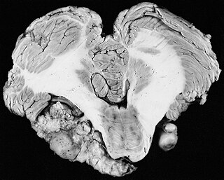
A vestibular schwannoma (VS), also called acoustic neuroma, is a benign tumor that develops on the vestibulocochlear nerve that passes from the inner ear to the brain. The tumor originates when Schwann cells that form the insulating myelin sheath on the nerve malfunction. Normally, Schwann cells function beneficially to protect the nerves which transmit balance and sound information to the brain. However, sometimes a mutation in the tumor suppressor gene, NF2, located on chromosome 22, results in abnormal production of the cell protein named Merlin, and Schwann cells multiply to form a tumor. The tumor originates mostly on the vestibular division of the nerve rather than the cochlear division, but hearing as well as balance will be affected as the tumor enlarges.

Lars Leksell (1907–1986) was a Swedish physician and Professor of Neurosurgery at the Karolinska Institute in Stockholm, Sweden. He was the inventor of radiosurgery.

<span class="mw-page-title-main">Radiosurgery</span> Surgical Specialty

Radiosurgery is surgery using radiation, that is, the destruction of precisely selected areas of tissue using ionizing radiation rather than excision with a blade. Like other forms of radiation therapy, it is usually used to treat cancer. Radiosurgery was originally defined by the Swedish neurosurgeon Lars Leksell as "a single high dose fraction of radiation, stereotactically directed to an intracranial region of interest".

<span class="mw-page-title-main">Stereotactic surgery</span> Medical procedure

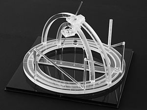
Stereotactic surgery is a minimally invasive form of surgical intervention that makes use of a three-dimensional coordinate system to locate small targets inside the body and to perform on them some action such as ablation, biopsy, lesion, injection, stimulation, implantation, radiosurgery (SRS), etc.

Image-guided surgery (IGS) is any surgical procedure where the surgeon uses tracked surgical instruments in conjunction with preoperative or intraoperative images in order to directly or indirectly guide the procedure. Image guided surgery systems use cameras, ultrasonic, electromagnetic or a combination or fields to capture and relay the patient's anatomy and the surgeon's precise movements in relation to the patient, to computer monitors in the operating room or to augmented reality headsets. This is generally performed in real-time though there may be delays of seconds or minutes depending on the modality and application.

<span class="mw-page-title-main">Neuroimaging</span> Set of techniques to measure and visualize aspects of the nervous system

Neuroimaging is the use of quantitative (computational) techniques to study the structure and function of the central nervous system, developed as an objective way of scientifically studying the healthy human brain in a non-invasive manner. Increasingly it is also being used for quantitative studies of brain disease and psychiatric illness. Neuroimaging is a highly multidisciplinary research field and is not a medical specialty.

<span class="mw-page-title-main">Fiducial marker</span> Reference point inserted in an image

A fiducial marker or fiducial is an object placed in the field of view of an imaging system that appears in the image produced, for use as a point of reference or a measure. It may be either something placed into or on the imaging subject, or a mark or set of marks in the reticle of an optical instrument.

Hypophysectomy is the surgical removal of the hypophysis. It is most commonly performed to treat tumors, especially craniopharyngioma tumors. Sometimes it is used to treat Cushing's syndrome due to pituitary adenoma or Simmond's disease It is also applied in neurosciences to understand the functioning of hypophysis. There are various ways a hypophysectomy can be carried out. These methods include transsphenoidal hypophysectomy, open craniotomy, and stereotactic radiosurgery.

Elekta is a global Swedish company that develops and produces radiation therapy and radiosurgery-related equipment and clinical management for the treatment of cancer and brain disorders. Elekta has a global presence in more than 120 countries, with over 40 offices around the world and about 4,700 employees.

John R. Adler is an American neurosurgeon.

Image-guided radiation therapy is the process of frequent imaging, during a course of radiation treatment, used to direct the treatment, position the patient, and compare to the pre-therapy imaging from the treatment plan. Immediately prior to, or during, a treatment fraction, the patient is localized in the treatment room in the same position as planned from the reference imaging dataset. An example of IGRT would include comparison of a cone beam computed tomography (CBCT) dataset, acquired on the treatment machine, with the computed tomography (CT) dataset from planning. IGRT would also include matching planar kilovoltage (kV) radiographs or megavoltage (MV) images with digital reconstructed radiographs (DRRs) from the planning CT.

Neuronavigation is the set of computer-assisted technologies used by neurosurgeons to guide or "navigate” within the confines of the skull or vertebral column during surgery, and used by psychiatrists to accurately target rTMS. The set of hardware for these purposes is referred to as a neuronavigator.

<span class="mw-page-title-main">Cyberknife (device)</span>

The CyberKnife System is a radiation therapy device manufactured by Accuray Incorporated. The CyberKnife System is the only radiation delivery system in the world that features a linear accelerator (linac) directly mounted on a robot to deliver high-energy x-rays or photons used in radiation therapy and combines real-time artificial intelligence (AI)-driven target tracking and treatment delivery. The platform is designed to deliver precise stereotactic radiosurgery (SRS) and stereotactic body radiation therapy (SBRT) for the treatment of benign tumors, malignant tumors, neurologic disorders and other medical conditions.

<span class="mw-page-title-main">Russell A. Brown</span> American physician and computer scientist

Russell A. Brown, an American physician and computer scientist, is the inventor of the N-localizer technology that enables guidance of stereotactic surgery or radiosurgery using medical images that are obtained via computed tomography (CT), magnetic resonance imaging (MRI), or positron emission tomography (PET).

<span class="mw-page-title-main">Computed tomography of the head</span> Cross-sectional X-rays of the head

Computed tomography of the head uses a series of X-rays in a CT scan of the head taken from many different directions; the resulting data is transformed into a series of cross sections of the brain using a computer program. CT images of the head are used to investigate and diagnose brain injuries and other neurological conditions, as well as other conditions involving the skull or sinuses; it used to guide some brain surgery procedures as well. CT scans expose the person getting them to ionizing radiation which has a risk of eventually causing cancer; some people have allergic reactions to contrast agents that are used in some CT procedures.

<span class="mw-page-title-main">Brain positron emission tomography</span> Form of positron emission tomography

Brain positron emission tomography is a form of positron emission tomography (PET) that is used to measure brain metabolism and the distribution of exogenous radiolabeled chemical agents throughout the brain. PET measures emissions from radioactively labeled metabolically active chemicals that have been injected into the bloodstream. The emission data from brain PET are computer-processed to produce multi-dimensional images of the distribution of the chemicals throughout the brain.

References

  1. Arle, J (2009). "Development of a Classic: The Todd-Wells Apparatus, the BRW, and the CRW Stereotactic Frames". In Lozano, AM; Gildenberg, PL; Tasker, RR (eds.). Textbook of Stereotactic and Functional Neurosurgery. Berlin: Springer-Verlag. pp. 456–460. doi:10.1007/978-3-540-69960-6. ISBN   978-3-540-69959-0. S2CID   58803140.
  2. "System Using Computed Tomography as for Selective Body Treatment". U.S. Patent 4608977. 1986.
  3. Galloway, RL Jr. (2015). "Introduction and Historical Perspectives on Image-Guided Surgery". In Golby, AJ (ed.). Image-Guided Neurosurgery. Amsterdam: Elsevier. pp. 2–4. doi:10.1016/B978-0-12-800870-6.00001-7. ISBN   978-0-12-800870-6.
  4. Thomas DG, Anderson RE, du Boulay GH (1984). "CT-guided stereotactic neurosurgery: experience in 24 cases with a new stereotactic system". Journal of Neurology, Neurosurgery & Psychiatry. 47 (1): 9–16. doi:10.1136/jnnp.47.1.9. PMC   1027634 . PMID   6363629.
  5. Heilbrun MP, Sunderland PM, McDonald PR, Wells TH Jr, Cosman E, Ganz E (1987). "Brown-Roberts-Wells stereotactic frame modifications to accomplish magnetic resonance imaging guidance in three planes". Applied Neurophysiology. 50 (1–6): 143–152. doi:10.1159/000100700. PMID   3329837.
  6. Maciunas RJ, Kessler RM, Maurer C, Mandava V, Watt G, Smith G (1992). "Positron emission tomography imaging-directed stereotactic neurosurgery". Stereotactic and Functional Neurosurgery. 58 (1–4): 134–140. doi:10.1159/000098986. PMID   1439330.
  7. Gildenberg, PL; Krauss, JK (2009). "History of Stereotactic Surgery". In Lozano, AM; Gildenberg, PL; Tasker, RR (eds.). Textbook of Stereotactic and Functional Neurosurgery. Berlin: Springer-Verlag. p. 23. doi:10.1007/978-3-540-69960-6. ISBN   978-3-540-69959-0. S2CID   58803140.
  8. Heilbrun MP, Roberts TS, Apuzzo ML, Wells TH, Sabshin JK (1983). "Preliminary experience with Brown-Roberts-Wells (BRW) computerized tomography stereotaxic guidance system". Journal of Neurosurgery. 59 (2): 217–222. doi:10.3171/jns.1983.59.2.0217. PMID   6345727.
  9. Goerss S, Kelly PJ, Kall B, Alker GJ Jr (1982). "A computed tomography stereotactic adaptation system". Neurosurgery. 10 (3): 375–379. doi:10.1227/00006123-198203000-00014. PMID   7041006.
  10. Leksell L, Leksell D, Schwebel J (1985). "Stereotaxis and nuclear magnetic resonance". Journal of Neurology, Neurosurgery & Psychiatry. 48 (1): 14–18. doi:10.1136/jnnp.48.1.14. PMC   1028176 . PMID   3882889.
  11. Couldwell WT, Apuzzo ML (1990). "Initial experience related to the Cosman-Roberts-Wells stereotactic instrument. Technical note". Journal of Neurosurgery. 72 (1): 145–8. doi:10.3171/jns.1990.72.1.0145. PMID   2403588. S2CID   1363168.
  12. Sedrak M, Alaminos-Bouza AL, Srivastava S (2020). "Coordinate systems for navigating stereotactic space: how not to get lost". Cureus. 12 (6): e8578. doi: 10.7759/cureus.8578 . PMC   7358954 . PMID   32670714.
  13. Tse, VCK; Kalani, MYS; Adler, JR (2015). "Techniques of Stereotactic Localization". In Chin, LS; Regine, WF (eds.). Principles and Practice of Stereotactic Radiosurgery. New York: Springer. pp. 25–32. doi:10.1007/978-1-4614-8363-2. ISBN   978-1-4614-8362-5.
  14. Sturm V, Pastyr O, Schlegel W, Scharfenberg H, Zabel HJ, Netzeband G, Schabbert S, Berberich W (1983). "Stereotactic computer tomography with a modified Riechert-Mundinger device as the basis for integrated stereotactic neuroradiological investigations". Acta Neurochirurgica. 68 (1–2): 11–17. doi:10.1007/BF01406197. PMID   6344559. S2CID   38864553.
  15. Krauss, JK (2009). "The Riechert/Mundinger Stereotactic Apparatus". In Lozano, AM; Gildenberg, PL; Tasker, RR (eds.). Textbook of Stereotactic and Functional Neurosurgery. Berlin: Springer-Verlag. pp. 487–493. doi:10.1007/978-3-540-69960-6. ISBN   978-3-540-69959-0. S2CID   58803140.
  16. Alaminos-Bouza AL, Brown RA (2020). "Comparative accuracies of the N-localizer and Sturm-Pastyr localizer in the presence of image noise". Cureus. 12 (7): e9137. doi: 10.7759/cureus.9137 . PMC   7364427 . PMID   32685325.
  17. Weaver K, Smith V, Lewis JD, Lulu B, Barnett CM, Leibel SA, Gutin P, Larson D, Phillips T (1990). "A CT-based computerized treatment planning system for I-125 stereotactic brain implants". International Journal of Radiation Oncology, Biology, Physics. 18 (2): 445–454. doi:10.1016/0360-3016(90)90114-Y. PMID   2406230.
  18. Dai J, Zhu Y, Qu H, Hu Y (2001). "An algorithm for stereotactic localization by computed tomography or magnetic resonance imaging". Physics in Medicine and Biology. 46 (1): N1–N7. doi:10.1088/0031-9155/46/1/401. PMID   11197682. S2CID   9196917.
  19. Sedrak M, Alaminos-Bouza AL, Bruna A, Brown RA (2021). "Monte Carlo simulation of errors for N-localizer systems in stereotactic neurosurgery: novel proposals for improvements". Cureus. 13 (2): e13393. doi: 10.7759/cureus.13393 . PMC   7977485 . PMID   33758694.
  20. Alaminos-Bouza AL, Brown RA (2021). "Improved accuracy for the Sturm-Pastyr localizer in the presence of image noise". Cureus. 13 (9): e17905. doi: 10.7759/cureus.17905 . PMC   8509111 . PMID   34660100.

Further reading